 Биология
Похожие презентации:


Human embryology. Progenesis. The initial period of embryonic development
1.
Practical lesson №2Human embryology. Progenesis. The
initial period of embryonic
development.
2.
MOTIVATIVE CHARACTERISTIC OF THE TOPIC• Knowledge of the features of the formation and structure
of male germ cells (progenesis) is a necessary part of the
study of the main stages of human embryonic
development. This creates the prerequisites for
understanding the patterns of the initial stages of
embryogenesis - fertilization, forms the basis for ideas
about some of the mechanisms and forms of development
of male infertility.
3.
PURPOSE OF THE LESSON1. Get an idea about progenesis - the formation of male and
female germ cells (spermatogenesis, ovogenesis).
2. Know the structure of the human sperm and egg, the
classification of eggs.
3. Understand the main stages of embryogenesis.
4. Know: the stages and processes of fertilization, cleavage in
humans, the structure of the human blastula.
5. Be able to identify on diagrams, preparations and
micrographs of the structure of germ cells and embryos in
the initial period of embryonic development
4.
NECESSARY BASIC LEVEL OF KNOWLEDGE• The structure of a eukaryotic cell.
•Meiosis.
•The concept of diploid and haploid genetic set of
chromosomes.
•The main stages of embryogenesis in human
embryonic development.
5.
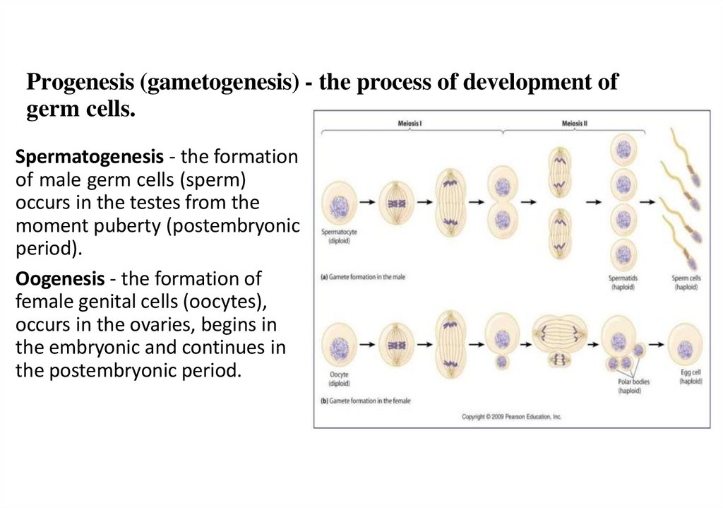
Progenesis (gametogenesis) - the process of development ofgerm cells.
Spermatogenesis - the formation
of male germ cells (sperm)
occurs in the testes from the
moment puberty (postembryonic
period).
Oogenesis - the formation of
female genital cells (oocytes),
occurs in the ovaries, begins in
the embryonic and continues in
the postembryonic period.
6.
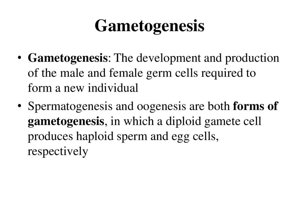
Gametogenesis• Gametogenesis: The development and production
of the male and female germ cells required to
form a new individual
• Spermatogenesis and oogenesis are both forms of
gametogenesis, in which a diploid gamete cell
produces haploid sperm and egg cells,
respectively
7.
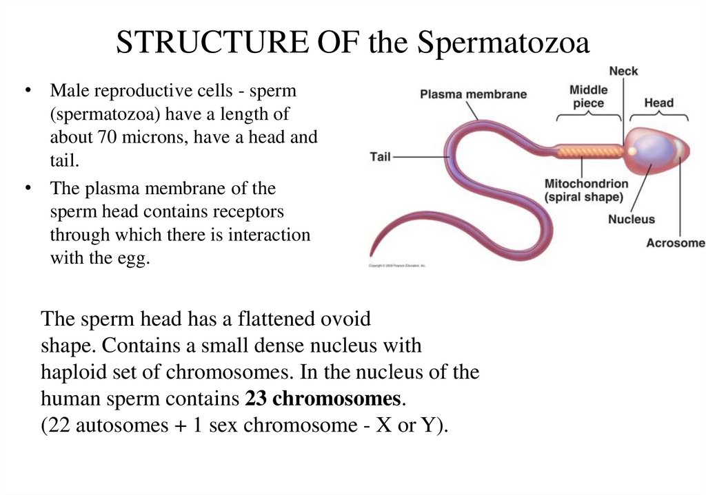
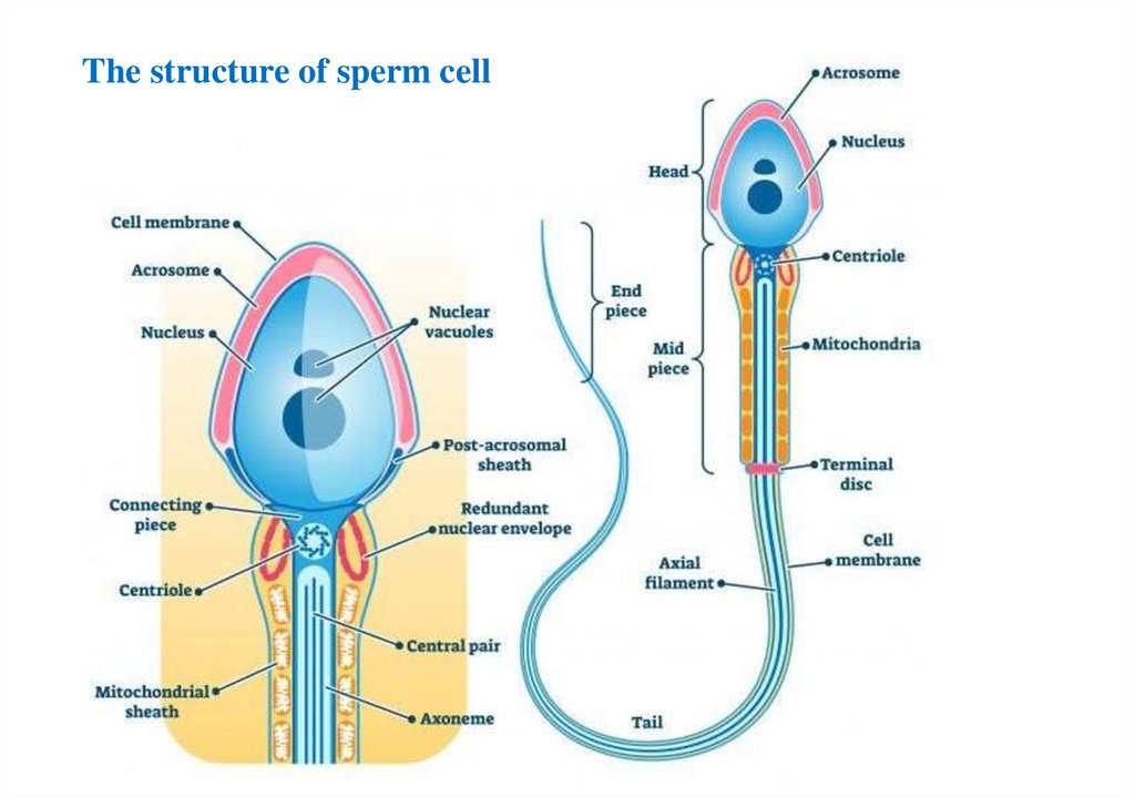
STRUCTURE OF the Spermatozoa• Male reproductive cells - sperm
(spermatozoa) have a length of
about 70 microns, have a head and
tail.
• The plasma membrane of the
sperm head contains receptors
through which there is interaction
with the egg.
The sperm head has a flattened ovoid
shape. Contains a small dense nucleus with
haploid set of chromosomes. In the nucleus of the
human sperm contains 23 chromosomes.
(22 autosomes + 1 sex chromosome - X or Y).
8.
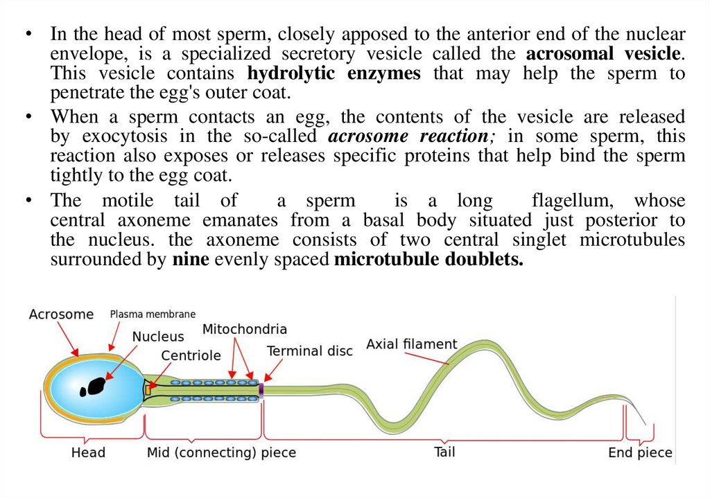
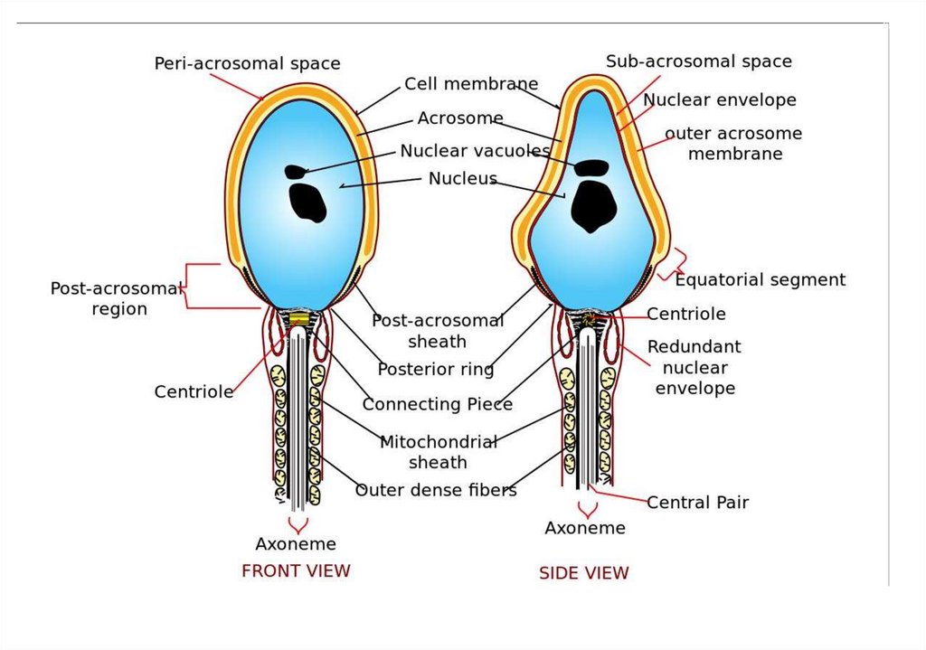
• In the head of most sperm, closely apposed to the anterior end of the nuclearenvelope, is a specialized secretory vesicle called the acrosomal vesicle.
This vesicle contains hydrolytic enzymes that may help the sperm to
penetrate the egg's outer coat.
• When a sperm contacts an egg, the contents of the vesicle are released
by exocytosis in the so-called acrosome reaction; in some sperm, this
reaction also exposes or releases specific proteins that help bind the sperm
tightly to the egg coat.
• The motile tail of
a sperm
is a long
flagellum, whose
central axoneme emanates from a basal body situated just posterior to
the nucleus. the axoneme consists of two central singlet microtubules
surrounded by nine evenly spaced microtubule doublets.
9.
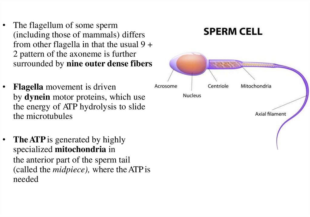
• The flagellum of some sperm(including those of mammals) differs
from other flagella in that the usual 9 +
2 pattern of the axoneme is further
surrounded by nine outer dense fibers
• Flagella movement is driven
by dynein motor proteins, which use
the energy of ATP hydrolysis to slide
the microtubules
• The ATP is generated by highly
specialized mitochondria in
the anterior part of the sperm tail
(called the midpiece), where the ATP is
needed
10.
11.
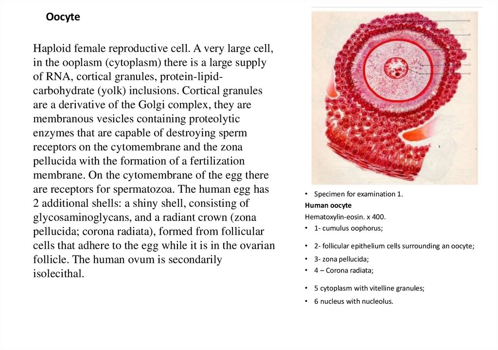
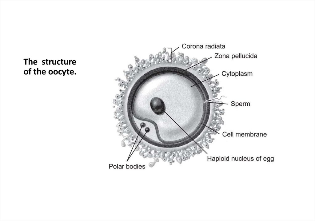
OocyteHaploid female reproductive cell. A very large cell,
in the ooplasm (cytoplasm) there is a large supply
of RNA, cortical granules, protein-lipidcarbohydrate (yolk) inclusions. Cortical granules
are a derivative of the Golgi complex, they are
membranous vesicles containing proteolytic
enzymes that are capable of destroying sperm
receptors on the cytomembrane and the zona
pellucida with the formation of a fertilization
membrane. On the cytomembrane of the egg there
are receptors for spermatozoa. The human egg has
2 additional shells: a shiny shell, consisting of
glycosaminoglycans, and a radiant crown (zona
pellucida; corona radiata), formed from follicular
cells that adhere to the egg while it is in the ovarian
follicle. The human ovum is secondarily
isolecithal.
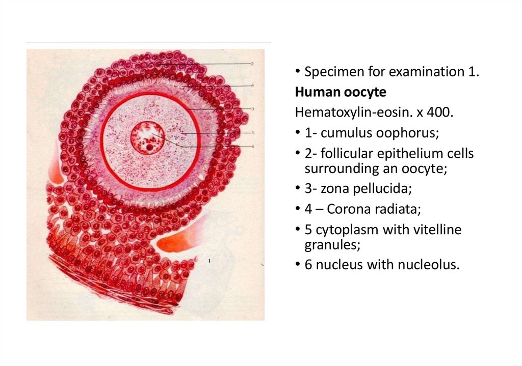
• Specimen for examination 1.
Human oocyte
Hematoxylin-eosin. x 400.
• 1- cumulus oophorus;
• 2- follicular epithelium cells surrounding an oocyte;
• 3- zona pellucida;
• 4 – Corona radiata;
• 5 cytoplasm with vitelline granules;
• 6 nucleus with nucleolus.
12.
CLASSIFICATIONS of oocytesby the number of yolk inclusions:
alecithal - almost no yolk inclusions (helminths) oligolecithal - few yolk inclusions
a) primary low-yolk oocytes (lanceolate)
b) b) secondarily * low-yolk eggs (mammals) polylecithal - many yolk inclusions (fish,
amphibians, oviparous)
according to the distribution of yolk inclusions in the cytoplasm of the oocytes
(ooplasm):
isolecithal - yolk inclusions are evenly distributed (mammals)
centrolecithal - yolk inclusions are concentrated in the center
mesolecithal - yolk inclusions occupy about half of the cell
telolecithal - yolk inclusions occupy almost the entire cell, and the organelles and the
nucleus are pushed to one pole
HUMANS HAVE OLIGOLECYTAL, SECONDARY ISOLECYTAL OOCYTES
13.
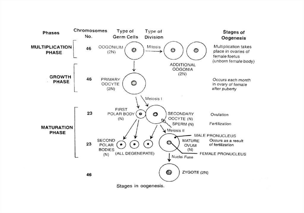
Oogenesis - the formation ofoocytes
• Female germ cells - oocytes - are formed in female reproductive
glands (ovaries). This process is cyclical, during the sexual cycle (24-28
days), as a rule, 1 egg matures.
• Oogenesis takes place in 3 stages:
- multiplication;
- growth;
- maturation.
14.
• The first stage is the multiplication period is carried out during theperiod of intrauterine oogonia - development, and in some species of
mammals and in the first months of postnatal life, when the division
of oogonia and the formation of primary follicles occurs in the ovary
of the embryo.
• The multiplication period ends when meiosis starts, - the beginning of
differentiation into the primary oocyte.
• Meiotic division stops at prophase, and at this stage the cells are
retained until the period of puberty.
15.
• The second stage - the growth period - takes place in a functioningmature ovary (after the puberty) and consists in the transformation of
the primary oocyte of the primary follicle into the primary oocyte in
the mature follicle. In the nucleus of a growing oocyte, conjugation of
chromosomes and the formation of tetrads occur, and yolk inclusions
accumulate in their cytoplasm.
16.
• The third (last) stage - the period of maturation - begins with the formation of asecondary oocyte and ends with its release from the ovary as a result of ovulation.
The maturation period, as during spermatogenesis, includes two divisions, moreover,
the second follows the first without interkinesis, which leads to a decrease
(reduction) in the number of chromosomes by half, and their set becomes haploid. At
the first division of maturation, the primary oocyte is divided into as a result, a
secondary oocyte and a small reduction body are formed. The secondary oocyte
receives almost the entire mass of the accumulated yolk and therefore it remains as
large in volume as the primary oocyte.
• The reduction body is a small cell with a small amount of cytoplasm receiving one
dyad of chromosomes from each tetrad of the primary oocyte nucleus. During the
second division of maturation, as a result of the division of the secondary oocyte, one
egg and a second reduction body are formed. The first reduction body is sometimes
also divided into two identical small cells. As a result of these transformations of
theprimary oocyte one ovum and two or three reduction (polar) bodies are formed of
this order.
• The stage of formation - unlike spermatogenesis, is absent in oogenesis.
17.
18.
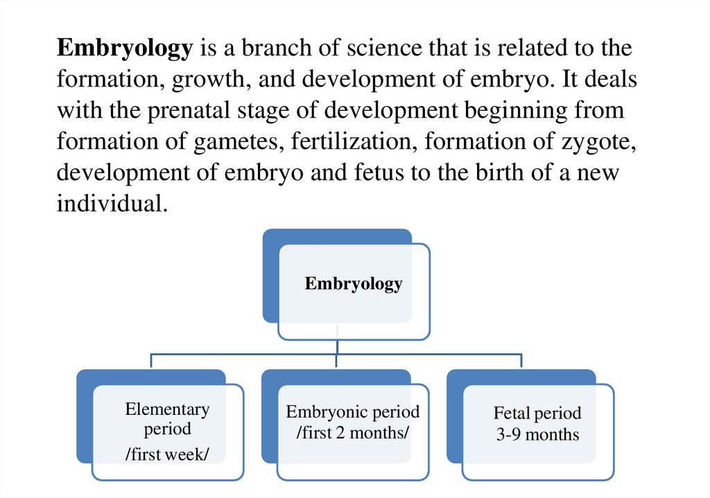
Embryology is a branch of science that is related to theformation, growth, and development of embryo. It deals
with the prenatal stage of development beginning from
formation of gametes, fertilization, formation of zygote,
development of embryo and fetus to the birth of a new
individual.
Embryology
Elementary
period
/first week/
Embryonic period
/first 2 months/
Fetal period
3-9 months
19.
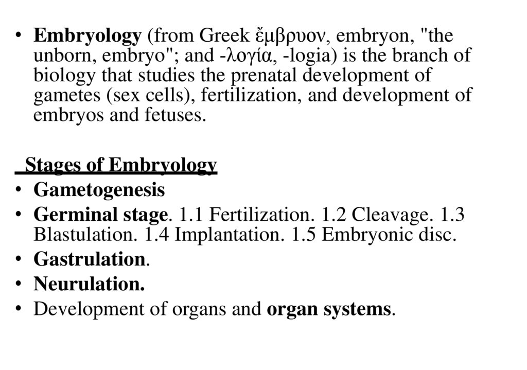
• Embryology (from Greek ἔμβρυον, embryon, "theunborn, embryo"; and -λογία, -logia) is the branch of
biology that studies the prenatal development of
gametes (sex cells), fertilization, and development of
embryos and fetuses.
Stages of Embryology
• Gametogenesis
• Germinal stage. 1.1 Fertilization. 1.2 Cleavage. 1.3
Blastulation. 1.4 Implantation. 1.5 Embryonic disc.
• Gastrulation.
• Neurulation.
• Development of organs and organ systems.
20.
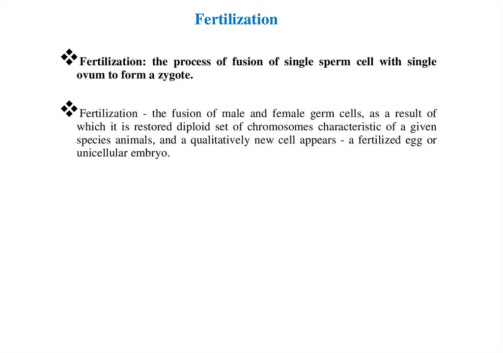
FertilizationFertilization: the process of fusion of single sperm cell with single
ovum to form a zygote.
Fertilization - the fusion of male and female germ cells, as a result of
which it is restored diploid set of chromosomes characteristic of a given
species animals, and a qualitatively new cell appears - a fertilized egg or
unicellular embryo.
21.
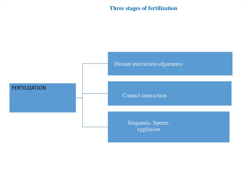
FertilizationThree stages of fertilization
Contact
interaction
Distant interaction
of gametes
Singamia- Sperm-egg fusion
Distant interaction ofgametes
FERTILIZATION
Contact interaction
Singamia- Spermeggfusion
22.
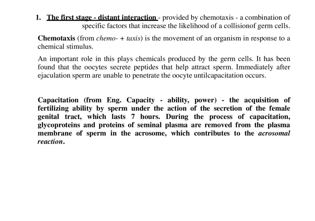
1. The first stage - distant interaction - provided by chemotaxis - a combination ofspecific factors that increase the likelihood of a collisionof germ cells.
Chemotaxis (from chemo- + taxis) is the movement of an organism in response to a
chemical stimulus.
An important role in this plays chemicals produced by the germ cells. It has been
found that the oocytes secrete peptides that help attract sperm. Immediately after
ejaculation sperm are unable to penetrate the oocyte untilcapacitation occurs.
Capacitation (from Eng. Capacity - ability, power) - the acquisition of
fertilizing ability by sperm under the action of the secretion of the female
genital tract, which lasts 7 hours. During the process of capacitation,
glycoproteins and proteins of seminal plasma are removed from the plasma
membrane of sperm in the acrosome, which contributes to the acrosomal
reaction.
23.
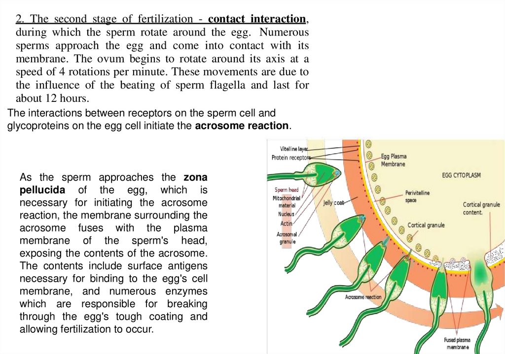
2. The second stage of fertilization - contact interaction,during which the sperm rotate around the egg. Numerous
sperms approach the egg and come into contact with its
membrane. The ovum begins to rotate around its axis at a
speed of 4 rotations per minute. These movements are due to
the influence of the beating of sperm flagella and last for
about 12 hours.
The interactions between receptors on the sperm cell and
glycoproteins on the egg cell initiate the acrosome reaction.
As the sperm approaches the zona
pellucida of the egg, which is
necessary for initiating the acrosome
reaction, the membrane surrounding the
acrosome fuses with the plasma
membrane of the sperm's head,
exposing the contents of the acrosome.
The contents include surface antigens
necessary for binding to the egg's cell
membrane, and numerous enzymes
which are responsible for breaking
through the egg's tough coating and
allowing fertilization to occur.
24.
1. Third stage- Sperm-egg fusion -The head and the intermediate part of the tailsection penetrate into the ooplasm. After the entry of the sperm into the
oocyte at the periphery of the ooplasm, it is compacted (zone reaction) and a
fertilization membrane is formed.
The cortical reaction is a process initiated during fertilization by the release
of cortical granules from the egg, which prevents polyspermy, the fusion of
multiple sperm with one egg. In contrast to the fast block of
polyspermy which immediately but temporarily blocks additional sperm from
fertilizing the egg, the cortical reaction gradually establishes a permanent
barrier to sperm entry and functions as the main part of the slow block of
polyspermy in many animals.
25.
CleavageAfter fertilization successfully activates the egg, the egg begins a series of rapid
cell divisions called cleavage. “Typical” cell division occurs every 18-24 hours, but
cleavage cell divisions can occur as frequently as every 10 minutes. During
cleavage, the cells divide without an increase in size (without growing); so the large
single-cell zygote divides into smaller cells called blastomeres.
26.
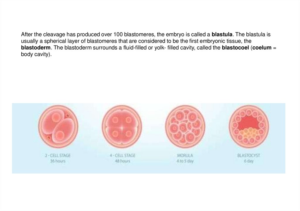
After the cleavage has produced over 100 blastomeres, the embryo is called a blastula. The blastula isusually a spherical layer of blastomeres that are considered to be the first embryonic tissue, the
blastoderm. The blastoderm surrounds a fluid-filled or yolk- filled cavity, called the blastocoel (coelum =
body cavity).
27.
Due to the actual absence of the G1 period, during which the growth of cellsformed as a result of division occurs, the cells are much smaller than the
maternal, therefore, the size of the embryo as a whole during this period,
regardless of the number of its constituent cells, does not exceed the size of
the original cell - the zygote. All this made it possible to call the described
process crushing (i.e., crushing), and the cells formed during the cleavage
process - blastomeres
28.
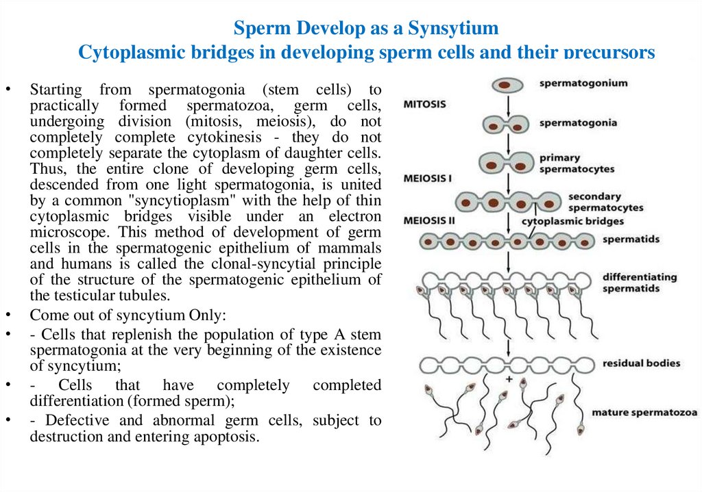
Sperm Develop as a SynsytiumCytoplasmic bridges in developing sperm cells and their precursors
Starting from spermatogonia (stem cells) to
practically formed spermatozoa, germ cells,
undergoing division (mitosis, meiosis), do not
completely complete cytokinesis - they do not
completely separate the cytoplasm of daughter cells.
Thus, the entire clone of developing germ cells,
descended from one light spermatogonia, is united
by a common "syncytioplasm" with the help of thin
cytoplasmic bridges visible under an electron
microscope. This method of development of germ
cells in the spermatogenic epithelium of mammals
and humans is called the clonal-syncytial principle
of the structure of the spermatogenic epithelium of
the testicular tubules.
Come out of syncytium Only:
- Cells that replenish the population of type A stem
spermatogonia at the very beginning of the existence
of syncytium;
- Cells that have completely completed
differentiation (formed sperm);
- Defective and abnormal germ cells, subject to
destruction and entering apoptosis.
29.
Diagnostics of histological slides30.
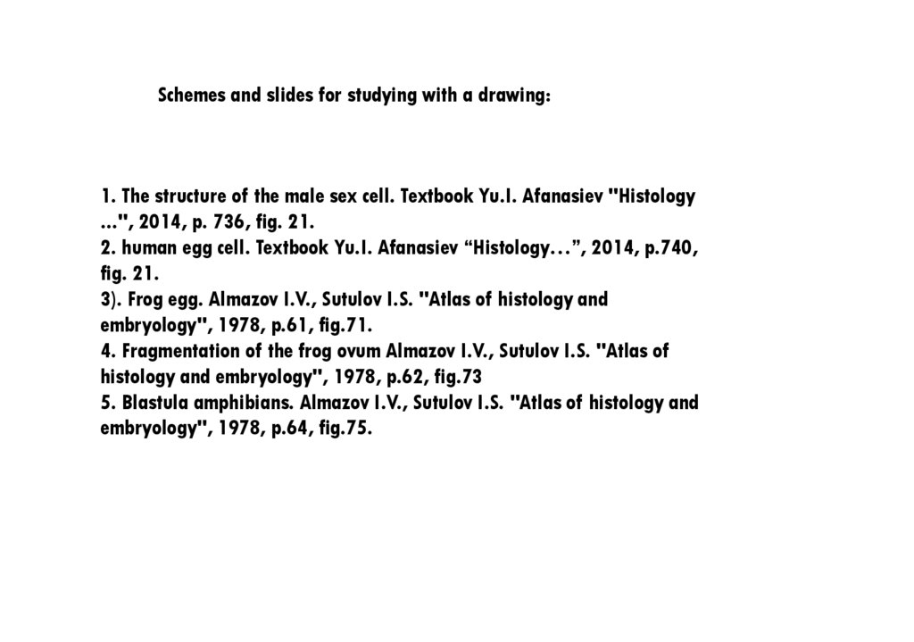
Schemes and slides for studying with a drawing:1. The structure of the male sex cell. Textbook Yu.I. Afanasiev "Histology
...", 2014, p. 736, fig. 21.
2. human egg cell. Textbook Yu.I. Afanasiev “Histology…”, 2014, p.740,
fig. 21.
3). Frog egg. Almazov I.V., Sutulov I.S. "Atlas of histology and
embryology", 1978, p.61, fig.71.
4. Fragmentation of the frog ovum Almazov I.V., Sutulov I.S. "Atlas of
histology and embryology", 1978, p.62, fig.73
5. Blastula amphibians. Almazov I.V., Sutulov I.S. "Atlas of histology and
embryology", 1978, p.64, fig.75.
31.
The structure of sperm cell32.
The structureof the oocyte.
33.
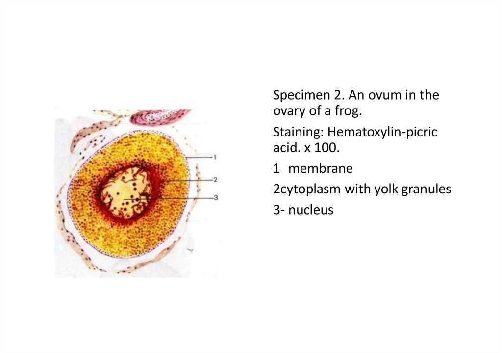
Specimen 2. An ovum in theovary of a frog.
Staining: Hematoxylin-picric
acid. x 100.
1 membrane
2cytoplasm with yolk granules
3- nucleus
34.
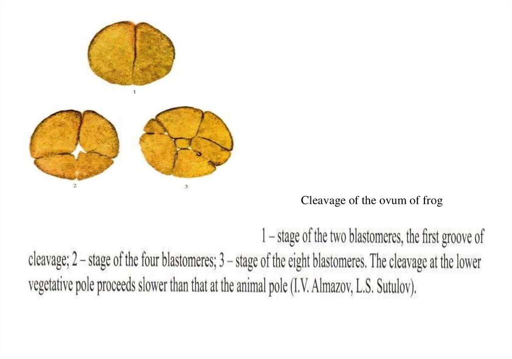
Cleavage of the ovum of frog35.
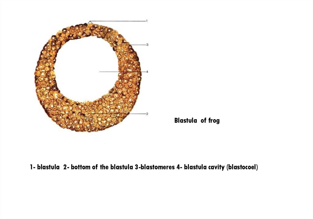
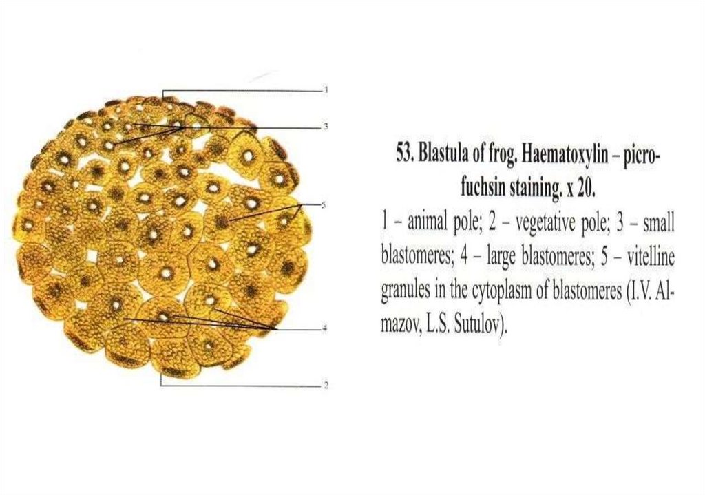
Blastula of frog1- blastula 2- bottom of the blastula 3-blastomeres 4- blastula cavity (blastocoel)
36.
37.
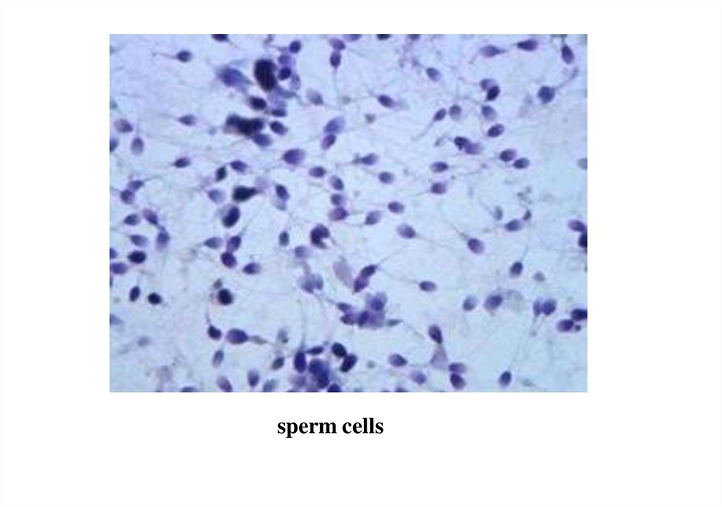
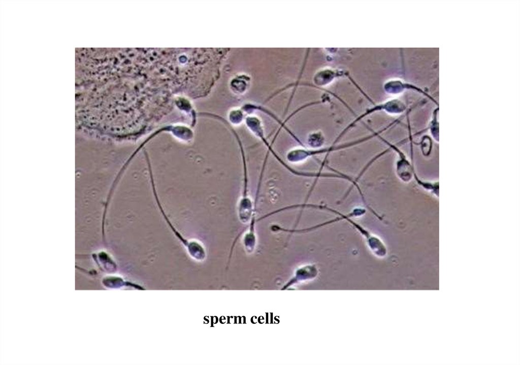
Diagnostics of histological slides38.
sperm cells39.
sperm cells40.
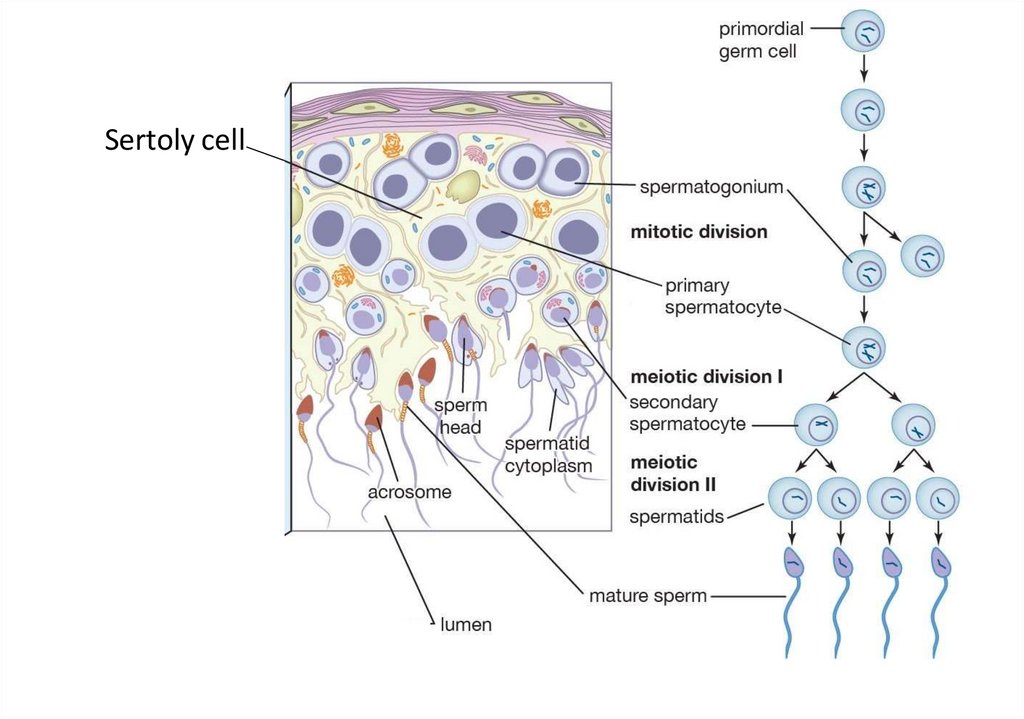
Sertoly cell41.
42.
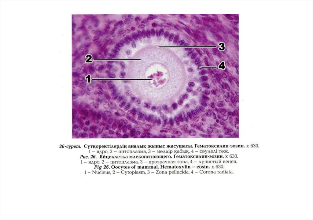
• Specimen for examination 1.Human oocyte
Hematoxylin-eosin. x 400.
• 1- cumulus oophorus;
• 2- follicular epithelium cells
surrounding an oocyte;
• 3- zona pellucida;
• 4 – Corona radiata;
• 5 cytoplasm with vitelline
granules;
• 6 nucleus with nucleolus.
43.
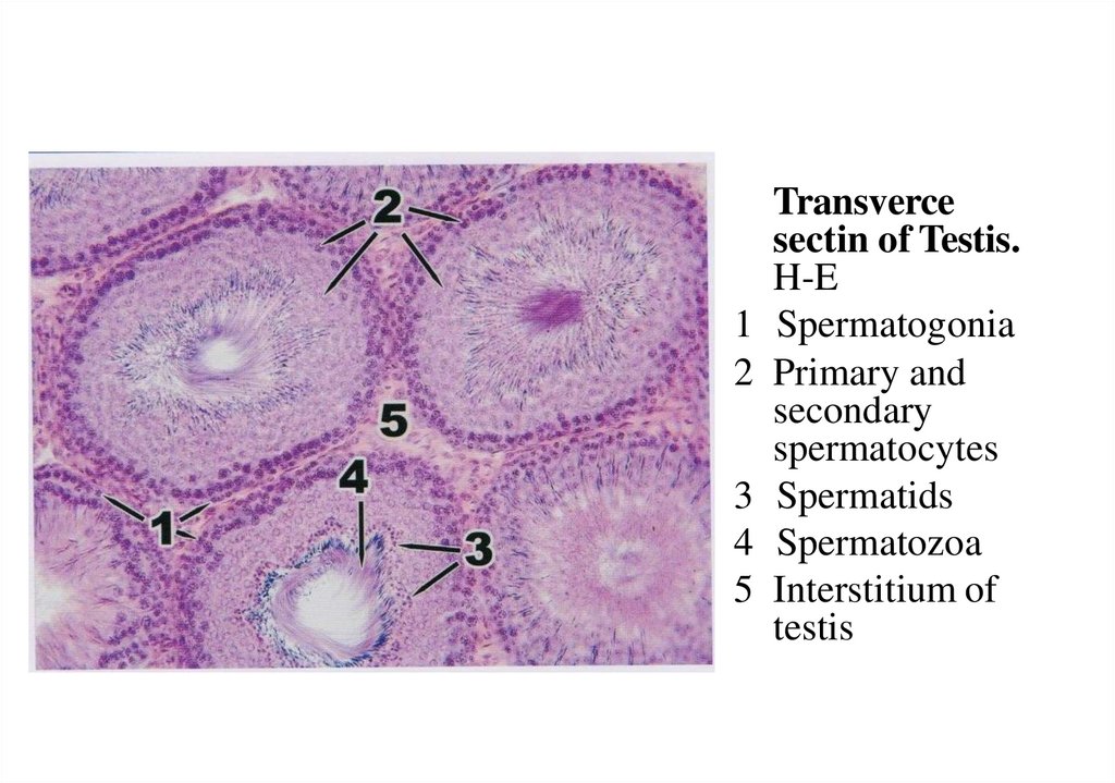
Transvercesectin of Testis.
H-E
1 Spermatogonia
2 Primary and
secondary
spermatocytes
3 Spermatids
4 Spermatozoa
5 Interstitium of
testis
44.
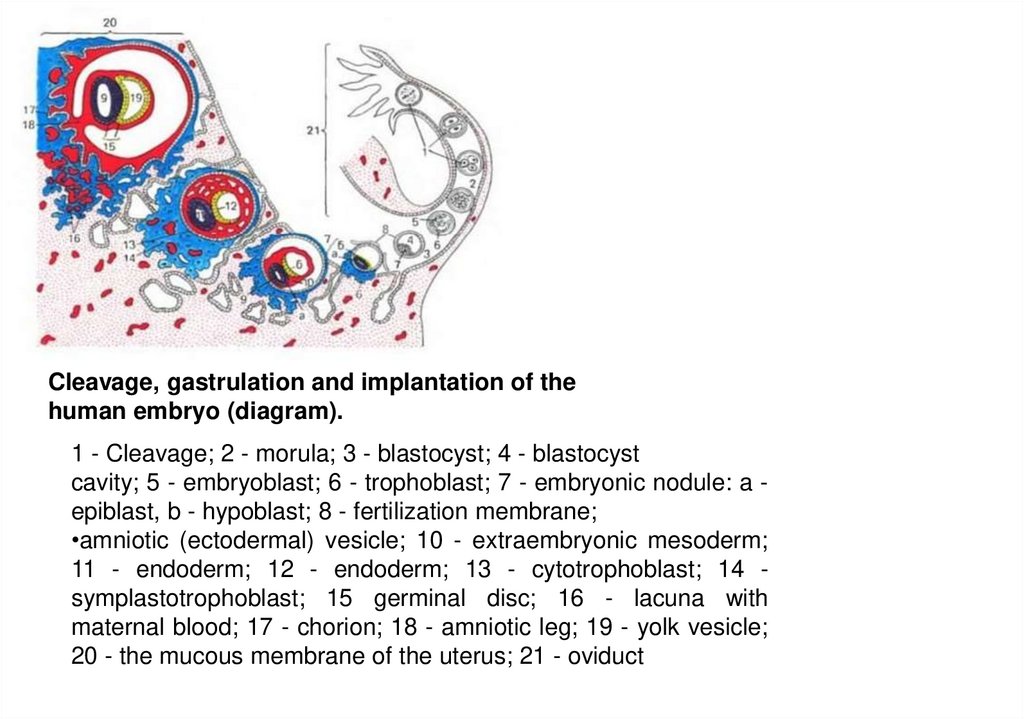
Cleavage, gastrulation and implantation of thehuman embryo (diagram).
1 - Cleavage; 2 - morula; 3 - blastocyst; 4 - blastocyst
cavity; 5 - embryoblast; 6 - trophoblast; 7 - embryonic nodule: a epiblast, b - hypoblast; 8 - fertilization membrane;
•amniotic (ectodermal) vesicle; 10 - extraembryonic mesoderm;
11 - endoderm; 12 - endoderm; 13 - cytotrophoblast; 14 symplastotrophoblast; 15 germinal disc; 16 - lacuna with
maternal blood; 17 - chorion; 18 - amniotic leg; 19 - yolk vesicle;
20 - the mucous membrane of the uterus; 21 - oviduct
45.
46.
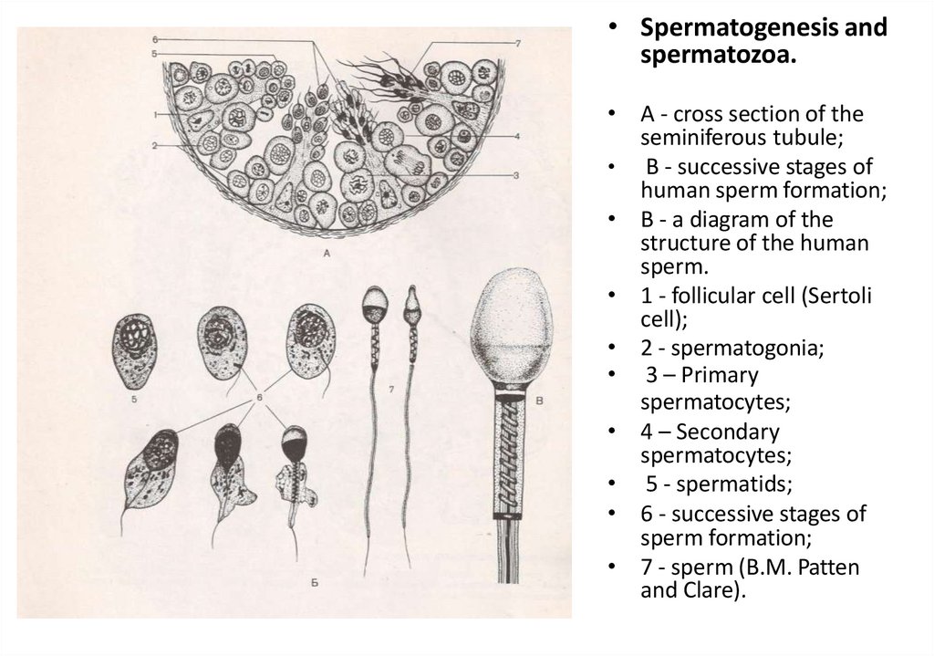
• Spermatogenesis andspermatozoa.
• A - cross section of the
seminiferous tubule;
• B - successive stages of
human sperm formation;
• B - a diagram of the
structure of the human
sperm.
• 1 - follicular cell (Sertoli
cell);
• 2 - spermatogonia;
• 3 – Primary
spermatocytes;
• 4 – Secondary
spermatocytes;
• 5 - spermatids;
• 6 - successive stages of
sperm formation;
• 7 - sperm (B.M. Patten
and Clare).
47.
Control questions1.
The concept of embryology as a science. The main stages of human embryonic
development.
2.
Progenesis. Some features of the formation of spermatozoa and eggs.
3.
The structure of the human sperm.
4.
Oocyte classification. Place the human ovum in the classifications. The structure of
the human egg.
5.
Fertilization in humans - localization, stages and processes.
6.
Characteristics of zygote cleavage. Features of crushing in humans, localization of
the embryo at the time of crushing.
7.
Features of the structure of the morula and blastula of mammals.
48.
Task №1• What is the name of the period of
spermatogenesis, in which mitotic reproduction of
spermatogonia occurs?
49.
Task 2• As a result of a genetic abnormality in a man, the
formation of sperm acrosome is impaired. Explain
why such a man is infertile?
50.
Task № 3• At what stage of spermatogenesis does crossing
over occur - an exchange of homologous regions
between the paternal and maternal chromosomes?