


 Биология
Похожие презентации:


Animal Development
1. Chapter 47
Animal DevelopmentPowerPoint® Lecture Presentations for
Biology
Eighth Edition
Neil Campbell and Jane Reece
Lectures by Chris Romero, updated by Erin Barley with contributions from Joan Sharp
Copyright © 2008 Pearson Education, Inc., publishing as Pearson Benjamin Cummings
2. Overview: A Body-Building Plan
• It is difficult to imagine that each of us beganlife as a single cell (fertilized egg) called a
zygote.
• A human embryo at about 6–8 weeks after
conception shows development of distinctive
features.
Copyright © 2008 Pearson Education, Inc., publishing as Pearson Benjamin Cummings
3.
1 mm4.
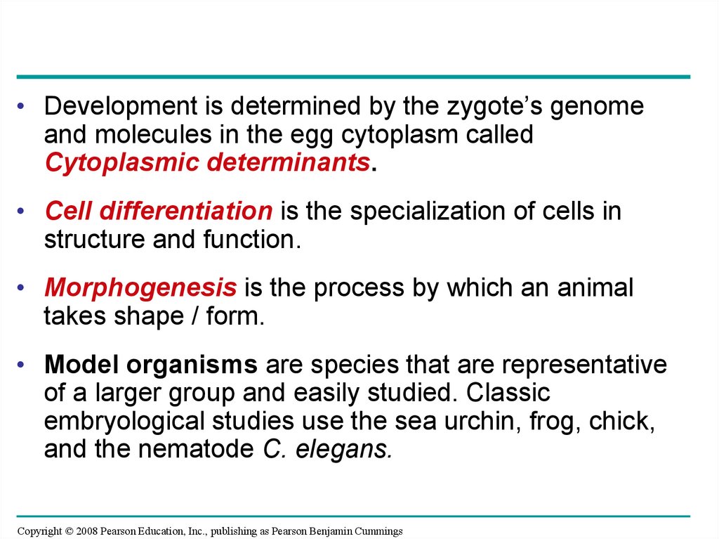
• Development is determined by the zygote’s genomeand molecules in the egg cytoplasm called
Cytoplasmic determinants.
• Cell differentiation is the specialization of cells in
structure and function.
• Morphogenesis is the process by which an animal
takes shape / form.
• Model organisms are species that are representative
of a larger group and easily studied. Classic
embryological studies use the sea urchin, frog, chick,
and the nematode C. elegans.
Copyright © 2008 Pearson Education, Inc., publishing as Pearson Benjamin Cummings
5. After fertilization, embryonic development proceeds through cleavage, gastrulation, and organogenesis
• Important events regulating development occurduring fertilization and the three stages that
build the animal’s body
– Cleavage: cell division creates a hollow ball of
cells called a blastula
– Gastrulation: cells are rearranged into a
three-layered gastrula
– Organogenesis: the three germ layers interact
and move to give rise to organs.
Copyright © 2008 Pearson Education, Inc., publishing as Pearson Benjamin Cummings
6. Fertilization: sperm + egg = zygote n + n = 2n
• Fertilization brings the haploid nuclei of spermand egg together, forming a diploid zygote.
• The sperm’s contact with the egg’s surface
initiates metabolic reactions in the egg that
trigger the onset of embryonic development:
• Acrosomal Reaction
• Cortical Reaction
Copyright © 2008 Pearson Education, Inc., publishing as Pearson Benjamin Cummings
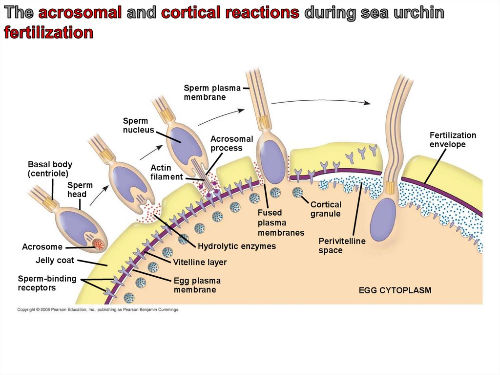
7. The Acrosomal Reaction
• The acrosomal reaction is triggered when thesperm meets the egg.
• The acrosome at the tip of the sperm releases
hydrolytic enzymes that digest material
surrounding the egg.
• Gamete contact and/or fusion depolarizes the
egg cell membrane and sets up a fast block to
polyspermy.
Copyright © 2008 Pearson Education, Inc., publishing as Pearson Benjamin Cummings
8.
Sperm plasmamembrane
Sperm
nucleus
Fertilization
envelope
Acrosomal
process
Basal body
(centriole)
Sperm
head
Acrosome
Jelly coat
Sperm-binding
receptors
Actin
filament
Cortical
Fused
granule
plasma
membranes
Perivitelline
Hydrolytic enzymes
space
Vitelline layer
Egg plasma
membrane
EGG CYTOPLASM
9. The Cortical Reaction
• Fusion of egg and sperm also initiates thecortical reaction:
• This reaction induces a rise in Ca2+ that
stimulates cortical granules to release their
contents outside the egg.
• These changes cause formation of a
fertilization envelope that functions as a slow
block to polyspermy.
Copyright © 2008 Pearson Education, Inc., publishing as Pearson Benjamin Cummings
10. Activation of the Egg
• The sharp rise in Ca2+ in the egg’s cytosolincreases the rates of cellular respiration and
protein synthesis by the egg cell.
• With these rapid changes in metabolism, the
egg is said to be activated.
• The sperm nucleus merges with the egg
nucleus and cell division begins.
Copyright © 2008 Pearson Education, Inc., publishing as Pearson Benjamin Cummings
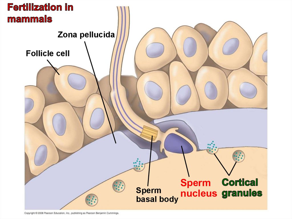
11. Fertilization in Mammals
• Fertilization in mammals and other terrestrial animalsis internal.
• In mammalian fertilization, the cortical reaction
modifies the zona pellucida, the extracellular matrix
of the egg, as a slow block to polyspermy.
• In mammals the first cell division occurs 12–36 hours
after sperm binding.
• The diploid nucleus forms after this first division of the
zygote.
Copyright © 2008 Pearson Education, Inc., publishing as Pearson Benjamin Cummings
12.
Zona pellucidaFollicle cell
Sperm
basal body
Sperm
nucleus
13. Cleavage = Rapid Mitosis / No Mass change
• Fertilization is followed by cleavage, a periodof rapid cell division without growth.
• Cleavage partitions the cytoplasm of one large
cell into many smaller cells called
blastomeres.
• The blastula is a ball of cells with a fluid-filled
cavity called a blastocoel.
Copyright © 2008 Pearson Education, Inc., publishing as Pearson Benjamin Cummings
14.
(a) Fertilized egg(b) Four-cell stage
(c) Early blastula
(d) Later blastula
15.
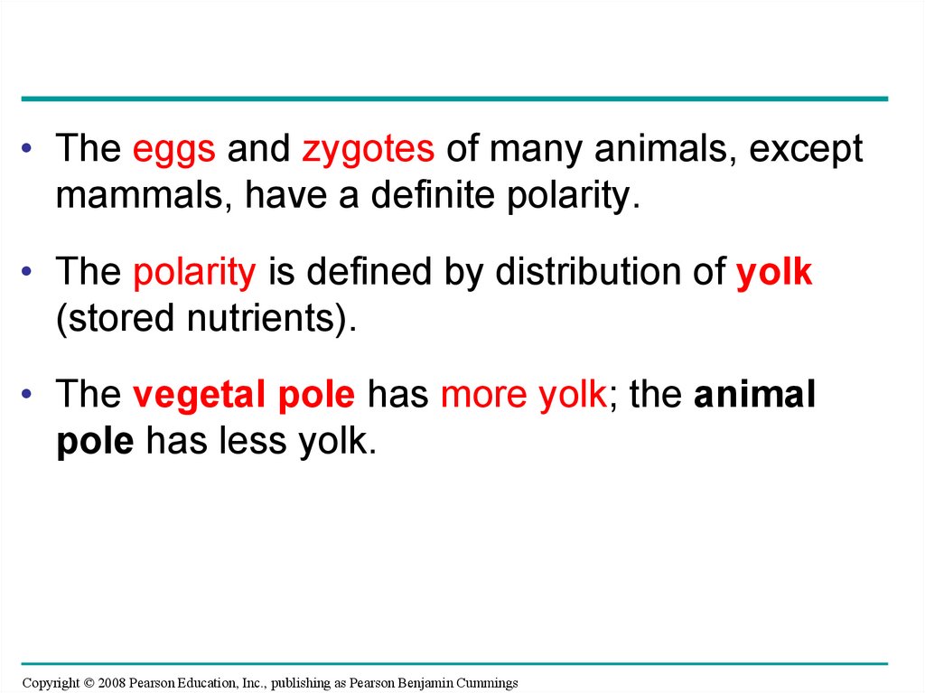
• The eggs and zygotes of many animals, exceptmammals, have a definite polarity.
• The polarity is defined by distribution of yolk
(stored nutrients).
• The vegetal pole has more yolk; the animal
pole has less yolk.
Copyright © 2008 Pearson Education, Inc., publishing as Pearson Benjamin Cummings
16.
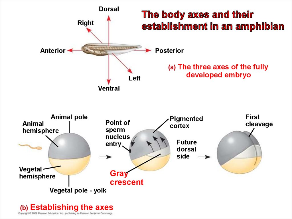
• The three body axes are established by theegg’s polarity and by a cortical rotation
following binding of the sperm.
• Cortical rotation exposes a gray crescent
opposite to the point of sperm entry.
Copyright © 2008 Pearson Education, Inc., publishing as Pearson Benjamin Cummings
17.
DorsalRight
Anterior
Posterior
(a) The three axes of the fully
Left
developed embryo
Ventral
Animal pole
Animal
hemisphere
Point of
sperm
nucleus
entry
Vegetal
hemisphere
Gray
crescent
Vegetal pole - yolk
(b) Establishing
the axes
Pigmented
cortex
Future
dorsal
side
First
cleavage
18.
• Cleavage planes usually follow a pattern that isrelative to the zygote’s animal and vegetal poles.
• Cell division is slowed by yolk. Yolk can cause
uneven cell division at the poles.
• Holoblastic cleavage, complete division of the egg,
occurs in species whose eggs have little or moderate
amounts of yolk, such as sea urchins and frogs.
• Meroblastic cleavage, incomplete division of the egg,
occurs in species with yolk-rich eggs, such as reptiles
and birds.
Copyright © 2008 Pearson Education, Inc., publishing as Pearson Benjamin Cummings
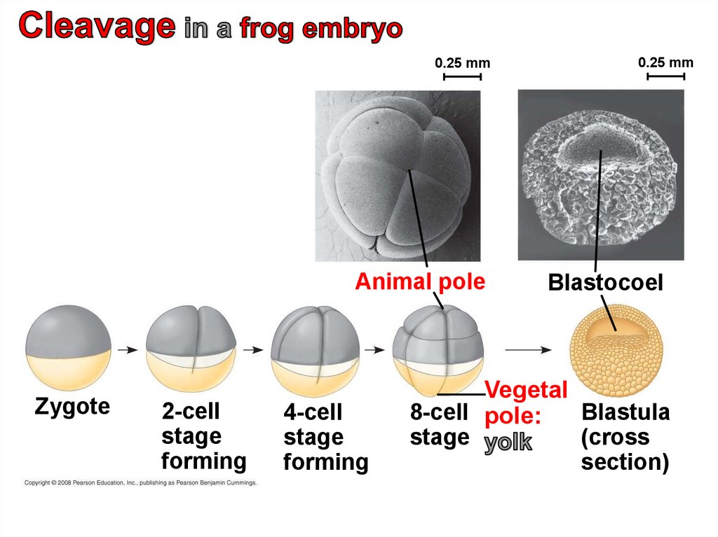
19.
0.25 mmAnimal pole
Zygote
2-cell
stage
forming
4-cell
stage
forming
0.25 mm
Blastocoel
Vegetal
8-cell pole:
Blastula
stage
(cross
section)
20. Gastrulation
• Gastrulation rearranges the cells of a blastulainto a three-layered embryo, called a gastrula,
which has a primitive gut.
• The three layers produced by gastrulation are
called embryonic germ layers:
– The ectoderm forms the outer layer
– The endoderm lines the digestive tract
– The mesoderm partly fills the space between
the endoderm and ectoderm.
Copyright © 2008 Pearson Education, Inc., publishing as Pearson Benjamin Cummings
21.
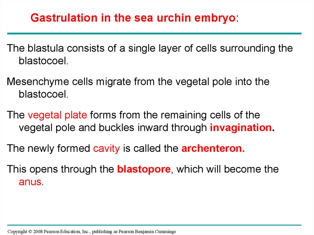
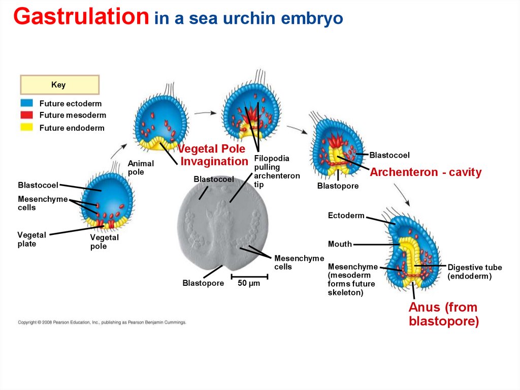
Gastrulation in the sea urchin embryo:The blastula consists of a single layer of cells surrounding the
blastocoel.
Mesenchyme cells migrate from the vegetal pole into the
blastocoel.
The vegetal plate forms from the remaining cells of the
vegetal pole and buckles inward through invagination.
The newly formed cavity is called the archenteron.
This opens through the blastopore, which will become the
anus.
Copyright © 2008 Pearson Education, Inc., publishing as Pearson Benjamin Cummings
22.
Gastrulation in a sea urchin embryoKey
Future ectoderm
Future mesoderm
Future endoderm
Animal
pole
Blastocoel
Vegetal Pole
Invagination
Blastocoel
Filopodia
pulling
archenteron
tip
Blastocoel
Archenteron - cavity
Blastopore
Mesenchyme
cells
Ectoderm
Vegetal
plate
Vegetal
pole
Mouth
Blastopore
50 µm
Mesenchyme
Mesenchyme
cells
(mesoderm
forms future
skeleton)
Digestive tube
(endoderm)
Anus (from
blastopore)
23.
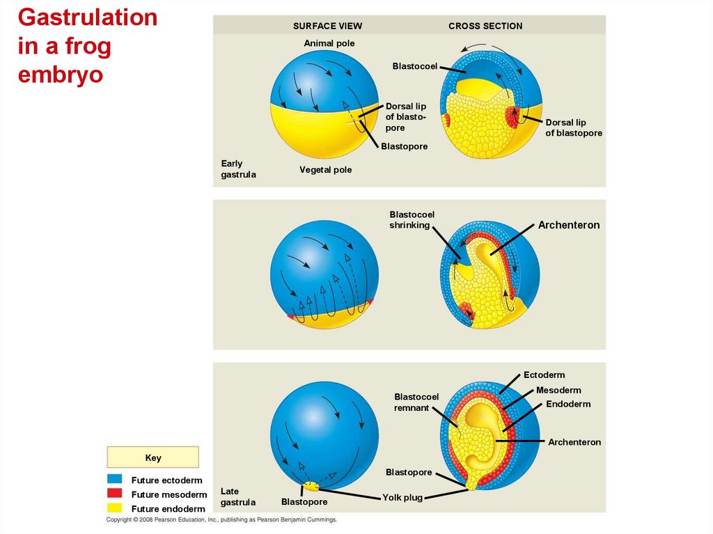
Gastrulation in the frog• The frog blastula is many cell layers thick. Cells of the
dorsal lip originate in the gray crescent and
invaginate to create the archenteron.
• Cells continue to move from the embryo surface into the
embryo by involution. These cells become the endoderm
and mesoderm.
– The blastopore encircles a yolk plug when
gastrulation is completed.
– The surface of the embryo is now ectoderm, the
innermost layer is endoderm, and the middle layer is
mesoderm.
Copyright © 2008 Pearson Education, Inc., publishing as Pearson Benjamin Cummings
24.
Gastrulationin a frog
embryo
SURFACE VIEW
CROSS SECTION
Animal pole
Blastocoel
Dorsal lip
of blastopore
Dorsal lip
of blastopore
Blastopore
Early
gastrula
Vegetal pole
Blastocoel
shrinking
Archenteron
Ectoderm
Blastocoel
remnant
Mesoderm
Endoderm
Archenteron
Key
Blastopore
Future ectoderm
Future mesoderm
Future endoderm
Late
gastrula
Blastopore
Yolk plug
25.
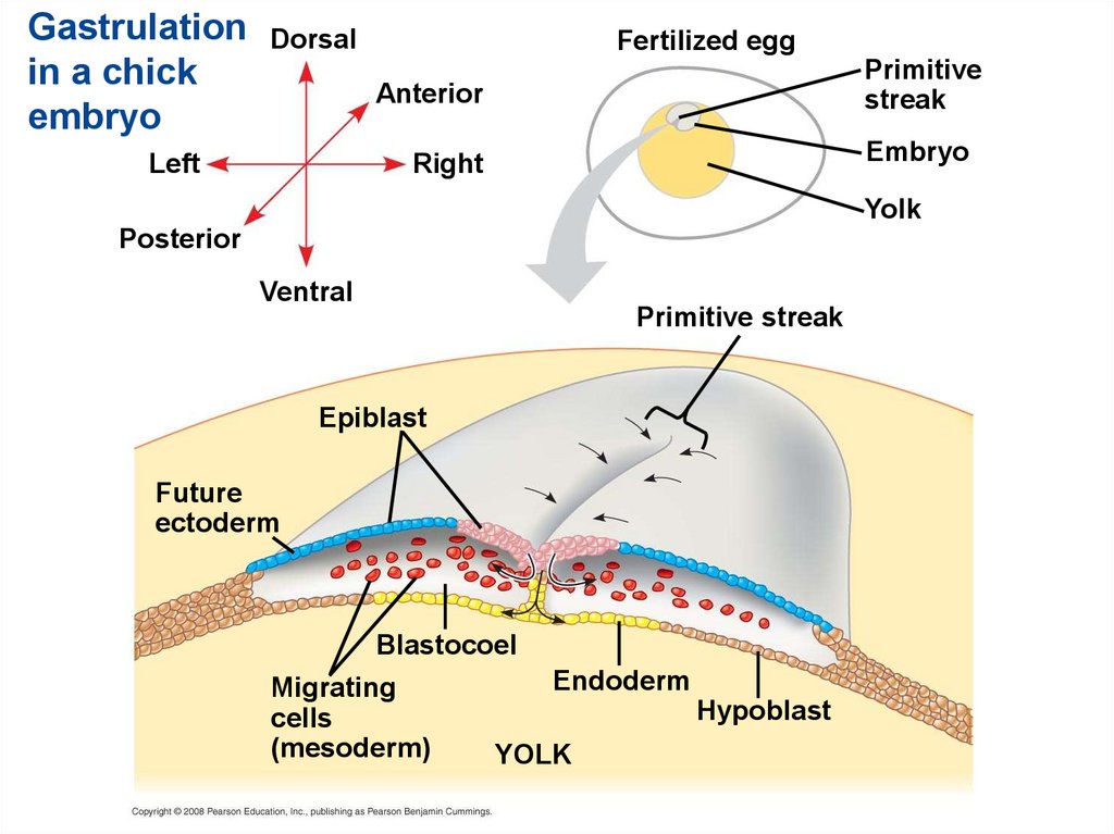
Gastrulation in the chick• The embryo forms from a blastoderm and sits on
top of a large yolk mass.
• During gastrulation, the upper layer of the
blastoderm (epiblast) moves toward the midline of
the blastoderm and then into the embryo toward
the yolk.
• The midline thickens and is called the primitive
streak.
• The movement of different epiblast cells gives rise
to the endoderm, mesoderm, and ectoderm.
Copyright © 2008 Pearson Education, Inc., publishing as Pearson Benjamin Cummings
26.
Gastrulationin a chick
embryo
Dorsal
Fertilized egg
Primitive
streak
Anterior
Left
Embryo
Right
Yolk
Posterior
Ventral
Primitive streak
Epiblast
Future
ectoderm
Blastocoel
Migrating
cells
(mesoderm)
Endoderm
Hypoblast
YOLK
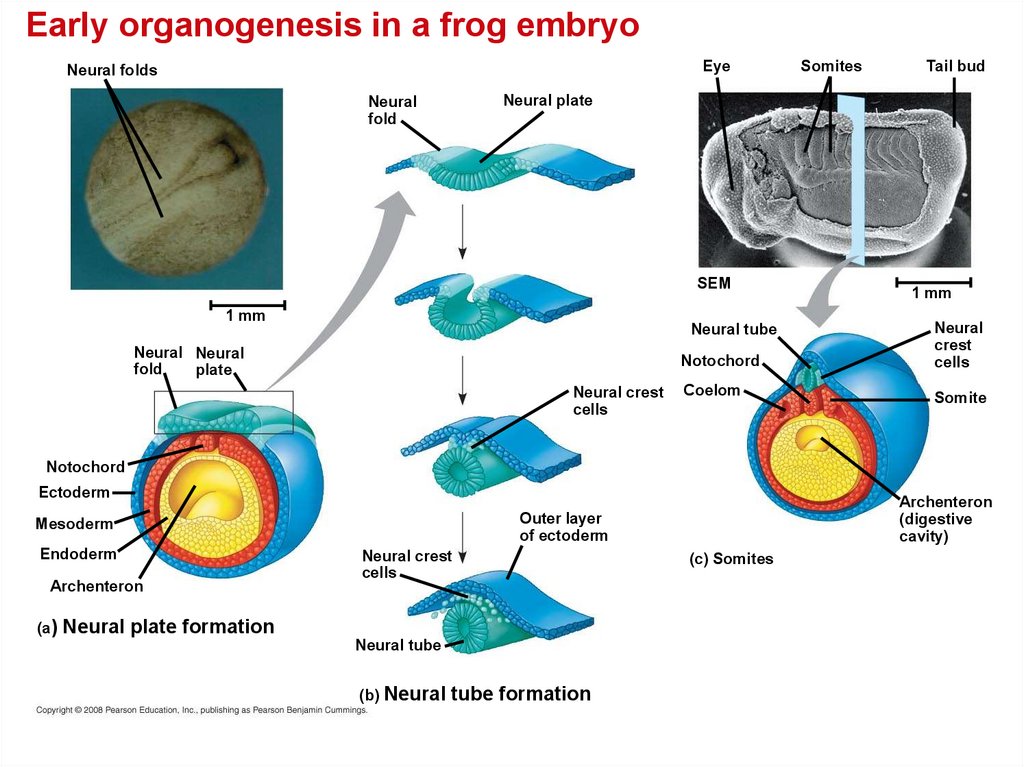
27. Organogenesis
• During organogenesis, various regions of thegerm layers develop into rudimentary organs.
• The frog is used as a model for organogenesis.
• Early in vertebrate organogenesis, the
notochord forms from mesoderm, and the
neural plate forms from ectoderm.
Copyright © 2008 Pearson Education, Inc., publishing as Pearson Benjamin Cummings
28.
Early organogenesis in a frog embryoEye
Neural folds
Somites
Tail bud
Neural plate
Neural
fold
SEM
1 mm
Notochord
Neural
crest
cells
Coelom
Somite
Neural tube
Neural Neural
fold
plate
Neural crest
cells
1 mm
Notochord
Ectoderm
Endoderm
Archenteron
(a)
Archenteron
(digestive
cavity)
Outer layer
of ectoderm
Mesoderm
Neural crest
cells
Neural plate formation
Neural tube
(b) Neural
tube formation
(c) Somites
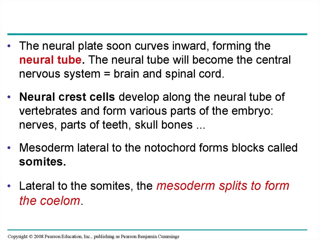
29.
• The neural plate soon curves inward, forming theneural tube. The neural tube will become the central
nervous system = brain and spinal cord.
• Neural crest cells develop along the neural tube of
vertebrates and form various parts of the embryo:
nerves, parts of teeth, skull bones ...
• Mesoderm lateral to the notochord forms blocks called
somites.
• Lateral to the somites, the mesoderm splits to form
the coelom.
Copyright © 2008 Pearson Education, Inc., publishing as Pearson Benjamin Cummings
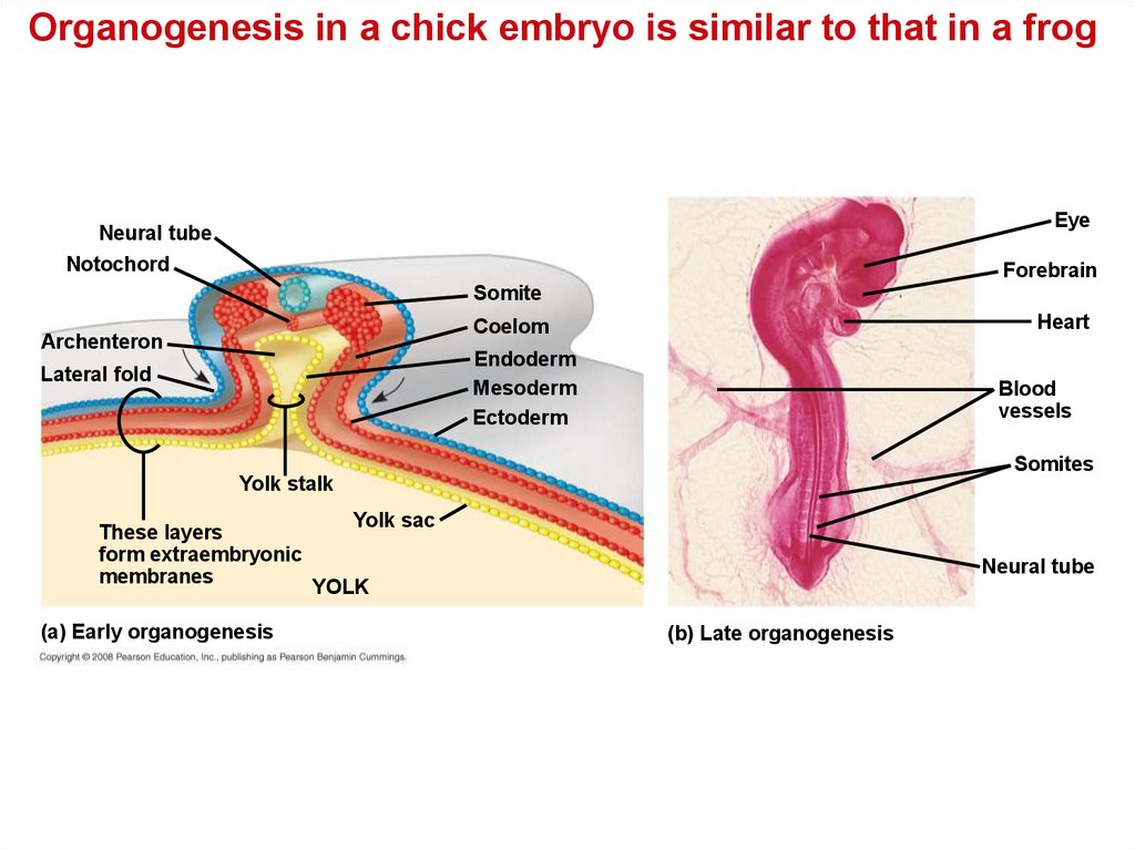
30.
Organogenesis in a chick embryo is similar to that in a frogEye
Neural tube
Notochord
Forebrain
Somite
Heart
Coelom
Archenteron
Endoderm
Mesoderm
Ectoderm
Lateral fold
Blood
vessels
Somites
Yolk stalk
These layers
form extraembryonic
membranes
(a) Early organogenesis
Yolk sac
Neural tube
YOLK
(b) Late organogenesis
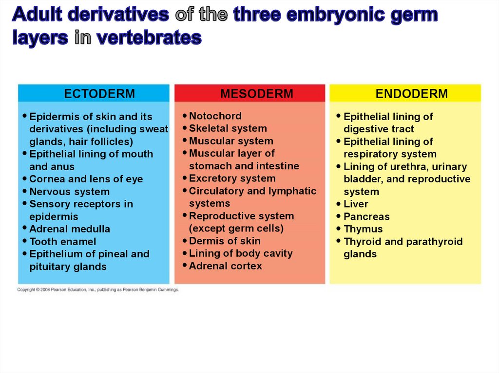
31.
ECTODERMEpidermis of skin and its
derivatives (including sweat
glands, hair follicles)
Epithelial lining of mouth
and anus
Cornea and lens of eye
Nervous system
Sensory receptors in
epidermis
Adrenal medulla
Tooth enamel
Epithelium of pineal and
pituitary glands
MESODERM
Notochord
Skeletal system
Muscular system
Muscular layer of
stomach and intestine
Excretory system
Circulatory and lymphatic
systems
Reproductive system
(except germ cells)
Dermis of skin
Lining of body cavity
Adrenal cortex
ENDODERM
Epithelial lining of
digestive tract
Epithelial lining of
respiratory system
Lining of urethra, urinary
bladder, and reproductive
system
Liver
Pancreas
Thymus
Thyroid and parathyroid
glands
32. Developmental Adaptations of Amniotes
• Embryos of birds, other reptiles, and mammalsdevelop in a fluid-filled sac in a shell or the
uterus.
• Organisms with these adaptations are called
amniotes.
• Amniotes develop extra-embryonic membranes
to support the embryo.
Copyright © 2008 Pearson Education, Inc., publishing as Pearson Benjamin Cummings
33.
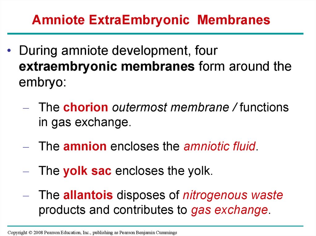
Amniote ExtraEmbryonic Membranes• During amniote development, four
extraembryonic membranes form around the
embryo:
– The chorion outermost membrane / functions
in gas exchange.
– The amnion encloses the amniotic fluid.
– The yolk sac encloses the yolk.
– The allantois disposes of nitrogenous waste
products and contributes to gas exchange.
Copyright © 2008 Pearson Education, Inc., publishing as Pearson Benjamin Cummings
34.
AmnionAllantois
Embryo
Amniotic
cavity
with
amniotic
fluid
Albumen
Shell
Yolk
(nutrients)
Chorion
Yolk sac
35. Mammalian Development
• The eggs of placental mammals– Are small yolk and store few nutrients
– Exhibit holoblastic cleavage
– Show no obvious polarity.
• Gastrulation and organogenesis resemble the
processes in birds and other reptiles.
• Early cleavage is relatively slow in humans and
other mammals.
Copyright © 2008 Pearson Education, Inc., publishing as Pearson Benjamin Cummings
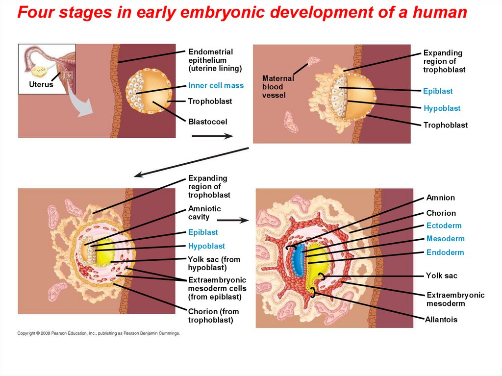
36.
• At completion of cleavage, the blastocystforms.
• A group of cells called the inner cell mass
develops into the embryo and forms the
extraembryonic membranes.
• The trophoblast, the outer epithelium of the
blastocyst, initiates implantation in the uterus,
and the inner cell mass of the blastocyst forms
a flat disk of cells.
• As implantation is completed, gastrulation
begins.
Copyright © 2008 Pearson Education, Inc., publishing as Pearson Benjamin Cummings
37.
Early embryonic development of a humanEndometrial
epithelium
(uterine lining)
Uterus
Inner cell mass
Trophoblast
Blastocoel
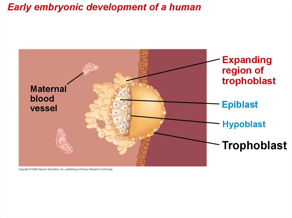
38.
Early embryonic development of a humanMaternal
blood
vessel
Expanding
region of
trophoblast
Epiblast
Hypoblast
Trophoblast
39.
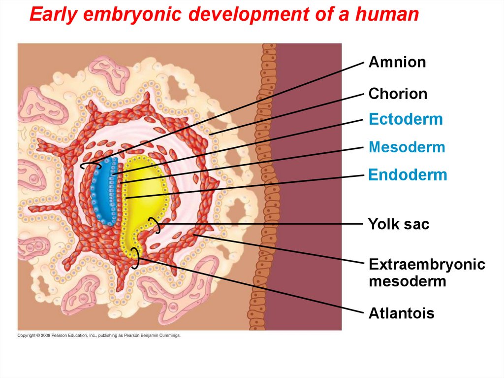
• The epiblast cells invaginate through a primitive streakto form mesoderm and endoderm.
• The placenta is formed from the trophoblast,
mesodermal cells from the epiblast, and adjacent
endometrial tissue.
• The placenta allows for the exchange of materials
between the mother and embryo.
• By the end of gastrulation, the embryonic germ layers
have formed. The extraembryonic membranes in
mammals are homologous to those of birds and other
reptiles and develop in a similar way.
Copyright © 2008 Pearson Education, Inc., publishing as Pearson Benjamin Cummings
40.
Early embryonic development of ahuman
Expanding
region of
trophoblast
Amniotic
cavity
Epiblast
Hypoblast
Yolk sac (from
hypoblast)
Extraembryonic
mesoderm cells
(from epiblast)
Chorion (from
trophoblast)
41.
Early embryonic development of a humanAmnion
Chorion
Ectoderm
Mesoderm
Endoderm
Yolk sac
Extraembryonic
mesoderm
Atlantois
42.
Four stages in early embryonic development of a humanEndometrial
epithelium
(uterine lining)
Uterus
Inner cell mass
Trophoblast
Expanding
region of
trophoblast
Maternal
blood
vessel
Epiblast
Hypoblast
Blastocoel
Expanding
region of
trophoblast
Amniotic
cavity
Epiblast
Hypoblast
Yolk sac (from
hypoblast)
Extraembryonic
mesoderm cells
(from epiblast)
Chorion (from
trophoblast)
Trophoblast
Amnion
Chorion
Ectoderm
Mesoderm
Endoderm
Yolk sac
Extraembryonic
mesoderm
Allantois
43. Morphogenesis in animals involves specific changes in cell shape, position, and adhesion
• Morphogenesis is a major aspect ofdevelopment in plants and animals.
• Only in animals does it involve the movement
of cells.
Copyright © 2008 Pearson Education, Inc., publishing as Pearson Benjamin Cummings
44. The Cytoskeleton, Cell Motility, and Convergent Extension
• Changes in cell shape usually involvereorganization of the cytoskeleton.
• Microtubules and microfilaments affect
formation of the neural tube.
Copyright © 2008 Pearson Education, Inc., publishing as Pearson Benjamin Cummings
45.
Change in cell shapeduring
morphogenesis
Ectoderm
Neural
plate
Microtubules
Actin filaments
Neural tube
46.
• The cytoskeleton also drives cell migration, orcell crawling, the active movement of cells.
• In gastrulation, tissue invagination is caused by
changes in cell shape and migration.
• Cell crawling is involved in convergent
extension, a morphogenetic movement in
which cells of a tissue become narrower and
longer.
Copyright © 2008 Pearson Education, Inc., publishing as Pearson Benjamin Cummings
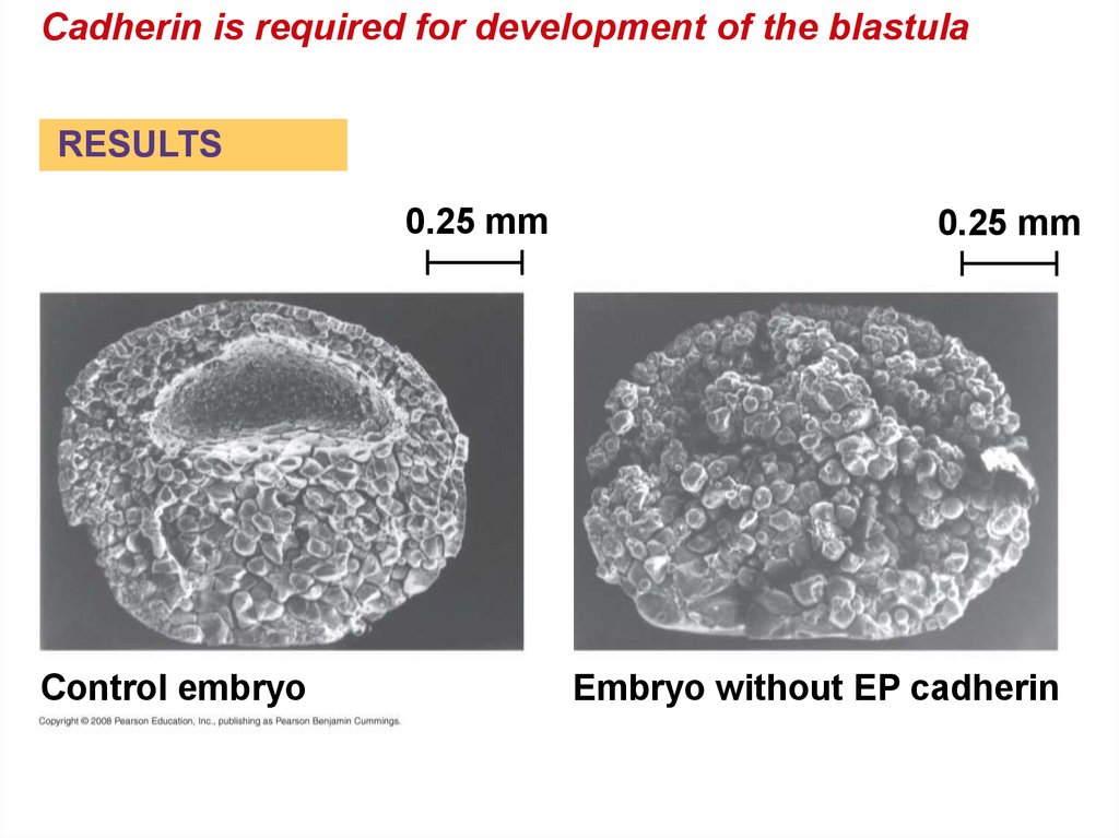
47. Role of Cell Adhesion Molecules and the Extracellular Matrix
• Cell adhesion molecules located on cellsurfaces contribute to cell migration and stable
tissue structure.
• One class of cell-to-cell adhesion molecule is
the cadherins, which are important in
formation of the frog blastula.
Copyright © 2008 Pearson Education, Inc., publishing as Pearson Benjamin Cummings
48.
Cadherin is required for development of the blastulaRESULTS
0.25 mm
Control embryo
0.25 mm
Embryo without EP cadherin
49. The developmental fate of cells depends on their history and on inductive signals
The developmental fate of cells depends on theirhistory and on
• Cells in a multicellular organism share the
same genome.
• Differences in cell types is the result of
differentiation, the expression of different
genes = differential gene expression.
Copyright © 2008 Pearson Education, Inc., publishing as Pearson Benjamin Cummings
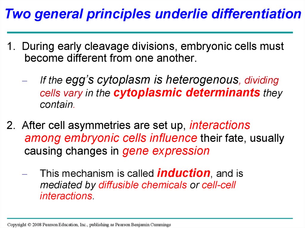
50.
Two general principles underlie differentiation1. During early cleavage divisions, embryonic cells must
become different from one another.
–
If the egg’s cytoplasm is heterogenous, dividing
cells vary in the cytoplasmic determinants they
contain.
2. After cell asymmetries are set up, interactions
among embryonic cells influence their fate, usually
causing changes in gene expression
–
This mechanism is called induction, and is
mediated by diffusible chemicals or cell-cell
interactions.
Copyright © 2008 Pearson Education, Inc., publishing as Pearson Benjamin Cummings
51.
Fate maps are general territorial diagrams of
embryonic development.
Classic studies using frogs indicated that cell lineage
in germ layers is traceable to blastula cells.
To understand how embryonic cells acquire their
fates, think about how basic axes of the embryo are
established.
Copyright © 2008 Pearson Education, Inc., publishing as Pearson Benjamin Cummings
52.
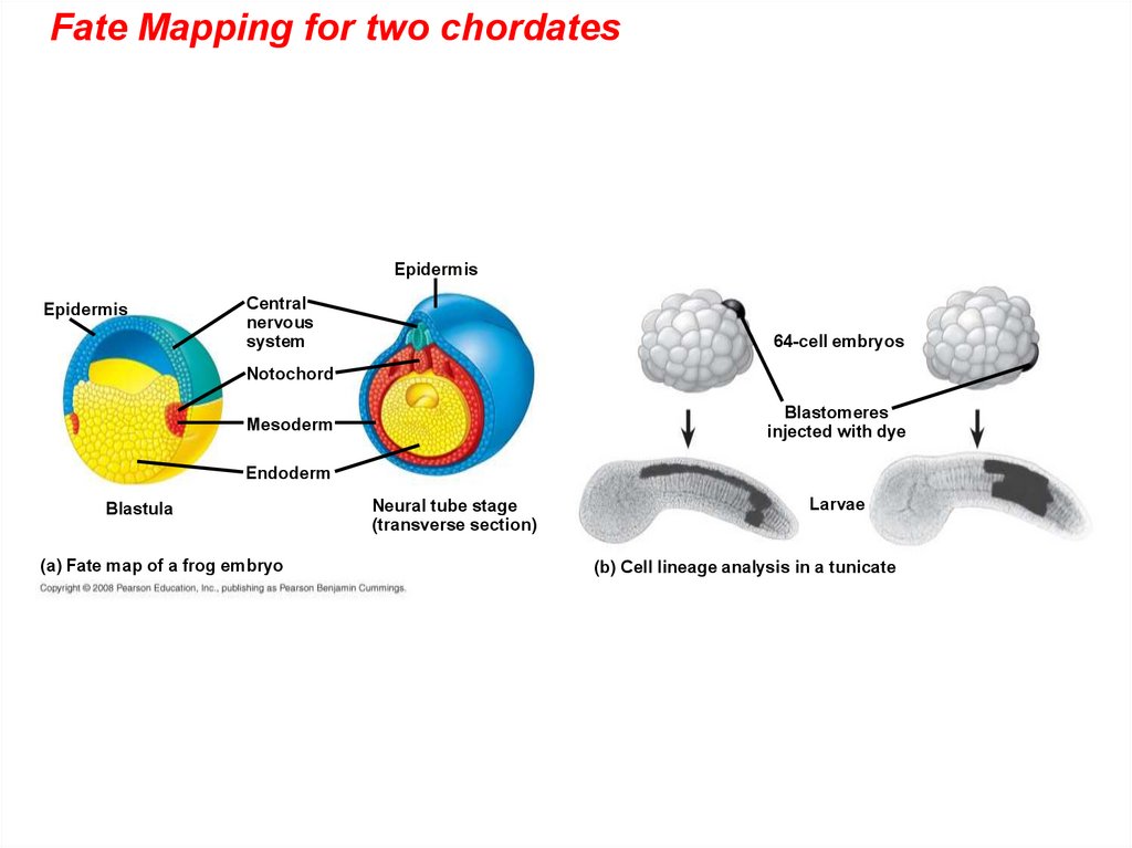
Fate Mapping for two chordatesEpidermis
Epidermis
Central
nervous
system
64-cell embryos
Notochord
Blastomeres
injected with dye
Mesoderm
Endoderm
Blastula
(a) Fate map of a frog embryo
Neural tube stage
(transverse section)
Larvae
(b) Cell lineage analysis in a tunicate
53. The Axes of the Basic Body Plan
• In nonamniotic vertebrates, basic instructions forestablishing the body axes are set down early during
oogenesis, or fertilization.
• In amniotes, local environmental differences play the
major role in establishing initial differences between
cells and the body axes.
• In many species that have cytoplasmic determinants,
only the zygote is totipotent.
• That is, only the zygote can develop into all the cell
types in the adult.
Copyright © 2008 Pearson Education, Inc., publishing as Pearson Benjamin Cummings
54.
• Unevenly distributed cytoplasmicdeterminants in the egg cell help establish the
body axes.
• These determinants set up differences in
blastomeres resulting from cleavage.
• As embryonic development proceeds, potency
of cells becomes more limited.
• After embryonic cell division creates cells that
differ from each other, the cells begin to
influence each other’s fates by induction
signals.
Copyright © 2008 Pearson Education, Inc., publishing as Pearson Benjamin Cummings
55.
EXPERIMENTHow does distribution of the gray crescent affect the
development potential of the two daughter cells?
Control egg
(dorsal view)
Experimental egg
(side view)
Gray
crescent
Gray
crescent
Thread
RESULTS
Normal
Belly piece
Normal
56. The Dorsal Lip = “Organizer” of Spemann and Mangold
• Based on their famous experiment, HansSpemann and Hilde Mangold concluded that
the blastopore’s dorsal lip is an organizer of
the embryo.
• The Spemann organizer initiates inductions
that result in formation of the notochord, neural
tube, and other organs.
Copyright © 2008 Pearson Education, Inc., publishing as Pearson Benjamin Cummings
57.
Can the dorsal lip of the blastopore induce cells in anotherpart of the amphibian embryo to change their developmental
fate?
EXPERIMENT
RESULTS
Dorsal lip of
blastopore
Pigmented gastrula
(donor embryo)
Nonpigmented gastrula
(recipient embryo)
Primary embryo
Secondary
(induced) embryo
Primary structures:
Neural tube
Notochord
Secondary structures:
Notochord (pigmented cells)
Neural tube (mostly nonpigmented cells)
58. Formation of the Vertebrate Limb
• Inductive signals play a major role in patternformation, development of spatial
organization.
• The molecular cues that control pattern
formation are called positional information.
• This information tells a cell where it is with
respect to the body axes.
• It determines how the cell and its descendents
respond to future molecular signals.
Copyright © 2008 Pearson Education, Inc., publishing as Pearson Benjamin Cummings
59.
• The wings and legs of chicks, like all vertebratelimbs, begin as bumps of tissue called limb
buds.
• The embryonic cells in a limb bud respond to
positional information indicating location along
three axes
– Proximal-distal axis
– Anterior-posterior axis
– Dorsal-ventral axis
Copyright © 2008 Pearson Education, Inc., publishing as Pearson Benjamin Cummings
60.
Vertebratelimb
development
Anterior
Limb bud
AER
ZPA
Limb buds
50 µm
Posterior
Apical
ectodermal
ridge (AER)
(a) Organizer regions
2
Digits
Anterior
4
3
Ventral
Distal
Proximal
Dorsal
Posterior
(b) Wing of chick embryo
61.
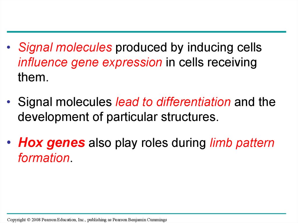
• Signal molecules produced by inducing cellsinfluence gene expression in cells receiving
them.
• Signal molecules lead to differentiation and the
development of particular structures.
• Hox genes also play roles during limb pattern
formation.
Copyright © 2008 Pearson Education, Inc., publishing as Pearson Benjamin Cummings
62.
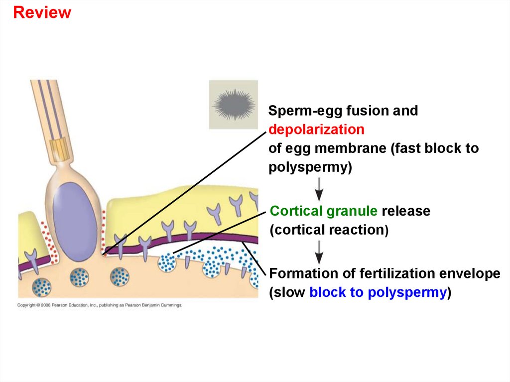
ReviewSperm-egg fusion and
depolarization
of egg membrane (fast block to
polyspermy)
Cortical granule release
(cortical reaction)
Formation of fertilization envelope
(slow block to polyspermy)
63.
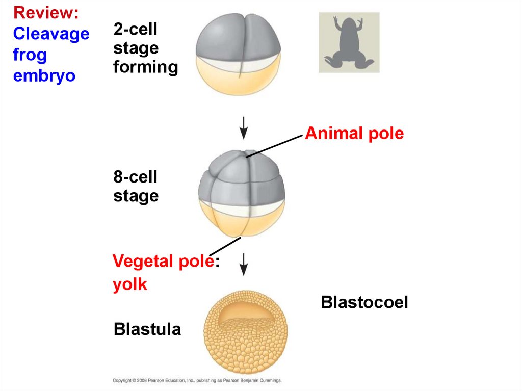
Review:Cleavage
frog
embryo
2-cell
stage
forming
Animal pole
8-cell
stage
Vegetal pole:
yolk
Blastocoel
Blastula
64.
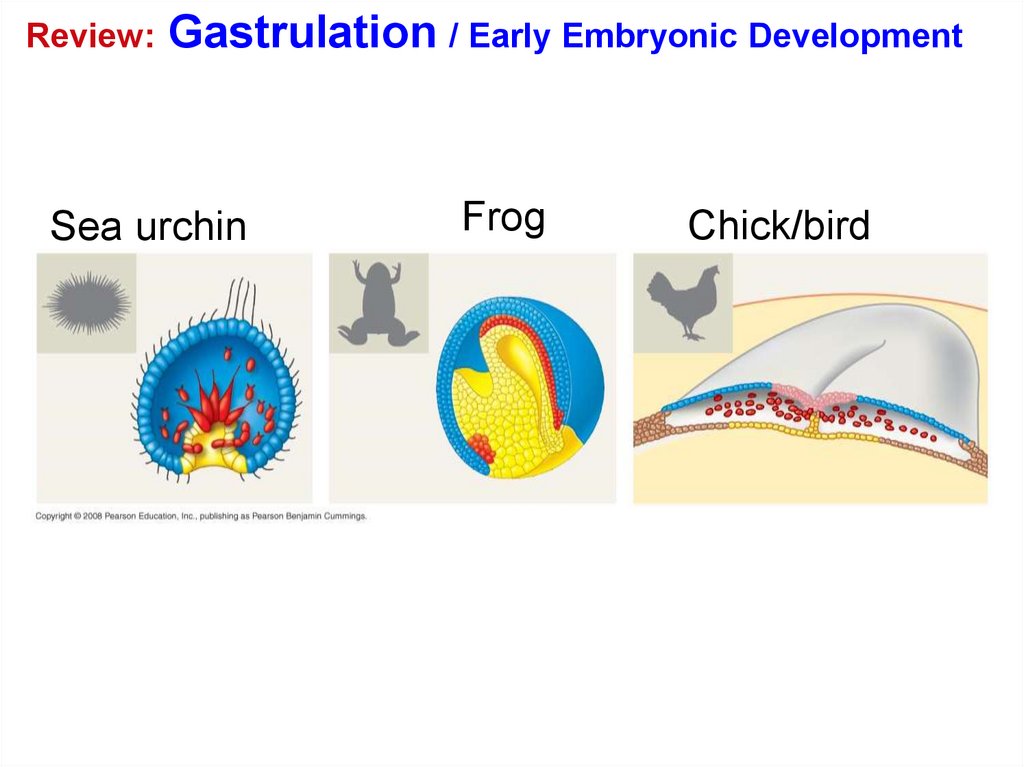
Review:Gastrulation / Early Embryonic Development
Sea urchin
Frog
Chick/bird
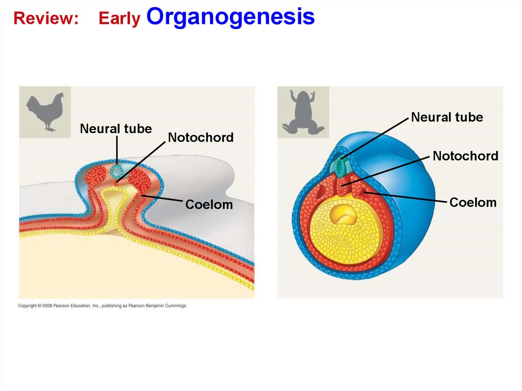
65.
Review:Early Organogenesis
Neural tube
Neural tube
Notochord
Notochord
Coelom
Coelom
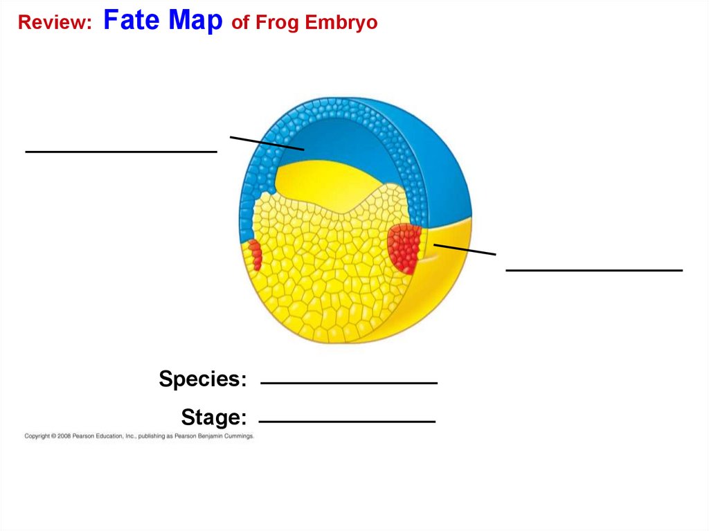
66.
Review:Fate Map of Frog Embryo
Species:
Stage:
67. You should now be able to:
1. Describe the acrosomal reaction.2. Describe the cortical reaction.
3. Distinguish among meroblastic cleavage and
holoblastic cleavage.
4. Compare the formation of a blastula and
gastrulation in a sea urchin, a frog, and a
chick.
5. List and explain the functions of the
extraembryonic membranes.
Copyright © 2008 Pearson Education, Inc., publishing as Pearson Benjamin Cummings
68.
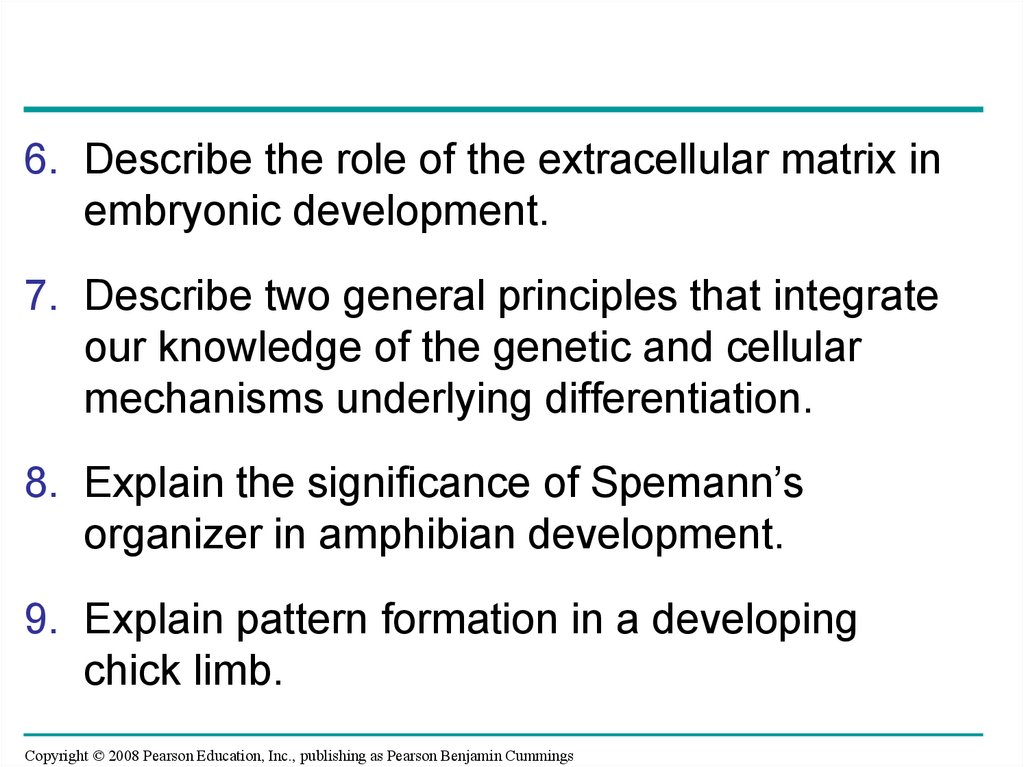
6. Describe the role of the extracellular matrix inembryonic development.
7. Describe two general principles that integrate
our knowledge of the genetic and cellular
mechanisms underlying differentiation.
8. Explain the significance of Spemann’s
organizer in amphibian development.
9. Explain pattern formation in a developing
chick limb.
Copyright © 2008 Pearson Education, Inc., publishing as Pearson Benjamin Cummings