


 Blastocyst consists of outer")


: Development of the Neural Tube - future nerve system - by the invagination of ectoderm:")


")


 --- brain, spinal cord, and the retina Neural crests --- Peripheral Nervous system, adrenal")


 Биология
Похожие презентации:


The First Three Weeks of Human Embryogenesis
1. The First Three Weeks of Human Embryogenesis
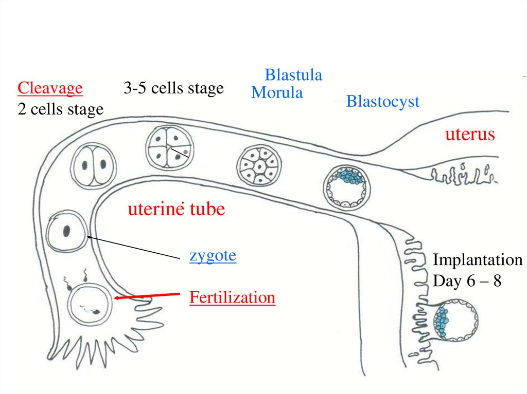
2. Week 1
• 1. Fertilization – is the fusion of the spermand ovum (male and female gametes) =
Zygote formation
(in the uterine tube) :
• - distant phase – sperms find ovum;
• - contact phase – 1 sperm fertilizes ovum.
3. Week 1
• Zygote – 1 cell embryo – starts to divide:• 2. Cleavage – is the division of the zygote
inside zona pellucida = Blastula formation
4.
Cleavage2 cells stage
3-5 cells stage
Blastula
Morula
.
Blastocyst
uterus
.
uterine tube
zygote
Fertilization
Implantation
Day 6 – 8
5. At the end of cleavage blastula is formed. Human blastula is called blastocyst (has cavity -cyst) Blastocyst consists of outer
At the end of cleavage blastula is formed.Human blastula is called blastocyst (has cavity cyst)
Blastocyst consists of outer cells (trophoblast),
inner cells (embryoblast) and cavity - Blastocoele.
Inner Cell Mass
(embryoblast)
Trophoblast
Blastocoele
6. At the 7-th day blastocyst sinks into the uterine wall due to activity of trophoblast – implantation.
TrophoblastEmbryoblast
7. Week 2: Beginning of 3. Gastrulation – formation of 3 germ layers Early Gastrulation take place by delamination, when
embryoblast dividesinto two germ layers - ectoderm and
endoderm, forming embryonic disc and two
sacs – ectoblast and endoblast
8. Result of early Delamination
TrophoblastEctoblast
Embryonic disc:
Ectoderm
Endoderm
Endoblast
9.
Late gastrulation – formation ofmesoderm – 3-d germ layer – take
place by cell migration:
cells which form mesoderm begin to
migrate from embryonic disc.
Mesoderm may be
extraembryonic and embryonic.
10.
1-st appear extraembryonic mesoderm:it surrounds upper and lower sacs,
and underly trophoblast
11.
ExtraembryonicMesoderm
Trophoblast
Extraembryonic
Mesoderm
Ectoderm
*
Endoderm
*
12.
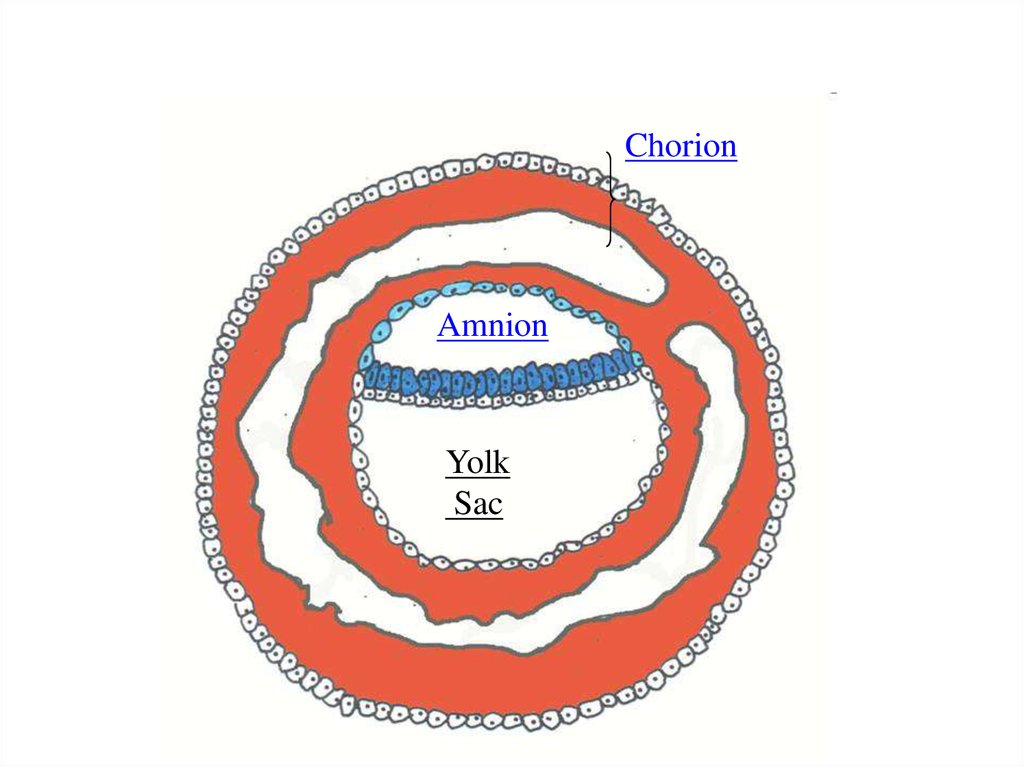
As a result appear so-calledextraembryonic organs amnion, yolk sac and chorion
13.
ChorionAmnion
Yolk
Sac
14.
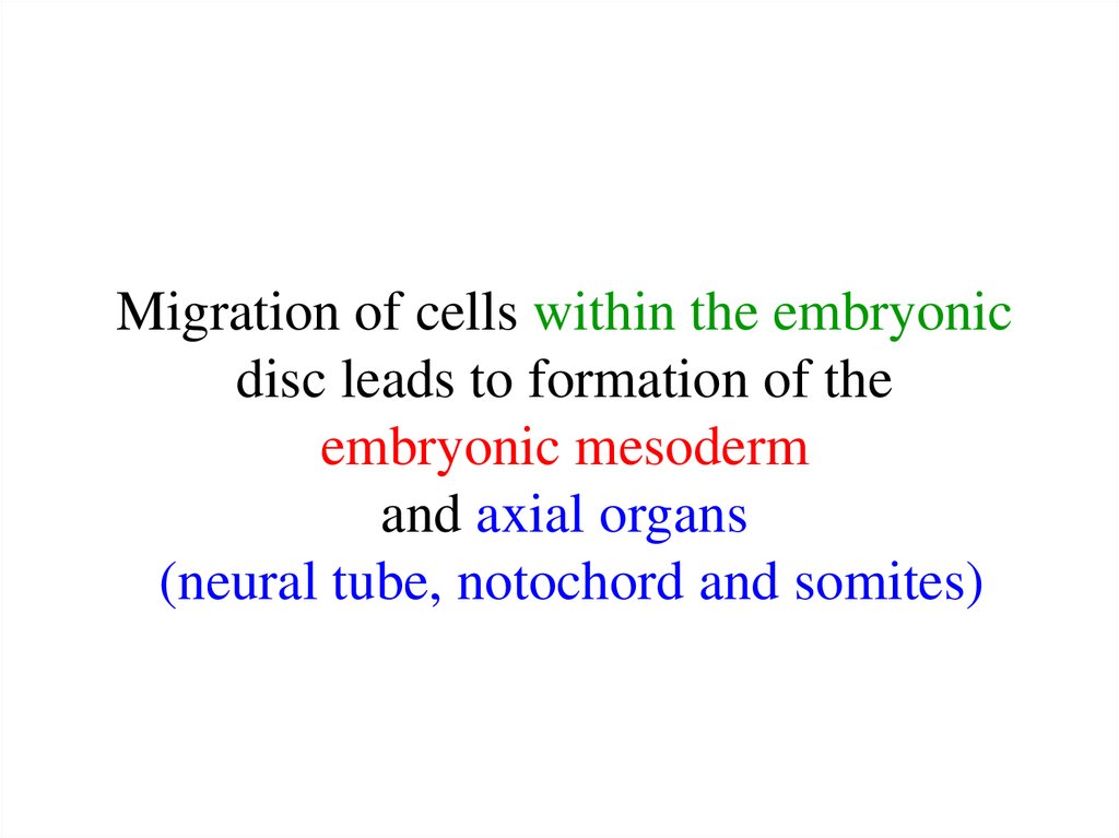
Migration of cells within the embryonicdisc leads to formation of the
embryonic mesoderm
and axial organs
(neural tube, notochord and somites)
15.
• Migration of cells withinembryonic disc leads to formation
of temporal cellular assemblage
between ectoderm and endoderm at
the caudal end of embryonic disc.
• It is a primitive streak.
16. Transverse section
Amniotic CavityEctoderm
Primitive streak
Yolk Sac
Endoderm
17. In front of primitive streak appears primitive knot. Cells of Primitive Streak begin to move laterally.
Primitive KnotMesoderm
18. Notochord appears by the primitive knot invagination. Mesoderm appears by migration of cells from primitive streak and
notochordectoderm
mesoderm
endoderm
notochord
19. 3-2.(next step): Development of the Neural Tube - future nerve system - by the invagination of ectoderm:
20. Neural plate in Surface Ectoderm forms Neural groove
21. Then - Neural Tube
22.
23. Development of the Neural Tube
Surface EctodermNeural Crest
Neural Tube
24. Neural tube formation
25. Neural tube formation
26. Gastrulation is finished with the formation of axial organs – neural tube, notochord, somites (mesoderm)
Neural tubeSomite
Notochord
27.
4. Formation of the embryo body(20-th day) by:
- body flexion,
- head and tail folds formation.
Result: separation of embryonic
organs from extra-embryonic organs
28. Body flexion
29. Differentiation of GERM LAYERS:
1. Differentiation of EctodermA. Surface Ectoderm
B. Neural Tube
2. Differentiation of Endoderm
A. G.I. Tract
B. Respiratory Tree
C. Endocrine glands
3. Differentiation of Mesoderm
A. Somites (have 3 part - dermatome, myotome, sclerotome)
B. Intermediate mesoderm - nephrotome
C. Lateral mesoderm -splanchnotome
D. Mesenchyme
30.
Differentiation of GERM LAYERS:Surface Ectoderm differentiates to
epithelium of skin, and its derivatives,
oral cavity epithelium,
rectal epithelium,
outer corneal epithelium, tooth enamel
31. Neural tube (neuroectoderm) --- brain, spinal cord, and the retina Neural crests --- Peripheral Nervous system, adrenal
medulla, melanocytes of skin, APUDsystem).32.
Endoderm differentiates to epitheliumof stomach, intestine, liver, pancreas,
respiratory system
33. Mesoderm
EctodermSomite
Amniotic Cavity
Intermediate mesoderm
(nephrotome)
Lateral plate mesoderm
(somatopleuric,
splanchnopleuric
mesoderm)
Endoderm
Yolk Sac
Notochord
34. Mesoderm
Nephrotomeurogenital system
including kydneys, gonads,
ducts, and accessory glands
dermatome - dermis
of skin
Somite myotome - muscles,
sclerotome skeleton
Lateral Mesoderm
serous membranes of pleura,
pericardium and peritoneum
Mesenchyme (loose part) –
connective tissue, smooth
muscle tissue, blood and
lymph cells, cardiovascular
and lymphatic systems
35. Late embryonic stages
• Histogenesis• Organogenesis
36. Summary: Week 1-3:
Early Stages:• 1. Fertilization – Zygote formation
• 2. Cleavage – Blastocyst formation
• 3. Gastrulation – Germ layers formation
Axial organs formation
• 4. Formation of the embryo body
• Late stages:
Histogenesis, Organogenesis – next lectures