


 Медицина
Похожие презентации:


Osgood-schlatter disease
1. OSGOOD-SCHLATTER DISEASE
Prepared by: Mailykanov O.A2.
Radiograph of
a patient who
is skeletally
mature. Note
that the tibial
tubercle is
enlarged and
there is an
ossicle. A bursa
was overlying
this.
3.
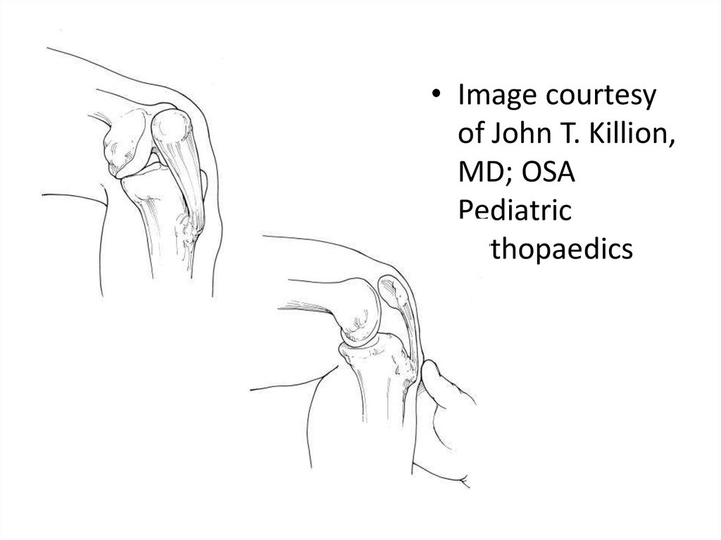
• Image courtesyof John T. Killion,
MD; OSA
Pediatric
Orthopaedics
4.
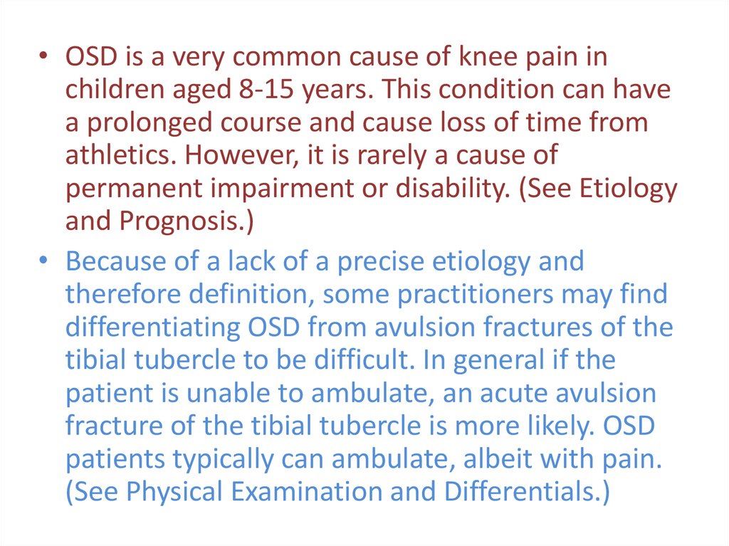
• OSD is a very common cause of knee pain inchildren aged 8-15 years. This condition can have
a prolonged course and cause loss of time from
athletics. However, it is rarely a cause of
permanent impairment or disability. (See Etiology
and Prognosis.)
• Because of a lack of a precise etiology and
therefore definition, some practitioners may find
differentiating OSD from avulsion fractures of the
tibial tubercle to be difficult. In general if the
patient is unable to ambulate, an acute avulsion
fracture of the tibial tubercle is more likely. OSD
patients typically can ambulate, albeit with pain.
(See Physical Examination and Differentials.)
5.
• The onset of OSD is usually gradual, with patientscommonly complaining of pain in the tibial tubercle
and/or patellar tendon region after repetitive activities.
Typically, running or jumping activities that significantly
stress the patellar tendon insertion upon the tibial
tubercle aggravate the patient's symptoms. They may
even have waxing and waning of symptoms that
correspond to variations in their athletic seasons. A
sudden onset of pain with no antecedent symptoms in
the region of the tibial tubercle should alert the
clinician to assess for a possible acute tibial tubercle
avulsion rather than OSD. (See History.)
6.
• OSD is a self-limiting condition. In a study by Krause etal, 90% of patients treated with conservative care were
relieved of all of their symptoms approximately 1 year
after onset of symptoms. [2] After skeletal maturity,
patients may continue to have problems kneeling. This
typically is due to tenderness over an unfused tibial
tubercle ossicle or a bursa that may require resection.
[3] Minimal association seems to exist between
residual anterior knee pain after OSD and patellar
stability, as was noted in the Krause study. The authors
also noted no cases of recurvatum from premature
closure of the proximal tibial physis. (See Prognosis.)
7. Development of Osgood-Schlatter disease
• The insertion of the patellar tendon at the tibial tubercleconsists of cartilaginous tissue in girls younger than age 11
years and in boys younger than age 13 years. The
secondary ossification center, or apophysis, of the tibial
tubercle develops when girls are aged 10-12 years and
when boys are aged 12-14 years. (During this stage of
skeletal development, the Osgood-Schlatter lesion may
occur.) (See Etiology.)
• By the end of the ensuing 2 stages of bony development
(eg, epiphyseal and bony stages), the primary growth plate
of the proximal tibia and the secondary ossification center
of the tibial tubercle fuse in males and females (usually
when aged 14-18 y), and the OSD usually subsides.
8.
• The most commonly accepted theory regardingthe development of OSD is that repeated traction
(traction apophysitis) on the anterior portion of
the developing ossification center leads to
multiple subacute microavulsion fractures and/or
tendinous inflammation, resulting in a benign,
self-limited disturbance manifested as pain,
swelling, and tenderness.
• The most common long-term ramifications of
OSD are pain on kneeling as an adult and the
cosmesis of a bony prominence on the anterior
knee. Less common complications are the
persistence of a painful ossicle requiring surgical
excision and a displaced avulsion of a tibial
tubercle.
9. Etiology
• The cause of Osgood-Schlatter disease (OSD)is unknown; however, theories suggest that
this condition is a result of repeated knee
extensor mechanism contraction that causes
partial microavulsions of the chondrofibroosseous tibial tubercle. This proposed
pathophysiology is supported by the repetitive
runner, jumper athletic patient population
OSD occurs most commonly.
10.
During running, jumping, gymnastics, and other sports requiring repeated
contractions of the quadriceps, an extra-articular osteochondral stress fracture or
microavulsion occurs. The proximal area of the patellar tendon insertion separates,
resulting in elevation of the tibial tubercle. During the reparative phase of this
stress fracture, new bone is laid down in the avulsion space, which may result in a
deviated and prominent tibial tubercle. When an individual with an injured tibial
tubercle continues to participate in sports, more and more microavulsions
develop, and the reparative process may result in a markedly pronounced
prominence of the tubercle, with longer-term cosmetic and functional
implications. A separated fragment may develop at the patellar tendon insertion
and may lead to chronic, nonunion-type pain.
In a magnetic resonance imaging (MRI) study of 20 patients with OSD, the patellar
tendon was noted to attach more proximally and in a broader area to the tibia in
patients with OSD. [4] Approximately 50% of patients with OSD relate a history of
precipitating trauma.
Histologic studies support a traumatic etiology.
Risk factors
Risk factors for OSD include the following:
• Age: female 8-12 years & male between 12-15 years
• Male sex (3:1)
• Rapid skeletal growth
• Repetitive sprinting and jumping sports
A study by Nakase et al found that quadriceps femoris muscle tightness and
strength during knee extension and flexibility of the hamstring muscles were risk
factors for incidence of Osgood-Schlatter disease
11. Epidemiology
• Incidence• One study found that Osgood-Schlatter disease (OSD) affected
approximately 21% of athletic adolescents surveyed, as compared with a
frequency of 4.5% in age-matched nonathletic controls. [6]
• One Finnish study found that OSD affected 13% of athletes.
• Sex predilection
• OSD occurs more frequently in boys, with a male-to-female ratio of 3:1.
• Age predilection
• OSD usually is seen in the adolescent years, after a patient has undergone
a rapid growth spurt the previous year.
• Girls who are affected are typically aged 10-11 years but can range from
age 8-12 years.
• Boys who are affected are typically aged 13-14 years but can range from
age 12-15 years.
12. Prognosis
• The prognosis in Osgood-Schlatter disease (OSD) is excellent. OSD isusually self-limiting and resolves by the time the patient is aged 18
years, when the tibial tubercle apophysis ossifies. In approximately
10% of patients, however, the symptoms continue unabated into
adulthood despite all conservative measures. [7] This may be from
residual enlargement of the tuberosity or from ossicle formation in
the patellar tendon.
• The likelihood for long-term sequelae increases in severe cases, in
cases in which treatment is not sought, or in cases in which the
patient demonstrates poor compliance with the physician's
recommendations.
• In the study by Krause et al, 90% of patients treated with
conservative care were relieved of all of their symptoms
approximately 1 year following symptom onset. [2]
• In some cases, however, discomfort may persist for 2-3 years until
the tibial growth plate closes.
13. Physical Examination
The physical examination is very specific, with point tenderness over the tibial tubercle. A firmmass may be palpable.
Other physical examination findings may include the following:
• Proximal tibial swelling and tenderness
• Enlargement or prominence of the tibial tubercle
• Reproducible and aggravated pain by direct pressure and jumping (quadriceps contraction)
• Pain with resisted knee extension (quadriceps contraction)
• Hamstring tightness
• Quadriceps atrophy
• Erythema of the tibial tuberosity
The following exam findings must be tested and confirmed to verify no concomitant or more
severe injury:
Full range of motion of the knee
No effusion or meniscal signs
Negative Lachman test (no knee instability)
Normal neurovascular examination
No abnormal findings in the hip and ankle joints
14. Diagnostic Considerations
The most significant pitfall is failing to diagnose another conditionthat could result in long-term permanent damage (eg, tumor,
osteochondritis dissecans). Most other conditions have a more
concerning clinical examination or history. Therefore, always obtain
radiographs and consider the possibility of a referred pain syndrome
from the hip.
Conditions to be considered in the differential diagnosis of OsgoodSchlatter disease (OSD), in addition to those in the next section,
include the following:
Sinding-Larson-Johansson syndrome
Tumor (bone or soft tissue)
Perthes disease (often presents with knee pain instead of hip
complaints)
Patellar tendon avulsion/rupture
Chondromalacia patellae (Patellofemoral syndrome)
Patellar tendonitis
Infectious apophysitis
Accessory ossification centers
Osteomyelitis of the proximal tibia
Hoffa's syndrome
Synovial plica injury
Tibial tubercle fracture
Differential Diagnoses
Femur Injuries and Fractures
Knee Osteochondritis Dissecans
Legg-Calve-Perthes Disease
Osteomyelitis
Patellar Tendon Rupture
Patellofemoral Joint Syndromes
Pes Anserine Bursitis
Prepatellar Bursitis
Tibia and Fibula Fracture in the ED
Tibial Tubercle Avulsion
15. Radiographs
Not all patients with Osgood-Schlatter disease (OSD) needradiography, since the diagnosis is clinical. However, plain
films are should be obtained at least once in the evaluation
and treatment to rule out other etiologies, such as neoplasm,
acute tibial apophyseal fracture, and infection.
• Radiographs may indicate:
• Superficial ossicle in the patellar tendon
• Irregular ossification of the proximal tibial tuberosity
• Calcification within the patellar tendon
• Thickening of the patellar tendon
• Soft-tissue edema proximal to the tibial tuberosity
The Osgood-Schlatter lesion is best seen on the lateral view,
with the knee in slight internal rotation of 10-20°.
The most common finding is that the knee films are normal,
especially if the child is in the preossification phase.
16.
• The acute phase of OSD may reveal a prominent and elevated tibialtubercle with anterior soft-tissue swelling.
• In severe cases, radiographs may reveal radiodense fragments or
ossicles separated from the tibial tuberosity. (An ossicle is seen in
the image below.)
• Radiograph of a patient who is skeletally mature.
• Radiograph of a patient who is skeletally mature. Note that the
tibial tubercle is enlarged and there is an ossicle. A bursa was
overlying this.
• View Media Gallery
• Occasionally, the radiographs may reveal irregularity, fragmentation
(seen below), or increased density of the ossification of the tibial
tubercle. This pattern may be a normal variant in asymptomatic
children.
17. Approach Considerations
• While there are no prospective studies evaluating thetreatment of OSD, including the recommended
conservative treatments, The American Academy of
Orthopaedic Surgeons and the American Academy of
Family Practice recommend the following for the
management of Osgood-Schlatter disease (OSD).
• Activity limitation
• Ice
• Anti-inflammatories
• Protective padding
• Quadriceps/hamstring strengthening
• Time
18. Medical Care
Medical Care
Treatment for Osgood-Schlatter disease (OSD) is conservative. Initial treatment includes the application
of ice for 20 minutes every 2-4 hours.
Analgesics and nonsteroidal anti-inflammatory drugs (NSAIDs) may be given for pain relief and reduction
of local inflammation. However, NSAIDs have not been shown to shorten the course of OSD.
Patellar tendon insertion injections have historically not been recommended. This is due in large part to
the possibility of subcutaneous tissue atrophy after corticosteroid injections. A recent study out of Japan
looked at lidocaine/dextrose injection vs. just lidocaine. No difference was seen between the two
injection groups however both groups reported symptom relief with no adverse outcomes. [13]
Long-term immobilization is typically contraindicated, because it may result in increased knee stiffness in
mild cases, thus predisposing the athlete to additional sports-related injuries. However, if a patient is
noncompliant, the clinician may recommend immobilization in a knee brace for a minimum of 6 weeks.
The brace should be removed daily, but only for stretching and strengthening exercises.
Inform the patient to avoid pain-producing activities (eg, sports that involve excess amounts of jumping).
Infrapatellar strap, pads, or braces may also be used for support, but none have any proven efficacy.
Once the acute symptoms have abated, quadriceps-stretching exercises, including hip extension for a
complete stretch of the extensor mechanism, may be performed to reduce tension on the tibial
tubercle. Stretching exercises for the hamstrings, which are commonly tight, may also be performed.
Other than the presence of an ossicle that causes pain with kneeling, there are no long-term disabilities
or problems associated with this condition.
19. Physical Therapy
The goal of rehabilitation is for the athlete to be able to return to his or her sport as quickly and
safely as possible. Since the main treatment is rest, ice, and NSAIDs, the role of physical therapy
is limited, if used at all. The pain may take up to 6-24 months to resolve. If an individual returns
to activity too soon, he or she may worsen the condition. Athletes need to work on improving
the flexibility and strength of the quadriceps and hamstring muscles throughout the course of
rehabilitation to ensure that they are ready to return to sports.
Acute phase
Several techniques may be recommended by the physical therapist to alleviate discomfort and
avert recurrence of the disease. Treatment recommendations are dependent upon the severity
of the condition.
An infrapatellar strap may be recommended during sports activity but has no proven efficacy.
Resting is recommended when pain arises.
Ice should be applied to the area for 20 minutes following activity.
Short-term rest and knee immobilization may be required.
On rare occasions, this author has casted a patient who has severe pain and is noncompliant
with conservative care. This is usually with a parent who is intent on relieving the pain. While a
brace can be recommended, it is doubtful that it will be used in a noncompliant patient.
Recovery phase
The following regimen recommendations for patients with OSD are taken from Meisterling, Wall,
and Meisterling.
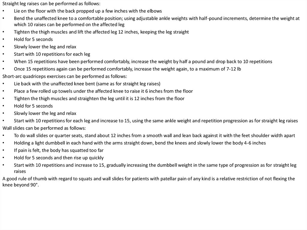
20.
Straight leg raises can be performed as follows:Lie on the floor with the back propped up a few inches with the elbows
Bend the unaffected knee to a comfortable position; using adjustable ankle weights with half-pound increments, determine the weight at
which 10 raises can be performed on the affected leg
Tighten the thigh muscles and lift the affected leg 12 inches, keeping the leg straight
Hold for 5 seconds
Slowly lower the leg and relax
Start with 10 repetitions for each leg
When 15 repetitions have been performed comfortably, increase the weight by half a pound and drop back to 10 repetitions
Once 15 repetitions again can be performed comfortably, increase the weight again, to a maximum of 7-12 lb
Short-arc quadriceps exercises can be performed as follows:
Lie back with the unaffected knee bent (same as for straight leg raises)
Place a few rolled up towels under the affected knee to raise it 6 inches from the floor
Tighten the thigh muscles and straighten the leg until it is 12 inches from the floor
Hold for 5 seconds
Slowly lower the leg and relax
Start with 10 repetitions for each leg and increase to 15, using the same ankle weight and repetition progression as for straight leg raises
Wall slides can be performed as follows:
To do wall slides or quarter seats, stand about 12 inches from a smooth wall and lean back against it with the feet shoulder width apart
Holding a light dumbbell in each hand with the arms straight down, bend the knees and slowly lower the body 4-6 inches
If pain is felt, the body has squatted too far
Hold for 5 seconds and then rise up quickly
Start with 10 repetitions and increase to 15, gradually increasing the dumbbell weight in the same type of progression as for straight leg
raises
A good rule of thumb with regard to squats and wall slides for patients with patellar pain of any kind is a relative restriction of not flexing the
knee beyond 90°.
21. Surgical Care
Surgery to treat Osgood-Schlatter disease (OSD) is rarely indicated.
Surgery in a skeletally immature patient is almost never indicated. Removal of
ossicle fragmentation in immature patients with an unfused apophysis can lead to
premature fusion of the tibial tubercle. [3]
In a study of the surgical treatment of unresolved Osgood-Schlatter disease (OSD),
Pihlajamäki et al concluded that in most young adults, good to excellent functional
outcomes can be achieved with surgical treatment of unresolved OSD. [14] The
investigators examined postsurgical clinical courses, radiographic characteristics,
and long-term outcomes of 107 military recruits (117 knees) who were operated
on for the condition. Functional outcome data were gathered from medical
records, interviews, questionnaires, and physical and radiographic examinations.
By the end of a (median) 10-year follow-up period, 93 patients (87%) reported that
they could participate without restriction in daily and work activities, and 80
patients (75%) had regained their preoperative sports activity level. In addition, 41
patients (38%) reported the ability to kneel without pain. Minor postoperative
complications occurred in 6 patients, and 2 patients required reoperation for OSD.
22.
• In a review of a series of patients who were treated operatively, Binazzi etal found that the most widely used procedure was excision of all
intratendinous ossicles, with or without removal of a portion of the
prominent tibial tubercle. [15] A comparison of 2 groups of individuals, 1
with 15 individuals treated with excision of ossicles and 1 with 11
individuals treated with various methods before 1975, clearly showed that
results of simple excision of the ossicles were better.
• A study looked to determine the outcomes of bursoscopic ossicle excision
in young, skeletally-mature, active patients with unresolved symptoms
from an ossicle related to prior Osgood-Schlatter disease. The study
concluded that bursoscopic ossicle excision showed satisfactory outcomes
in selective young, skeletally-mature, and active patients with persistent
symptoms and the presence of an ossicle. However, the authors added
that bursoscopic surgery showed limitation in reducing the prominence of
the tibial tuberosity. [16]
• In another study, patients treated operatively were found to be no more
likely than conservatively treated patients to be relieved of pain or to have
improvement of cosmetic appearance.
• If a true tibial tubercle avulsion occurs due to the contracture of the
extensor mechanism. Open reduction and internal fixation (ORIF) usually
is recommended, depending on the size and displacement of the fragment
as well as the phase of apophyseal closure.
23.
Indications for surgeryOccasionally, adults have a large ossicle and an overlying
bursa, which may cause pain with kneeling. If so,
treatment consists of excision of the bursa, ossicle, and
any prominence.
Contraindications for surgery
The real question is whether or not surgery is ever
indicated in the growing child, as OSD is self-limiting. Trail
reviewed 2 groups of symptomatic patients with this
condition with 4-5 years of follow-up. [18] One group was
treated surgically with tibial sequestrectomy, and the
other was managed conservatively. Surgery was found to
offer no significant benefit over conservative care. In
addition, a significant complication rate was identified
with tibial sequestrectomy.
24. Complications
• While the typical conservative management will relieve the painassociated once skeletal maturity is reached, continued tibial tubercle
prominence and pain upon kneeling can be a problem into adulthood.
• Complications following resection of an ossicle can include:
• surgical wound infection/dehiscence
• poor cosmesis
• unsightly scar
• peri-incisional numbness
• growth disturbance (skeletally immature)
• Trail et al showed 55% of patients had an obvious bony prominence
postoperatively. One third of these were quite apparent and troublesome
and 3 required repeat procedures to deal with associated discomfort.
25. List of references
• http://emedicine.medscape.com/article/1993268-treatment#d3