


 Медицина
Похожие презентации:


Diseases of the veins
1.
Diseases of the veinsDepartment of Surgery № 2
2.
3.
4.
5.
Venous return►Muscle
pump ( peripheral hearts)
►- ve intra thoracic pressure
►Arterial pulsation
►Vise at ergo
6.
7.
8.
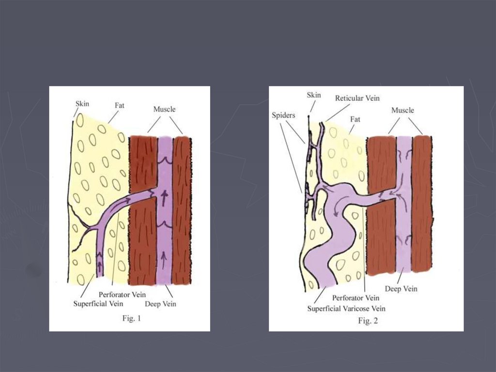
Varicose Veins►Dilated,
elongated & tortuous
vein of the LL
►problem comes from
incompetent calve.
►10 -20 % of worlds population
have varicose veins.
9.
Causes of varicos veins in lowerlimbs.
Secondary
Obstruction of venous outflow.
Pregnancy.
Fibroids
Ovarian cysts.
Abdominal lymphadenopathy
Pelvic cancer (cervical, uterus, ovary, rectum)
Ascites
Illiac vein thrombosis.
Retroperitoneal fibrosis
Valve destruction.
DVT
High flow and pressure:
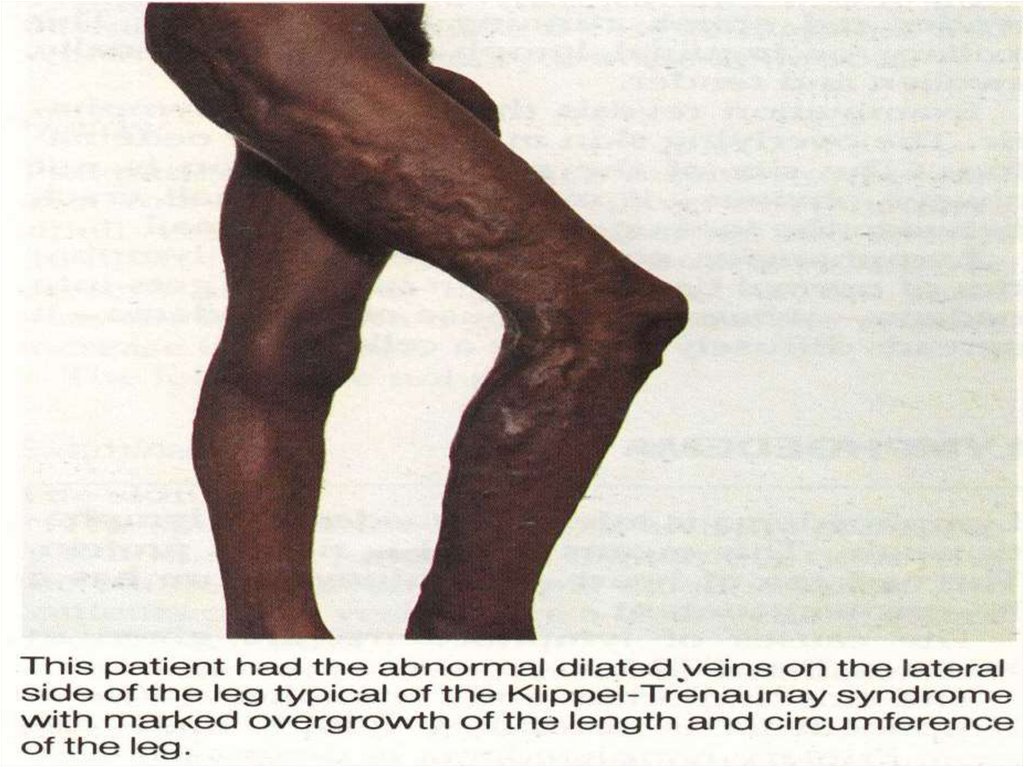
Arteriovenous fistula ( esp the aquired traumatic variety)eg.Klippel-Trenaunay
syndrome (which is one form of congenital AV malformation syndrome)
Primary:
Cause not known. Often familial.Probably weakness of vein wall that
permits valve ring to dilate.
Congenital abscence of valves very rare.
10.
Varicose Veins (Etiology)►Primary
Hereditary
Occupational
Pregnancy
obesity
Secondary
Venous obstruction
Venous compression
A/V fistula
11.
Varicose Veins (Etiology)► Obstruction
of venous outflow.
Pregnancy.
Fibroids
Ovarian cysts.
Abdominal lymphadenopathy
Pelvic cancer (cervical, uterus, ovary, rectum)
Ascites
Illiac vein thrombosis.
Retroperitoneal fibrosis
► Valve
destruction.
DVT
► High
flow and pressure:
Arteriovenous fistula ( esp the aquired traumatic
variety)
Klippel-Trenaunay syndrome (which is one form
of congenital AV malformation syndrome
12.
Varicose VeinsPrimary V V
► History:
young and middle aged
women most commonly
affected.1:10 men
:women
Aggrevating factors
associated with
increased incidence of
varicose veins:
female sex,
► parity,
► clothing,
► prolonged standing,
► marked obesity,
Secondary V V
History:
Any age most commonly
affect men
Aggrevating factors
associated with
increased incidence of
varicose veins:
DVT
Trauma
Compression
fracture
13.
Varicose Veins (symptoms)Disfiguring effects of the veins
usually principle complaint
Pain,
dull ache, and heaviness
felt in calves and lower leg
worse during day esp. on
standing up,
relieved by lying down for 1530 min.
Edema (swelling around ankle)
Aggregated by standing
Relived by recumbency
night cramps
Disfiguring effects of the veins
usually principle complaint
Pain,
dull ache, and heaviness
felt in calves and lower leg
worse during day esp. on standing
up and walking
relieved by lying down for 15-30
min.
Edema (swelling around ankle)
Aggregated by standing
Relived by recumbency
night cramps
Post phlebitic syndrome
dilated veins, venous stars,
pigmentation, eczema and
ulceration.
14.
Varicose Veins (signs)Dilated elongated tortuous
veins
Types of varices
Tubular with dilated LSV or
SSV
Saccular incompetent
perforator ( blow out)
Signs of PPS - ve
Special testes
Modified perthe’s
Cough impulse
Trendelenburg’s test
Multiple tourniquet test
Shwartz test
Dilated elongated tortuous
veins
Types of varices
Serpintine dilated tributeries
Spider indicate A/ V fistula
Signs of PPS + ve
Special testes
Modified perthe’s
Cough impulse
Trendelenburg’s test
Multiple tourniquet test
Shwartz test
15.
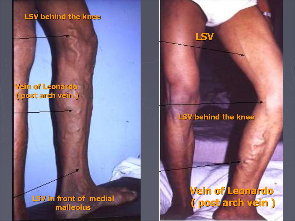
LSV behind the kneeLSV
Vein of Leonardo
( post arch vein )
LSV behind the knee
LSV in front of medial
malleolus
Vein of Leonardo
( post arch vein )
16.
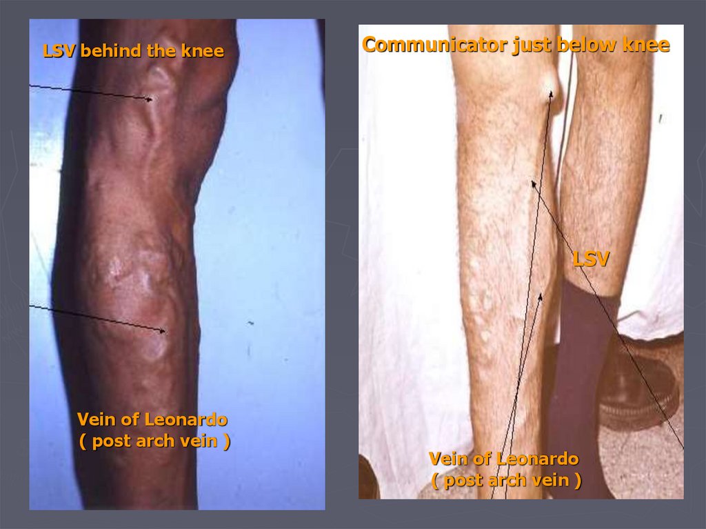
LSV behind the kneeCommunicator just below knee
LSV
Vein of Leonardo
( post arch vein )
Vein of Leonardo
( post arch vein )
17.
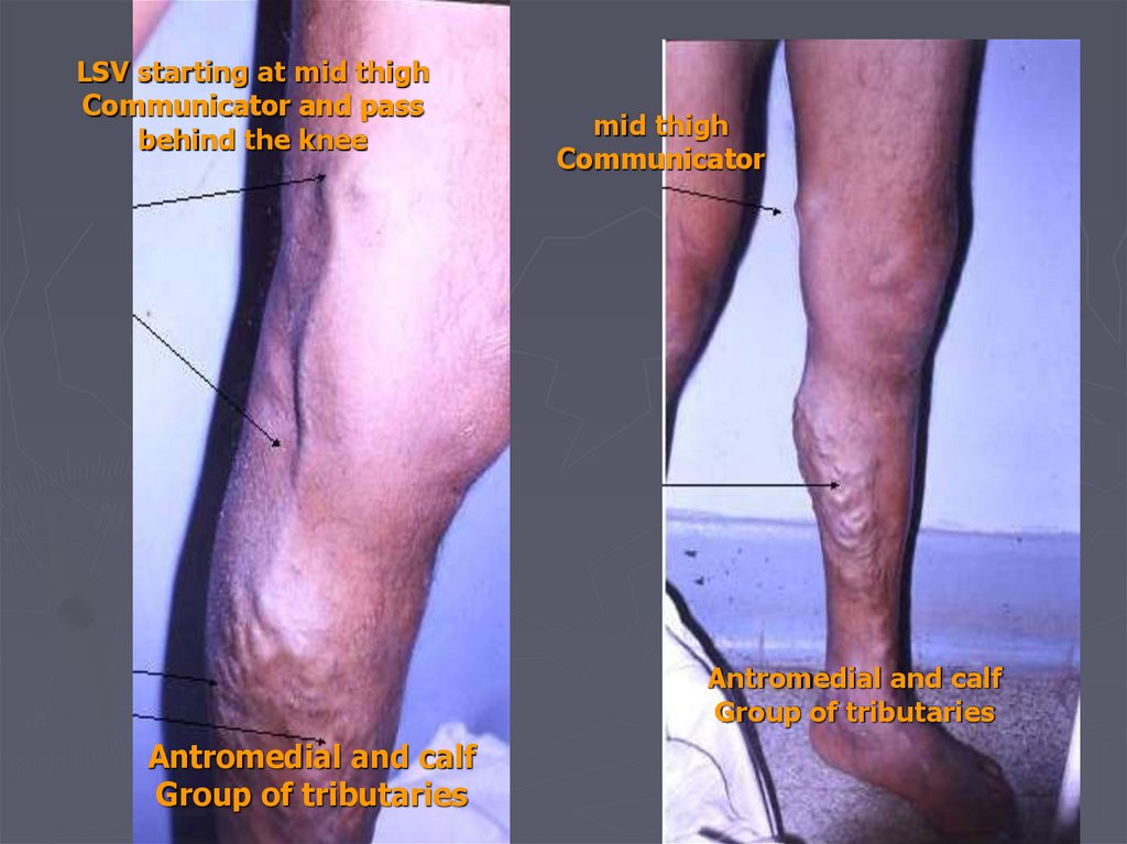
LSV starting at mid thighCommunicator and pass
behind the knee
mid thigh
Communicator
Antromedial and calf
Group of tributaries
Antromedial and calf
Group of tributaries
18.
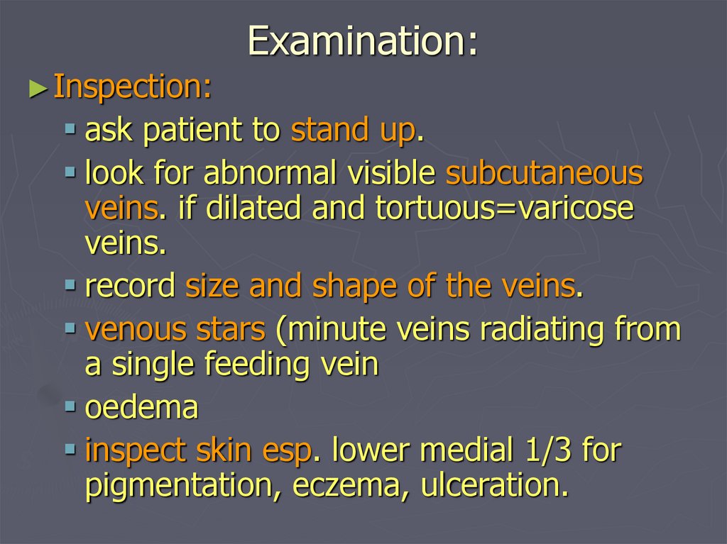
Examination:► Inspection:
ask patient to stand up.
look for abnormal visible subcutaneous
veins. if dilated and tortuous=varicose
veins.
record size and shape of the veins.
venous stars (minute veins radiating from
a single feeding vein
oedema
inspect skin esp. lower medial 1/3 for
pigmentation, eczema, ulceration.
19.
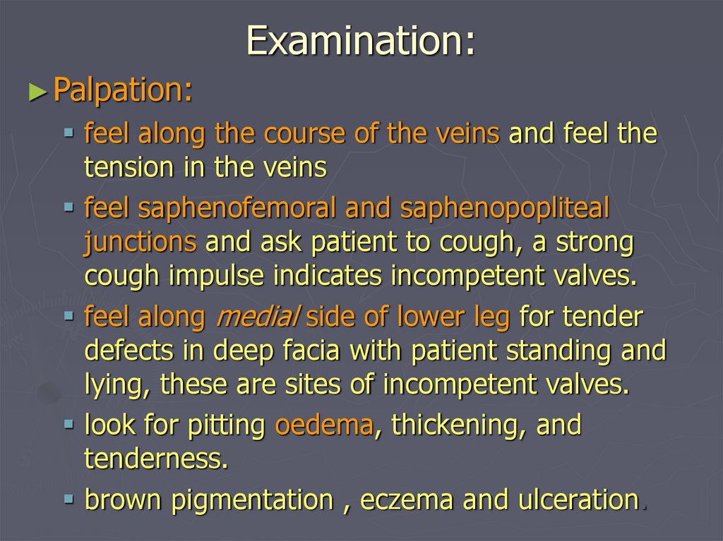
Examination:► Palpation:
feel along the course of the veins and feel the
tension in the veins
feel saphenofemoral and saphenopopliteal
junctions and ask patient to cough, a strong
cough impulse indicates incompetent valves.
feel along medial side of lower leg for tender
defects in deep facia with patient standing and
lying, these are sites of incompetent valves.
look for pitting oedema, thickening, and
tenderness.
brown pigmentation , eczema and ulceration.
20.
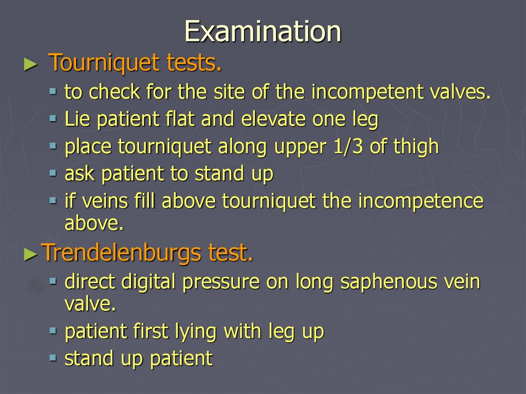
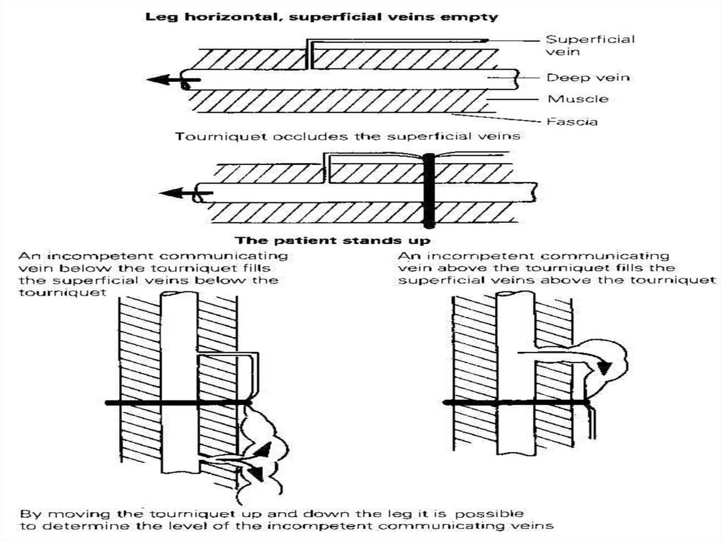
ExaminationTourniquet tests.
to check for the site of the incompetent valves.
Lie patient flat and elevate one leg
place tourniquet along upper 1/3 of thigh
ask patient to stand up
if veins fill above tourniquet the incompetence
above.
► Trendelenburgs
test.
direct digital pressure on long saphenous vein
valve.
patient first lying with leg up
stand up patient
21.
22.
ExaminationPercussion:
transmission of percussion waves downward
implies incompetent valves ( Shwartz test).
Place fingers of one hand on lower limit of
visible vein and tap top.
► Auscultation:
listen over clusters of veins especially if they
remain distended when patient lies down may
be arteriovenous fistula.
23.
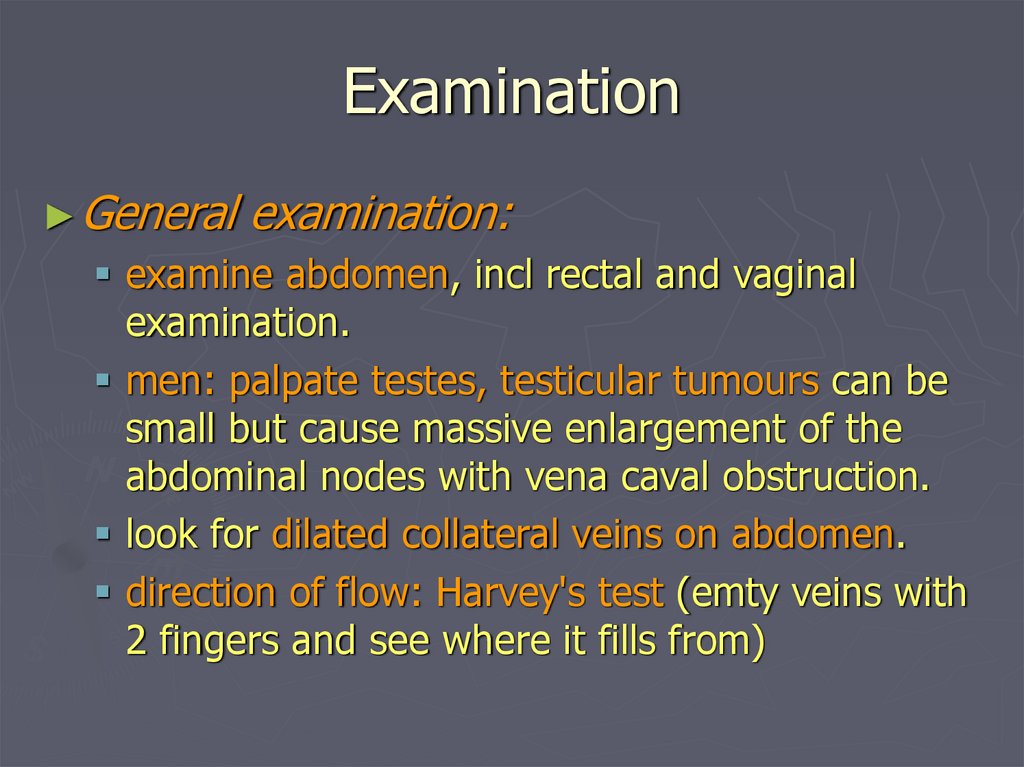
Examination► General
examination:
examine abdomen, incl rectal and vaginal
examination.
men: palpate testes, testicular tumours can be
small but cause massive enlargement of the
abdominal nodes with vena caval obstruction.
look for dilated collateral veins on abdomen.
direction of flow: Harvey's test (emty veins with
2 fingers and see where it fills from)
24.
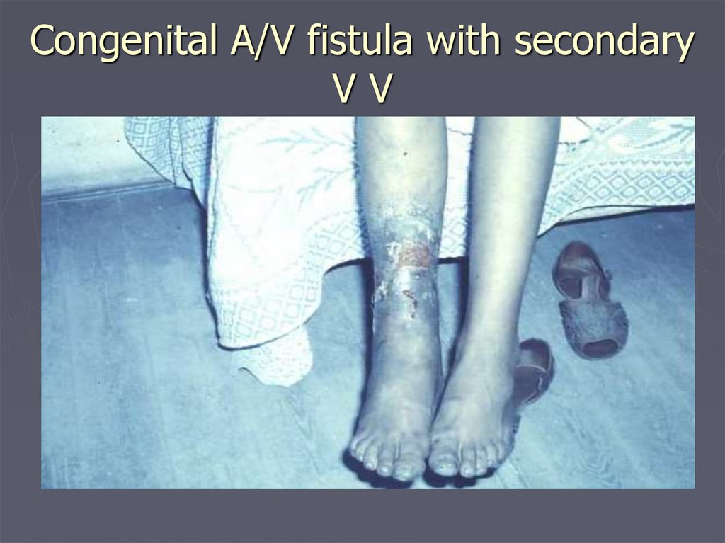
Congenital A/V fistula with secondaryVV
25.
26.
Traumatic A/V fistula with secondaryVV
27.
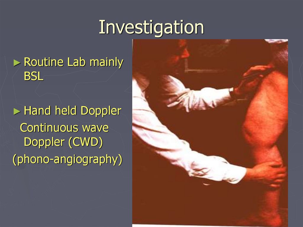
Investigation► Routine
BSL
► Hand
Lab mainly
held Doppler
Continuous wave
Doppler (CWD)
(phono-angiography)
28.
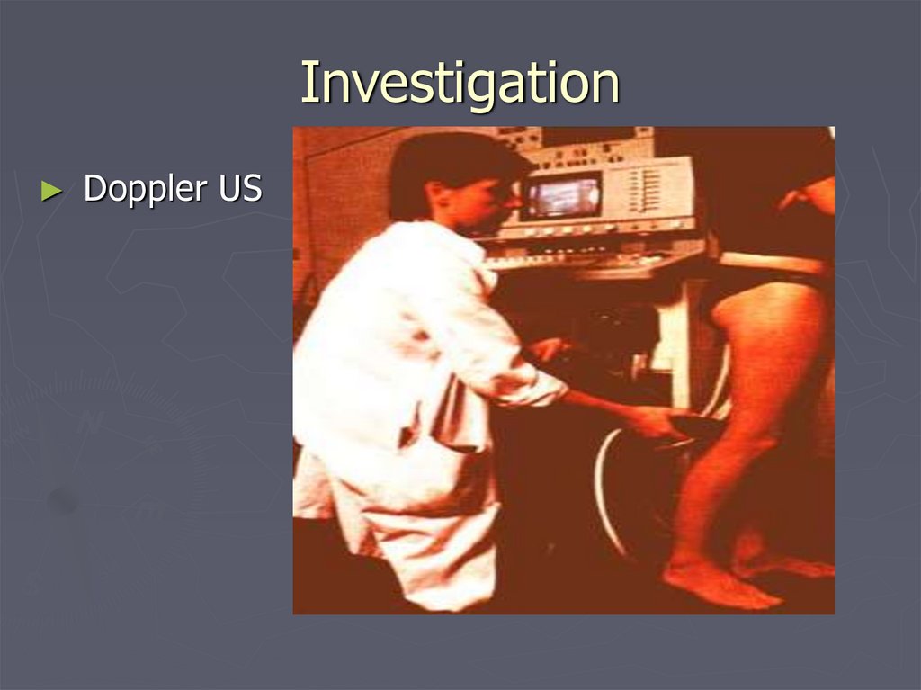
InvestigationDoppler US
29.
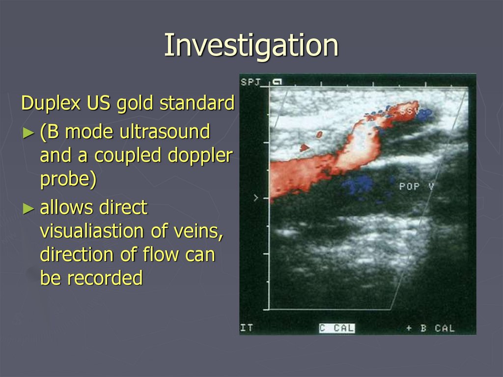
InvestigationDuplex US gold standard
► (B mode ultrasound
and a coupled doppler
probe)
► allows direct
visualiastion of veins,
direction of flow can
be recorded
30.
Investigation► Plethysmography
and Venography are
obsolete
► Venous pressure
► Radio-active isotope scanning
► Arteriograpgy if A/V fistula
31.
Complications:► Haemorrhage
► Oedema
► Skin
pigmentation
► Lipodermatosclerosis
► Varicose eczema
► Venous ulceration
► Thrombophlebitis
► Atrophie blanche
► Marjolin ulcer
► Equinous deformity
32.
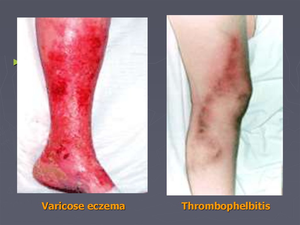
Varicose eczema
Thrombophelbitis
33.
Treatment► A.
Non- operative management.
walking should be encouraged and
prolonged sitting and standing should
be forbidden
patient should elevate leg as
frequently as possible to reduce
venous pressure.
elastic stockings. extending from
distal metatarsals to just below the
knee
34.
TreatmentCompression
sclerotherapy.
permanent fibrotic
occlusion of collapsed
veins.
patient is recumbent
and veins collapsed,
a small amout of 0.5 ml
of sclerosing solution
(3% sodium tetradecyl
sulfate) is injected into
each varix
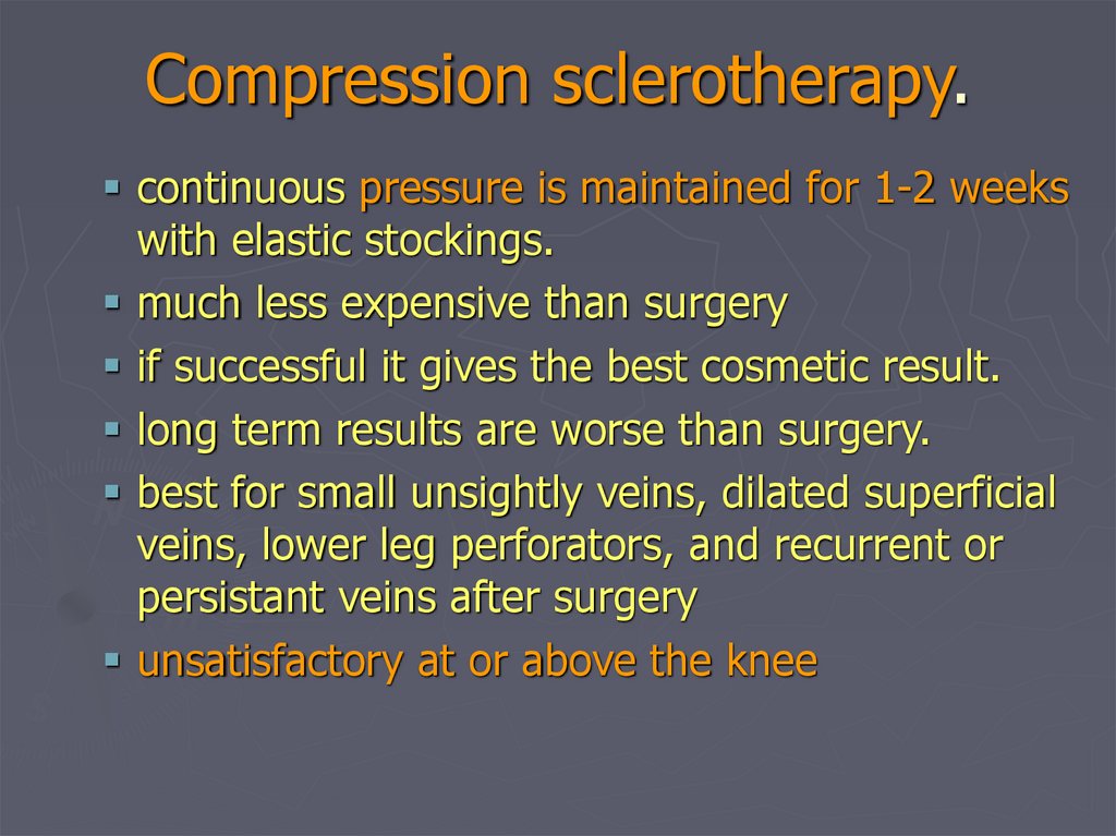
35.
Compression sclerotherapy.continuous pressure is maintained for 1-2 weeks
with elastic stockings.
much less expensive than surgery
if successful it gives the best cosmetic result.
long term results are worse than surgery.
best for small unsightly veins, dilated superficial
veins, lower leg perforators, and recurrent or
persistant veins after surgery
unsatisfactory at or above the knee
36.
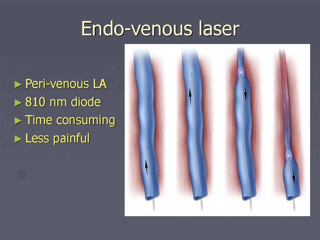
Endo-venous laser► Peri-venous
LA
► 810 nm diode
► Time consuming
► Less painful
37.
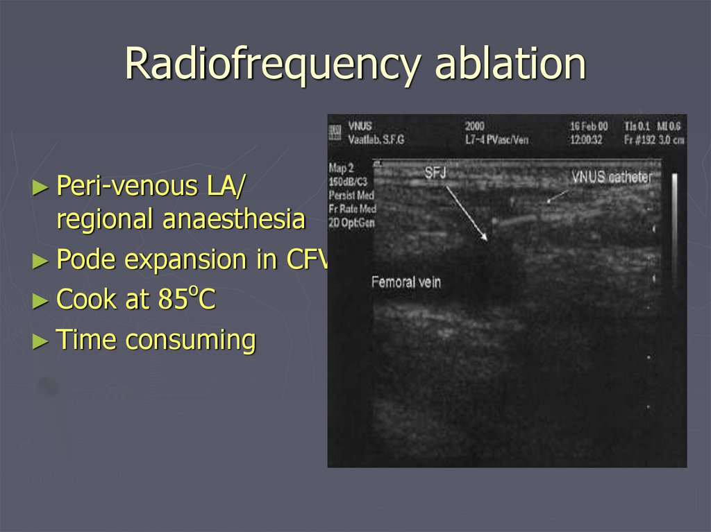
Radiofrequency ablation► Peri-venous
LA/
regional anaesthesia
► Pode expansion in CFV
o
► Cook at 85 C
► Time consuming
38.
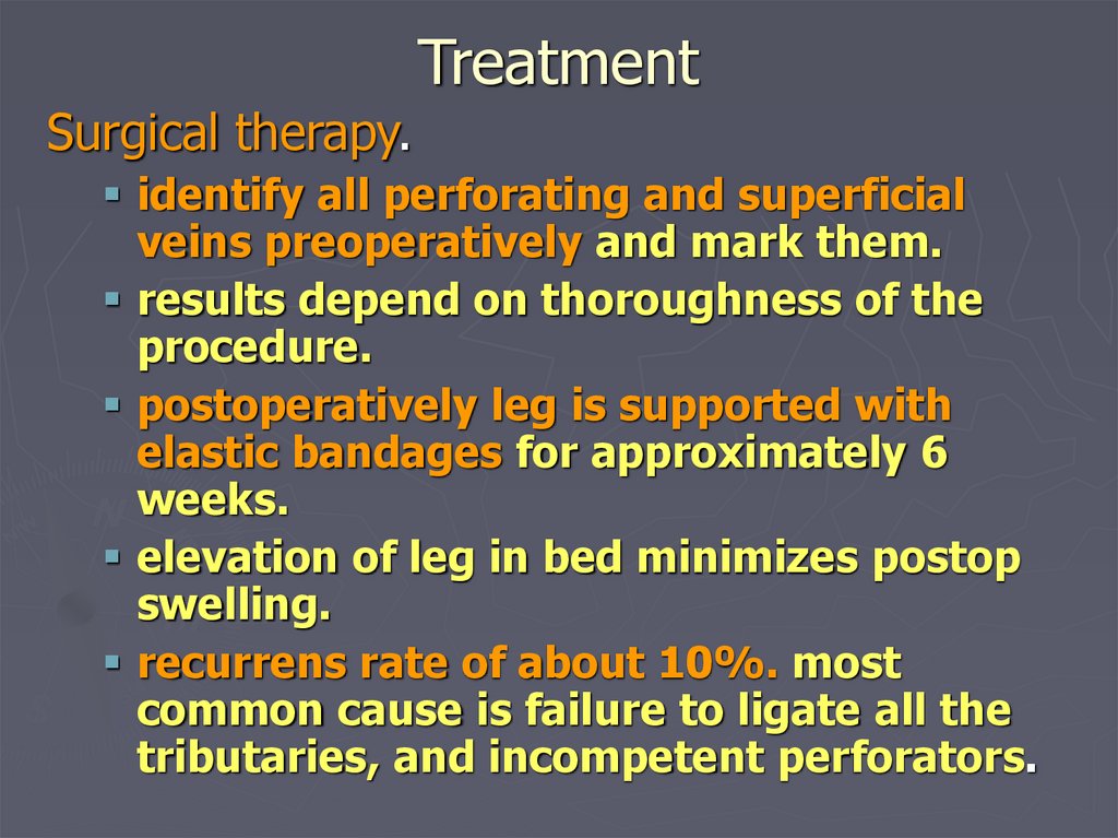
TreatmentSurgical therapy.
Indications:
►severe symptoms
►very large varices
►attacks of superficial phlebitis
►haemorrhage from rupturd
varices.
►ulceration from venous stasis.
►cosmetic reasons.
39.
TreatmentSurgical therapy.
identify all perforating and superficial
veins preoperatively and mark them.
results depend on thoroughness of the
procedure.
postoperatively leg is supported with
elastic bandages for approximately 6
weeks.
elevation of leg in bed minimizes postop
swelling.
recurrens rate of about 10%. most
common cause is failure to ligate all the
tributaries, and incompetent perforators.
40.
41.
42.
43.
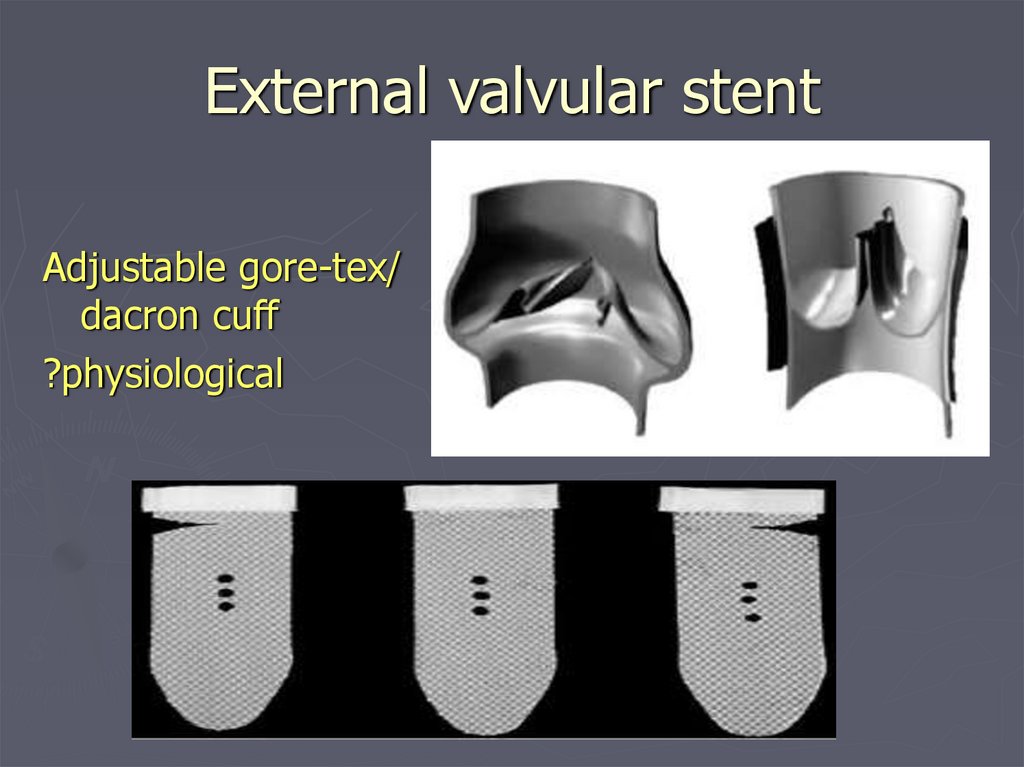
External valvular stentAdjustable gore-tex/
dacron cuff
?physiological
44.
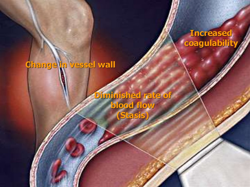
Deep Vein Thrombosis► Only
1/3 of DVT's cause symptoms and signs.
► predisposition to thrombosis is predicted with
Virchow's triad.
Change in vessel wall; distention, injury,
inflammation, trauma.
Diminished rate of blood flow; during and after
operations (postop rare before 40years, most common
operations;obesity, operations for cancer,prostate and
hip), debilitating diseases
Increased coagulability of the blood; infections,
after haemorrhage, visceral cancers,during pregnancy,
hypercoagulable states( congenital abnormalities of
protein C and S, antithrombin III), deficiencies in the
fibrinolytic system
45.
Increasedcoagulability
Change in vessel wall
Diminished rate of
blood flow
(Stasis)
46.
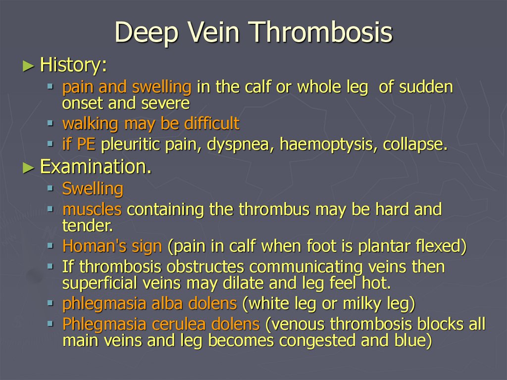
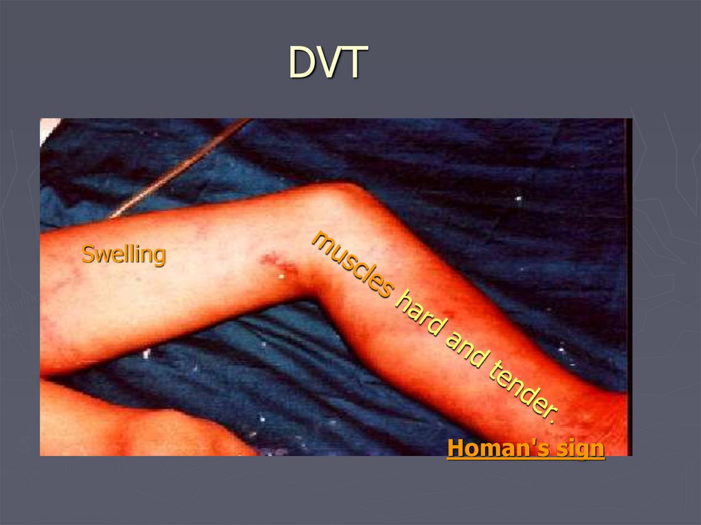
Deep Vein Thrombosis► History:
pain and swelling in the calf or whole leg of sudden
onset and severe
walking may be difficult
if PE pleuritic pain, dyspnea, haemoptysis, collapse.
► Examination.
Swelling
muscles containing the thrombus may be hard and
tender.
Homan's sign (pain in calf when foot is plantar flexed)
If thrombosis obstructes communicating veins then
superficial veins may dilate and leg feel hot.
phlegmasia alba dolens (white leg or milky leg)
Phlegmasia cerulea dolens (venous thrombosis blocks all
main veins and leg becomes congested and blue)
47.
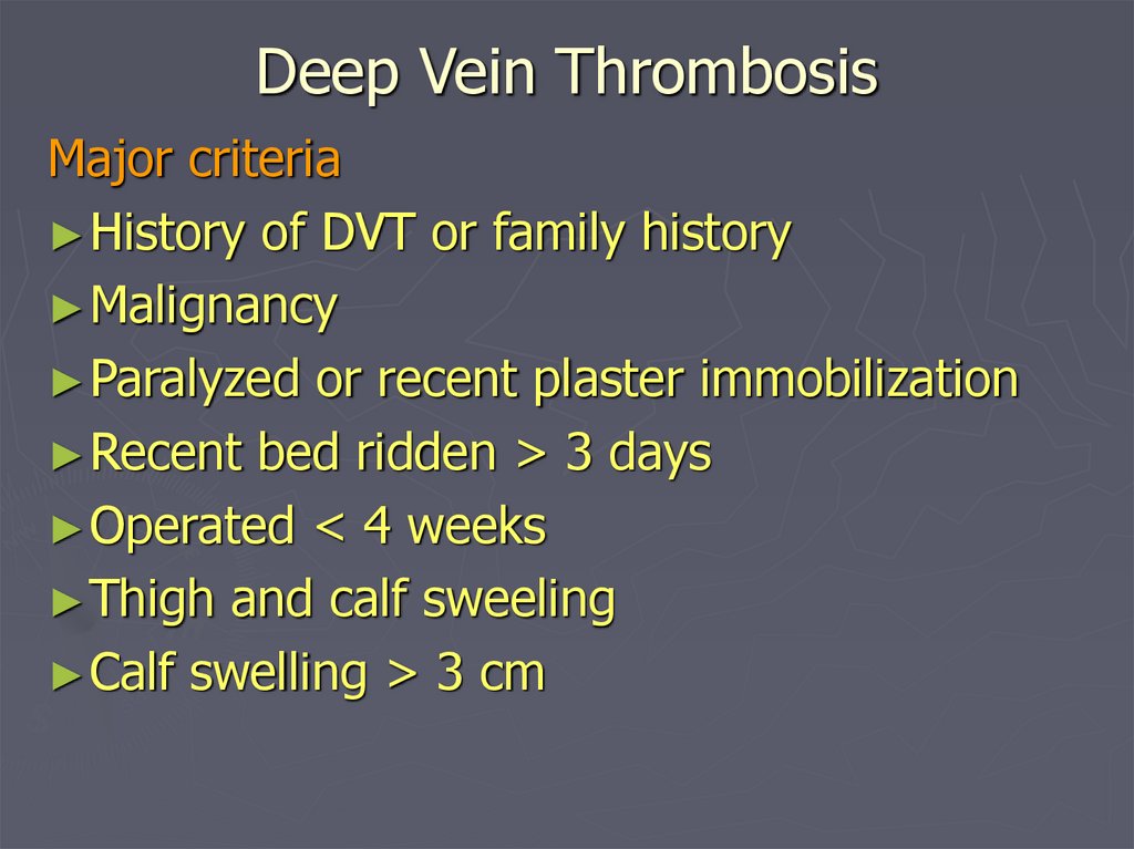
Deep Vein ThrombosisMajor criteria
► History of DVT or family history
► Malignancy
► Paralyzed or recent plaster immobilization
► Recent bed ridden > 3 days
► Operated < 4 weeks
► Thigh and calf sweeling
► Calf swelling > 3 cm
48.
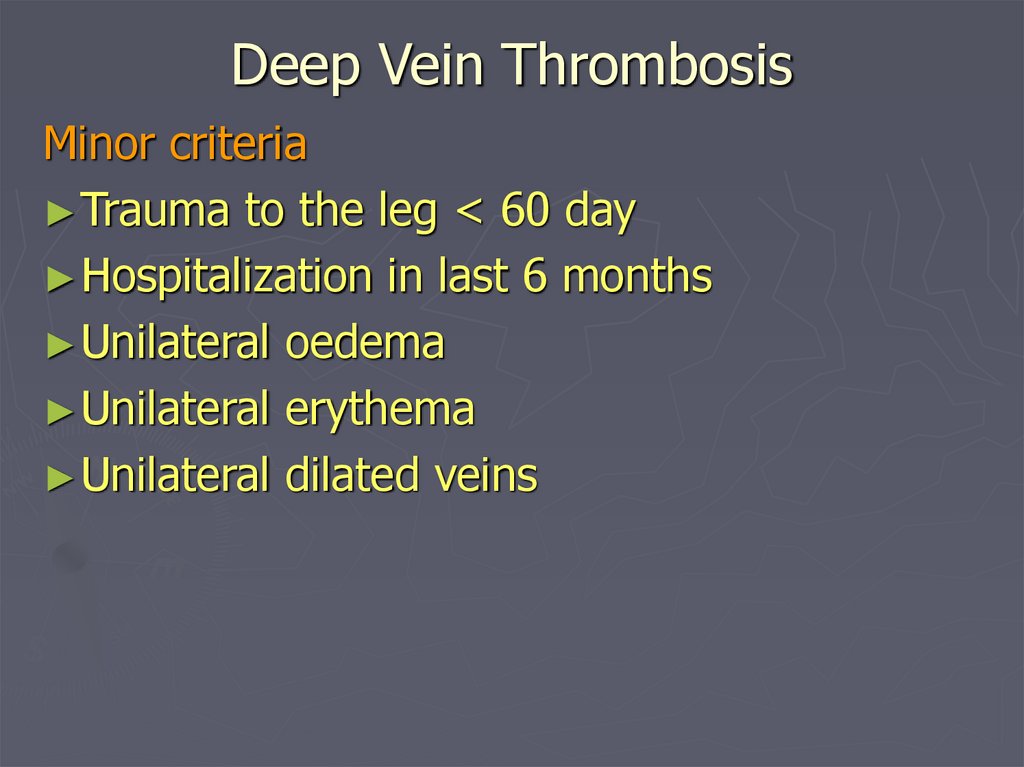
Deep Vein ThrombosisMinor criteria
► Trauma to the leg < 60 day
► Hospitalization in last 6 months
► Unilateral oedema
► Unilateral erythema
► Unilateral dilated veins
49.
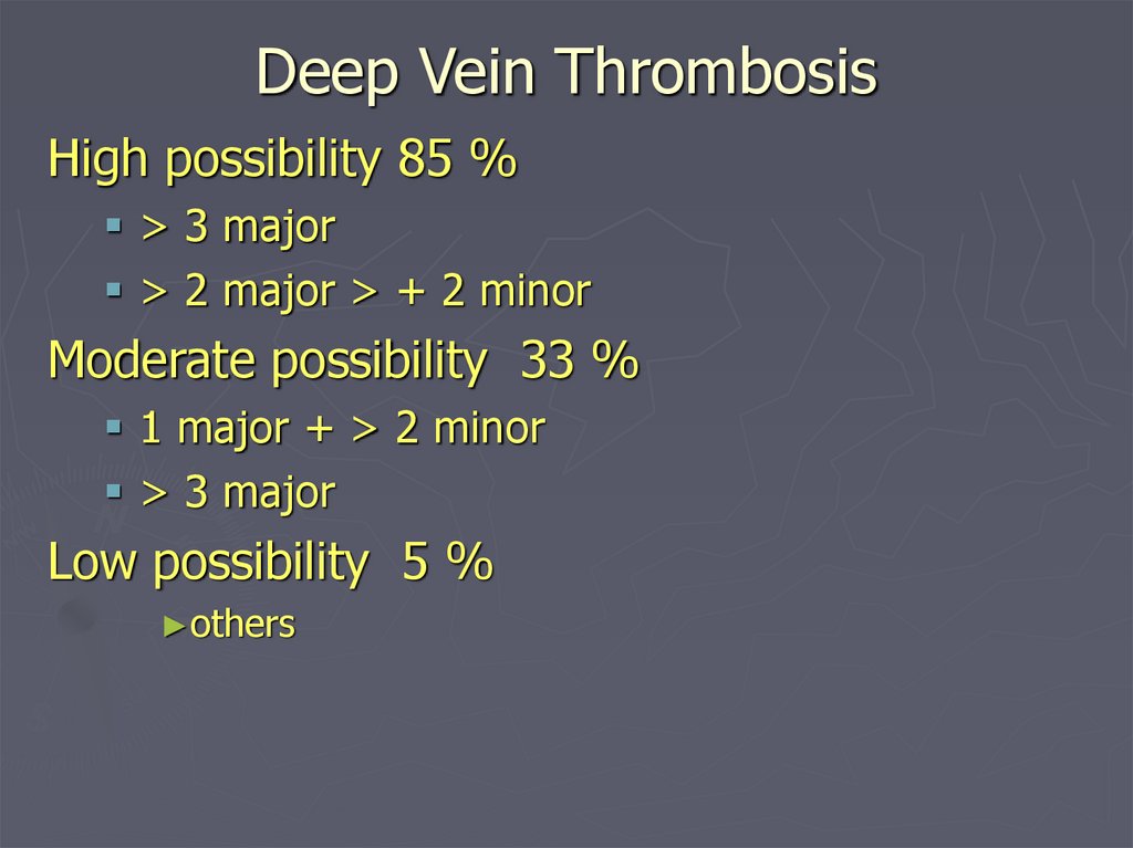
Deep Vein ThrombosisHigh possibility 85 %
> 3 major
> 2 major > + 2 minor
Moderate possibility 33 %
1 major + > 2 minor
> 3 major
Low possibility 5 %
►others
50.
DVTSwelling
Homan's sign
51.
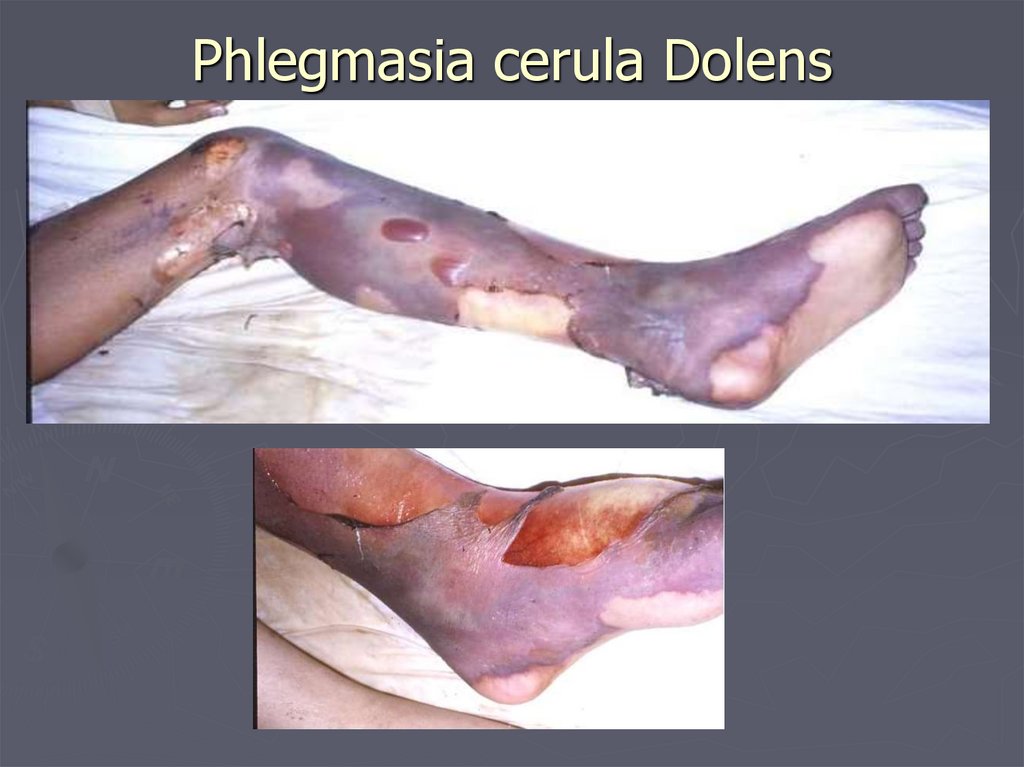
Phlegmasia cerula Dolens52.
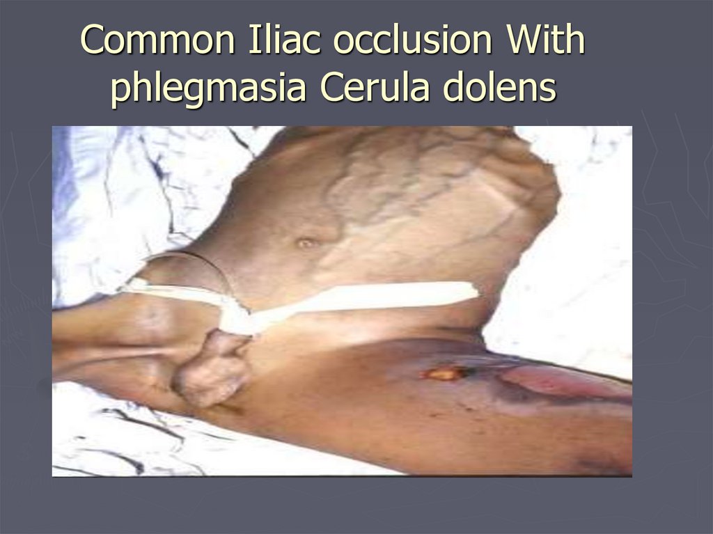
Common Iliac occlusion Withphlegmasia Cerula dolens
53.
IVC occlusion54.
Prevention of DVT► Before
operation: Stop pill ( if possible 6 weeks
before), grossly overweight patients should reduce
weight, those over 40 should have increased
activity 2-3 weeks at home, low dose heparin
► During operation: prevent pressure on venous
system(elevate leg on sand bag), graduated
compression stocking or intermittent pneumatic
compression, after operation elevate and massage
the leg.
► After operation: Massage, leg movements,
graduated stockings (TED stockings), low dose
heparin, adequate hydration, early ambulation,
Patients should not sit with their legs dependant
often better to have in bed then sitting in a chair.
55.
Prevention of DVTMethods of prevention:
Mechanical: assisting venous return by; Graduated static
compression elastic stockings (Kendall's Thrombo Embolic
Deterrent-TED) may reduce incidence of DVT to below
10% (20% in hip surgery), electronic stimulation of calf
muscles, Pneumatic compression.
► Low dose heparin 5000 units subcutaneously 2h before
operation and continued twice daily until patient is fully
ambulating, avoid if operation will leave bleeding areas or
if bleeding in cosed space may be disastrous.
► Low molecular weight heparins; reduced risk of bleeding
but as effective.
► (Dextran '70'. inhibits platelet adhesion 500ml iv during
operation and 500 ml following 24 h)
56.
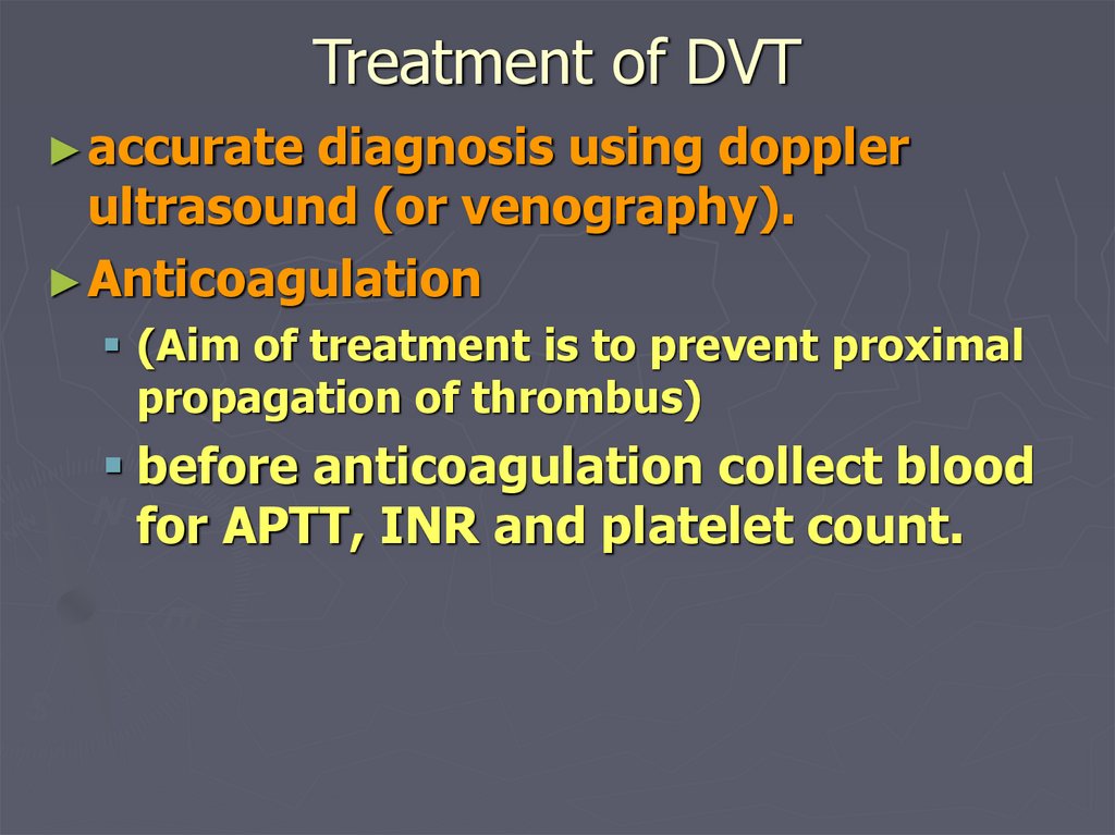
Treatment of DVT► accurate
diagnosis using doppler
ultrasound (or venography).
► Anticoagulation
(Aim of treatment is to prevent proximal
propagation of thrombus)
before anticoagulation collect blood
for APTT, INR and platelet count.
57.
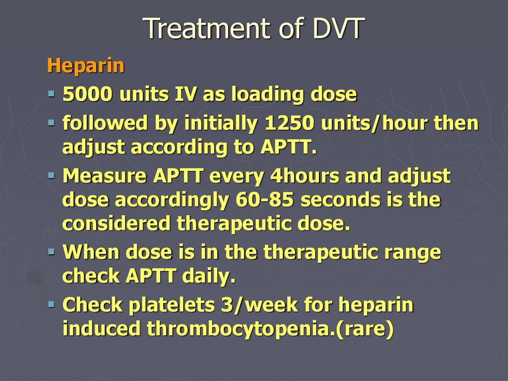
Treatment of DVTHeparin
5000 units IV as loading dose
followed by initially 1250 units/hour then
adjust according to APTT.
Measure APTT every 4hours and adjust
dose accordingly 60-85 seconds is the
considered therapeutic dose.
When dose is in the therapeutic range
check APTT daily.
Check platelets 3/week for heparin
induced thrombocytopenia.(rare)
58.
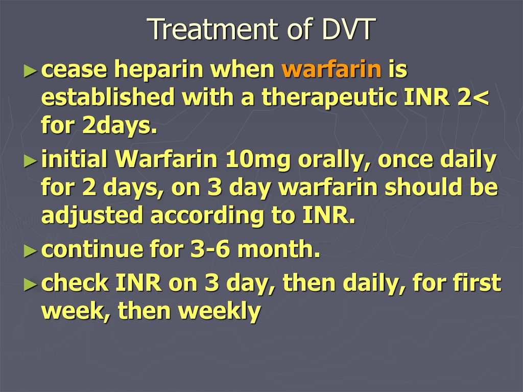
Treatment of DVT► cease
heparin when warfarin is
established with a therapeutic INR 2<
for 2days.
► initial Warfarin 10mg orally, once daily
for 2 days, on 3 day warfarin should be
adjusted according to INR.
► continue for 3-6 month.
► check INR on 3 day, then daily, for first
week, then weekly
59.
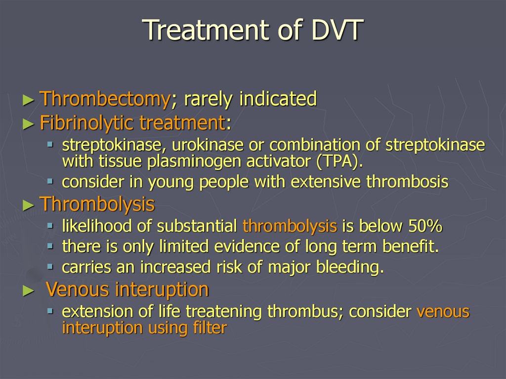
Treatment of DVT► Thrombectomy;
rarely indicated
► Fibrinolytic treatment:
streptokinase, urokinase or combination of streptokinase
with tissue plasminogen activator (TPA).
consider in young people with extensive thrombosis
► Thrombolysis
likelihood of substantial thrombolysis is below 50%
there is only limited evidence of long term benefit.
carries an increased risk of major bleeding.
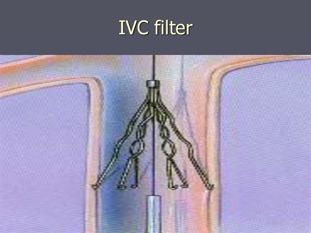
Venous interuption
extension of life treatening thrombus; consider venous
interuption using filter
60.
IVC filter61.
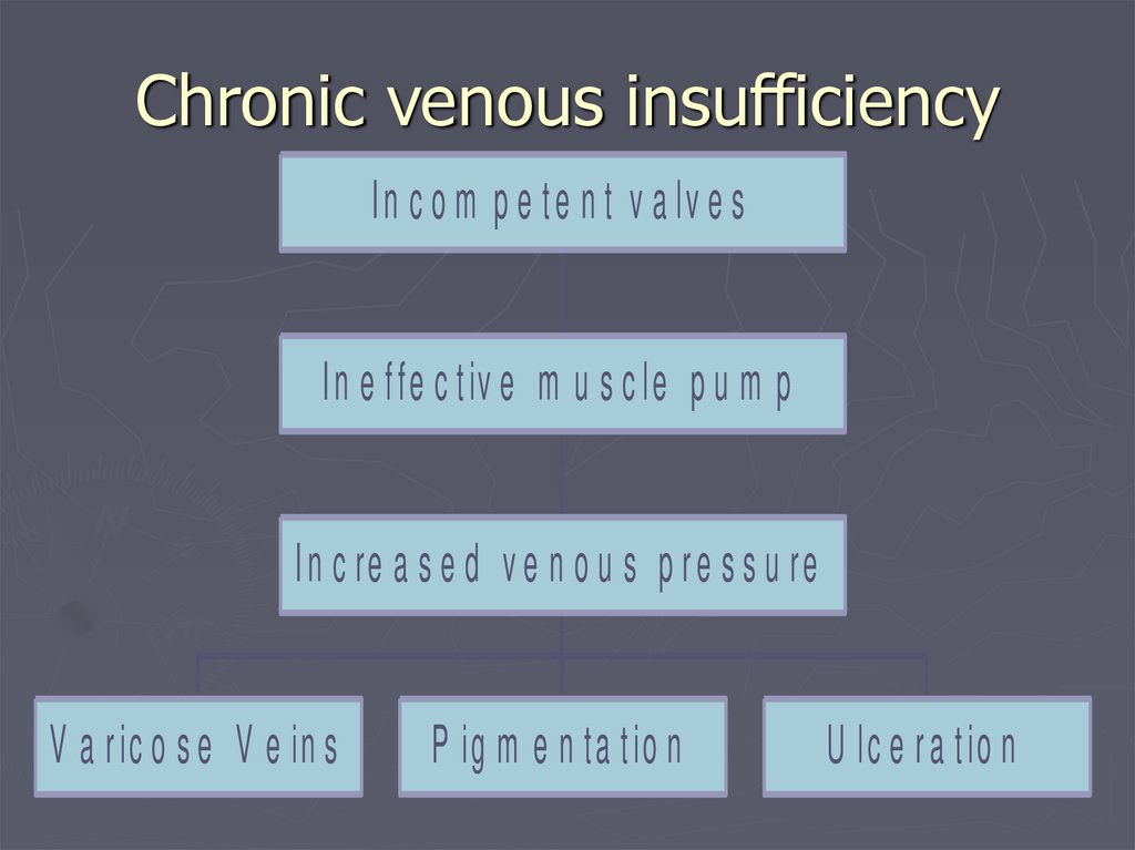
Chronic venous insufficiencyI n c o m p e t e n t v a lv e s
I n e f fe c t iv e m u s c le p u m p
I n c re a s e d v e n o u s p r e s s u re
V a r ic o s e V e in s
P ig m e n ta t io n
U lc e r a t io n
62.
Chronic venous insufficiencyMacro- circulation Changes
Muscle dysfunction
Increase AVP
Increase perforator
incompetence
SVI 40 %
Reflux
Primary
Secondary
To DVT
Obstruction
Physical Functional
Combined SVI+
DVI 40%
Perforator Incompetence isolated in 2-4%
DVI 10 %
63.
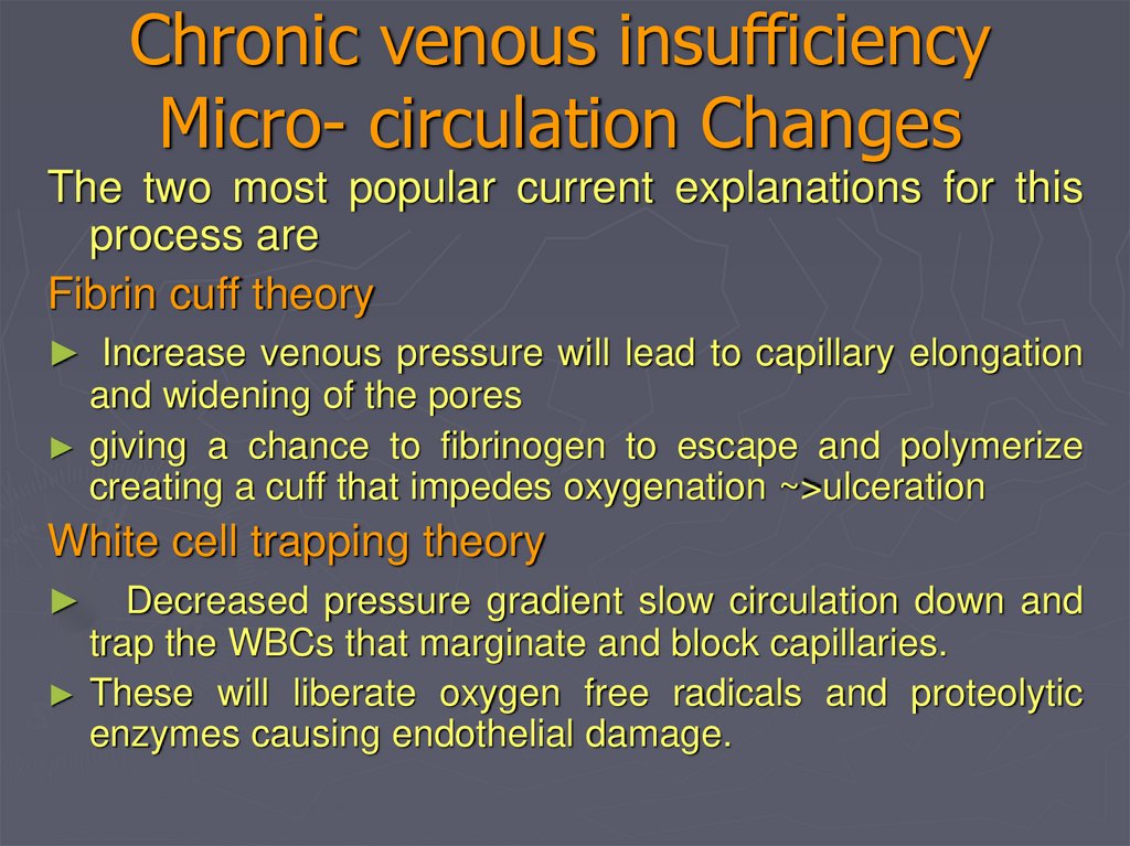
Chronic venous insufficiencyMicro- circulation Changes
The two most popular current explanations for this
process are
Fibrin cuff theory
► Increase venous pressure will lead to capillary elongation
and widening of the pores
giving a chance to fibrinogen to escape and polymerize
creating a cuff that impedes oxygenation ~>ulceration
White cell trapping theory
Decreased pressure gradient slow circulation down and
trap the WBCs that marginate and block capillaries.
These will liberate oxygen free radicals and proteolytic
enzymes causing endothelial damage.
64.
Chronic venous insufficiencyMicro- circulation Changes
Arterio-venous communications
Some suggest the presence arteriovenous shunts
further depriving the skin from oxygen
Trap hypothesis
Some suggest that macromolecules exuded can trap
growth factors and cells rendering them unavailable
for regular tissue repairs
Tissue pressure
The benefit of elevation, elastic stocking and corrective
venous surgery reduces the tissue pressures and heal
the ulcers
Cutaneous iron overload
The accumulation of ferritin can induce production of
oxygen free radilces causing tissue destruction.
65.
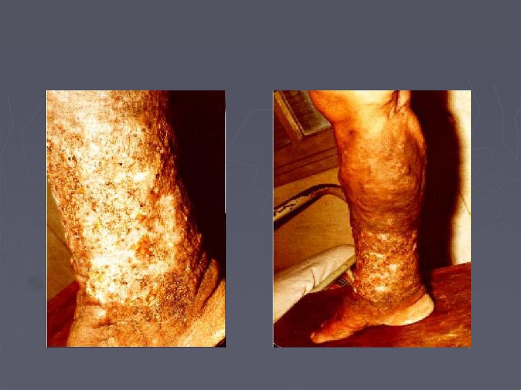
Chronic venous insufficiency► Oedema
► Skin
pigmentation
► Lipodermatosclerosis
► Varicose eczema
► Venous ulceration
► Thrombophlebitis
► Atrophie blanche
► Marjolin ulcer
► Equinous deformity
66.
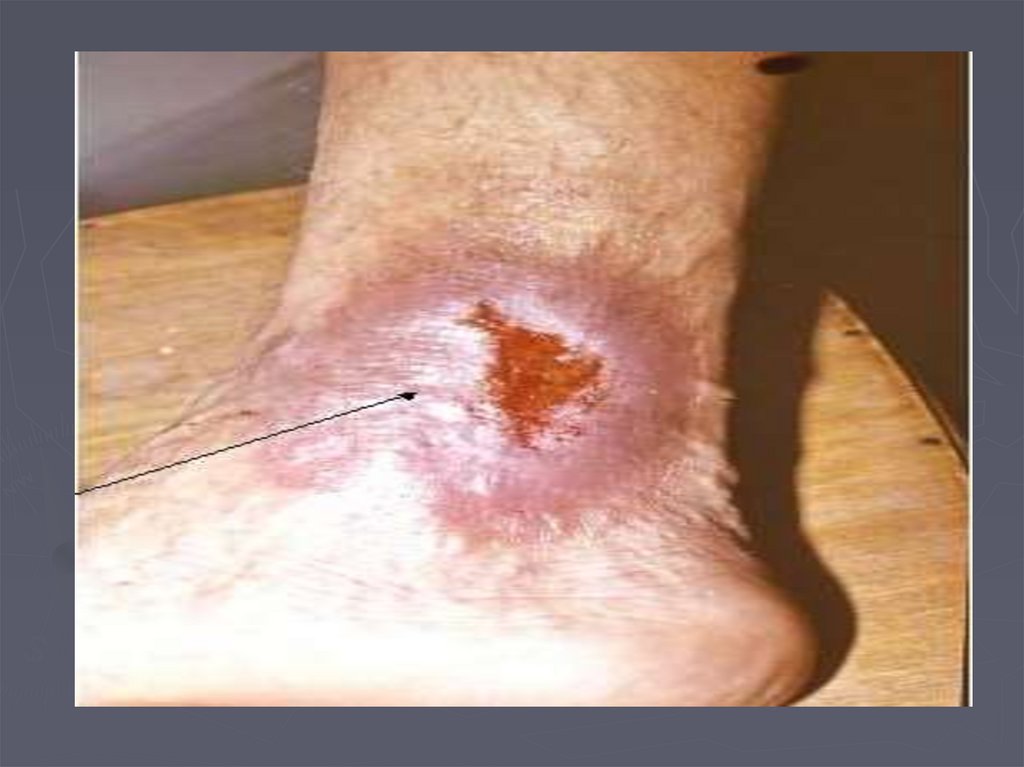
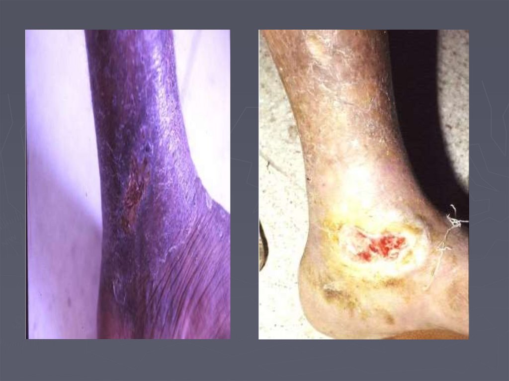
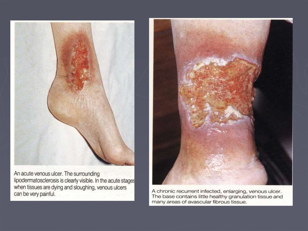
Venous Ulcer► The
ulcer
Gaiter area lower leg (medial lower 1/3)
edge sloping and pale purple-blue in colour.
base ping granulation tissue.
tendons and bones may be exposed.
seropurulent discharge, heavy infection and pus is not
common.
shallow and flat.
surrounding tissues show signs of venous hypertension
(pigmentation, warmth, redness and tenderness)
scars from previous ulcers, scar tissue may interfere
with movement of foot.
lymph nodes should not be enlarged.
67.
68.
LSVCVI
Ankle flare
LSV
CVI
Ankle flare
69.
70.
71.
72.
73.
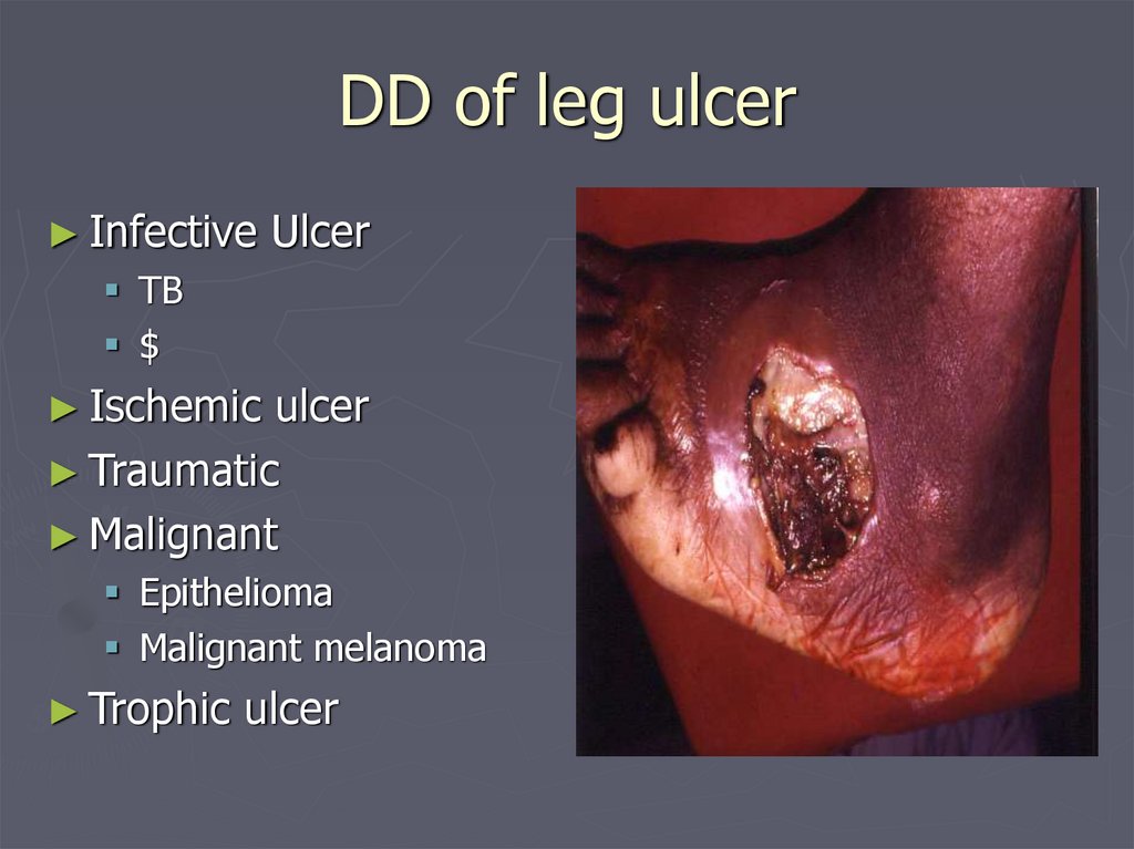
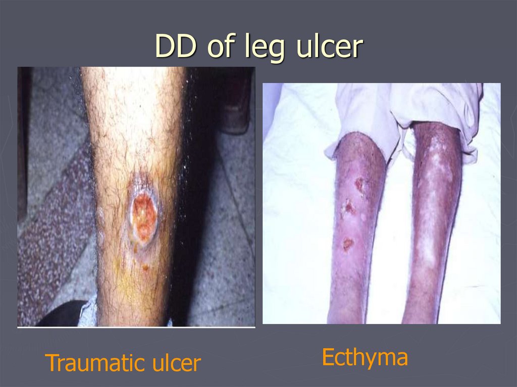
DD of leg ulcer► Infective
Ulcer
TB
$
► Ischemic
ulcer
► Traumatic
► Malignant
Epithelioma
Malignant melanoma
► Trophic
ulcer
74.
DD of leg ulcerTraumatic ulcer
Ecthyma
75.
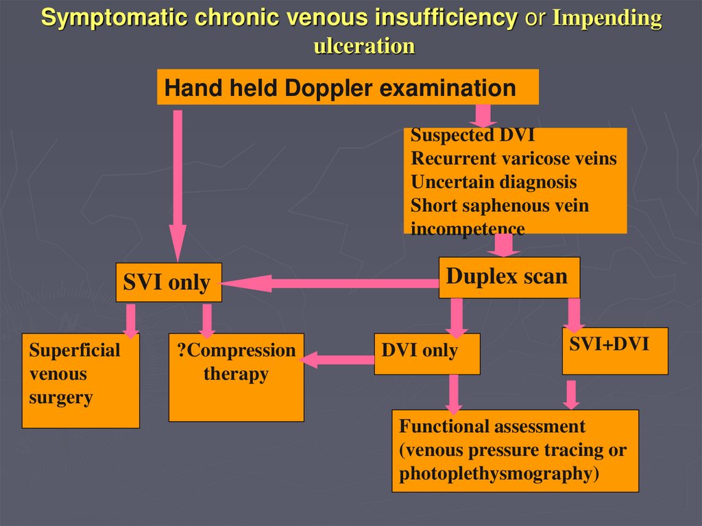
Symptomatic chronic venous insufficiency or Impendingulceration
Hand held Doppler examination
Suspected DVI
Recurrent varicose veins
Uncertain diagnosis
Short saphenous vein
incompetence
SVI only
Superficial
venous
surgery
?Compression
therapy
Duplex scan
DVI only
SVI+DVI
Functional assessment
(venous pressure tracing or
photoplethysmography)
76.
Symptomatic chronic venous insufficiency or Impendingulceration
Functional assessment
(venous pressure tracing or
photoplethysmography)
No improvement
with superficial
venous occlusion
Compression
therapy
Significant improvement
in refilling time with
superficial venous
occlusion
Superficial
venous
surgery
77.
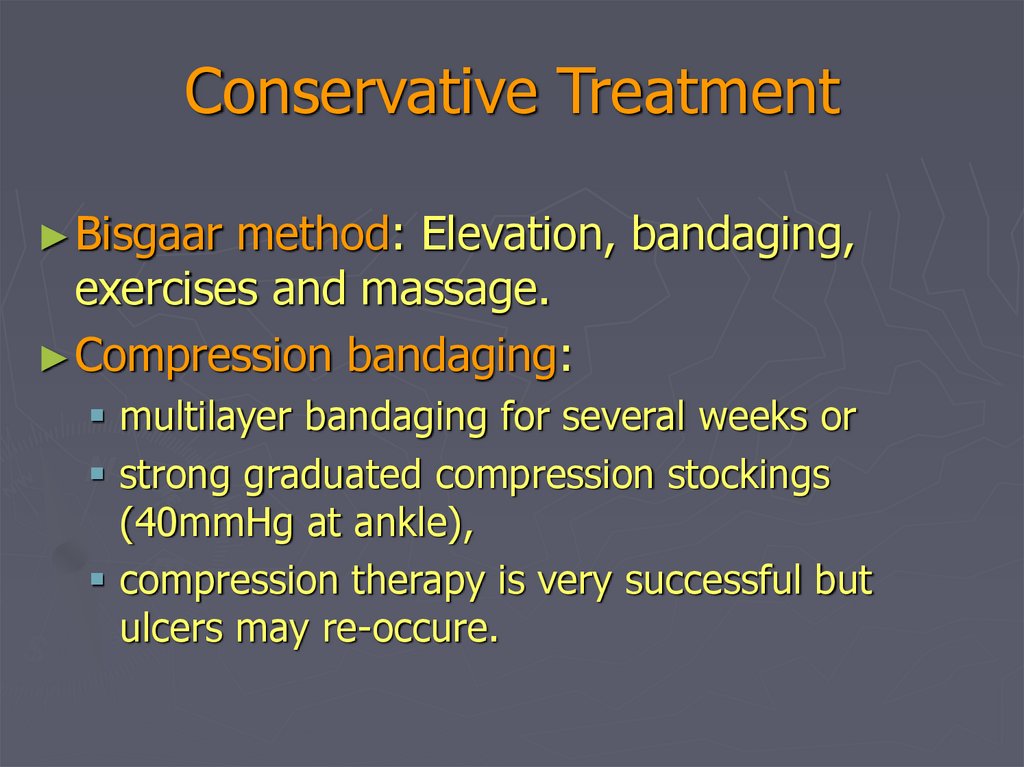
Conservative Treatment► Bisgaar
method: Elevation, bandaging,
exercises and massage.
► Compression bandaging:
multilayer bandaging for several weeks or
strong graduated compression stockings
(40mmHg at ankle),
compression therapy is very successful but
ulcers may re-occure.
78.
Surgical Treatment► Ligation
and division of incompetent perforating
veins
to prevent hydrodynamic forces generated in the
muscular compartment from reaching the skin
(surgical or endoscopic)
► Stripping
of incompetent main superficial systems
if they are contributing to the high AVP significantly
► Plastic surgery: grafting.
79.
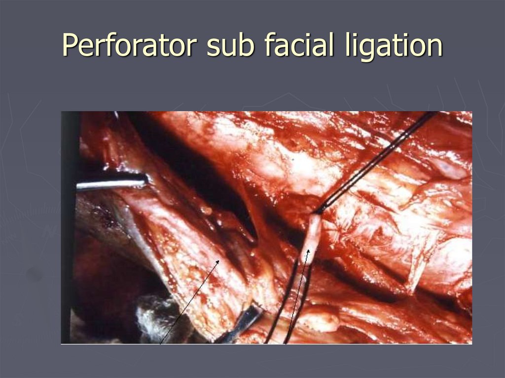
Perforator sub facial ligation80.
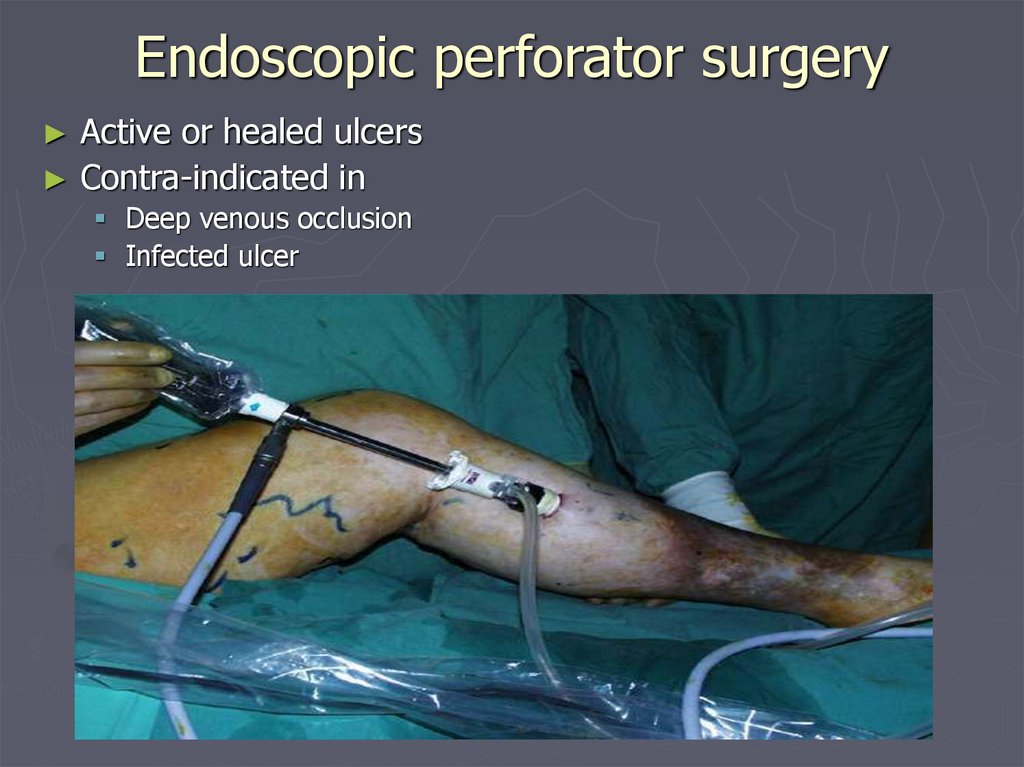
Endoscopic perforator surgeryActive or healed ulcers
Contra-indicated in
Deep venous occlusion
Infected ulcer
81.
TreatmentDeep venous reconstruction
► Not yet standard treatment
► Can correct primary deep veins reflux but not postthombotic reflux or obstruction
► Most commonly repaired veins are femoral and
popliteal
► Done from within
Valvuloplasty
Valve transposition or coursing
Valve transplantaton
82.
Kistner type valve repair for deepvein incompetence
83.
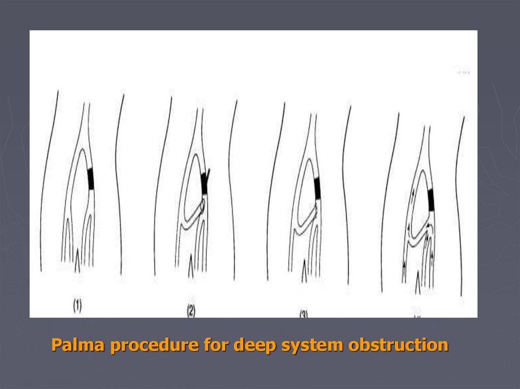
Palma procedure for deep system obstruction84.
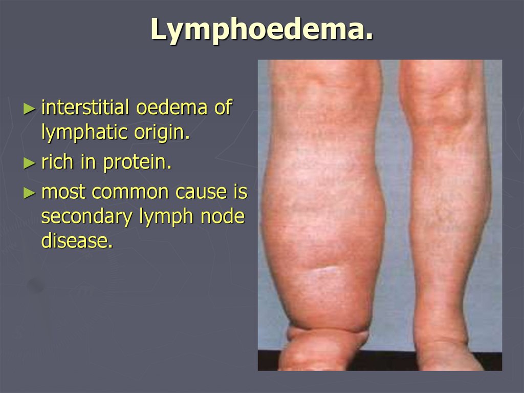
Lymphoedema.► interstitial
oedema of
lymphatic origin.
► rich in protein.
► most common cause is
secondary lymph node
disease.
85.
Causes► Primary:
Congenital or acquired deficiency of the
lymphatics ( aplasia or Hypoplasia)
Dilation and incompetence of the lymphatic
(Hyperplasia).
According to age of onset
►Congenita
since birth 10 %
►Precox adolescent (15- 35) 75 %
►Tarda > 35 y 15 %
86.
Causes► Secondary:
Neoplastic infiltration of lymph nodes.
► secondary
carcinoma
► Primary reticuloses.
Infection
► Filariasis
(parasite Wuchereria bancrofti) found in tropical and
subtropical climates.This is a cause of severe lymphoedema
(elephantiasis)
► lymphogranuloma inguinale
► TB
► Recurrent non-specific infection.
Iatrogenic
► surgical
excision
irradiation of lymph nodes.
87.
Clinical Classification► Sub
clinical with histological abnormalities of LN
and lymphatic
► Grade I
Oedema pit on pressure
Swelling disappear on elevation or bed rest
► Grade
II
Oedema does not pit on pressure
Swelling not disappear on elevation or bed rest
► Grade
III
Oedema
Irreversible skin changes ( fibrosis or papillae)
88.
Lymphoedema.History.
females>males.
slowly progressive swelling of the limb or genitelia.
lower limb most often affected often history of trauma
several years ago.
not painful and no discomfort.
commonly complicated by athlete's foot (tinea pedis)
and episodes of cellulitis.
Vesicles may appear on the skin that leak clear-coloured
fluid.
symptoms of underlying cause
very rare complication of lymphangiosarcoma.
oedema does not respond to leg elevation.
89.
Examination.► oedema
all oedema pits (clasically sayed to be non-pitting).
lymphoedema of the lower limb affect the toes much
more than other oedemas, if it has been present for
long time the toes become squared-off.
Examine the whole patient esp. cardia, renal and
abdomen, as well as local (venous congestion, venous
thrombosis) as diagnosis of lymphoedema is done after
everything else has been excluded
► SC
fibrosis
the skin on the dorsum of the foot can not be pinched
Stemmer’s sign
90.
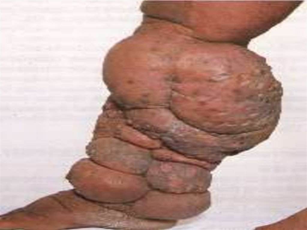
Examination.► In
advanced cases
Chronic eczema
Fungal skin infection ( Dermatophtosis)
Fungal nail infection ( Dermatomycosis)
skin gets thick and hyperkeratotic.
thick scales grow outward and look like warts.
► Ulceration
esp if associated CVI
► Rare
Lymphangectasia ( megalymphatics)
lymphangiosarcoma
91.
92.
Investigation► Laboratory
► Pathology
► Radiology
Contrast lymphangiography
Isotope lymphangiography
CT scan
MRI
93.
Management:►goals
of treatment is to control the
oedema and to prevent recurrent
infection.
►early treatment gives the best
results before fibrosis developes
and health of skin and
subcutaneous tissues are
compromised.
94.
Management:Non-operative Management:
► Physical methods
reduce lymph formation; elevation of the limb.
external compression; custom fitted, elastic stockings
worn threwout the day.
sequential air compression devices.
► Pharmacotherapy
restrict dietary sodium
diuretics when oedema is being actively treated.
instruction about foot care and hygiene to prevent
recurrent cellulitis.
prophylactic antibiotics may be recuired
Antifungal
95.
Management:► Surgical
Treatment:
Only needed in a small number of
patients.(16%)
Indications for surgery.
►impaired
function.
►pain
►recurrent
cellulitis and lymphangitis
►lymphangiosarcoma
►cosmetic although the result will not be a normal
looking limb.
96.
ManagementBypass
► Microsurgery: axial pattern and
mycocutaneous flaps and lymphaticlymphatic and lymphatico-venous
anastomoses.
► some procedures try to relieve the
obstruction by transplanting lymph channels
from normal areas
97.
ManagementReduction procedures
► excisional procedures removing skin and
lymphoedematous subcutaneous tissues and
usually requires extensive skin grafts.
complications: scarring, sensory loss,
recurrent swelling.
► Thompson procedure:
► Sistrunk
► Homan
► charles