


 Медицина
Похожие презентации:


Cardiac arrhythmias
1.
CARDIAC ARRHYTHMIASSergey Yalonetsky, MD
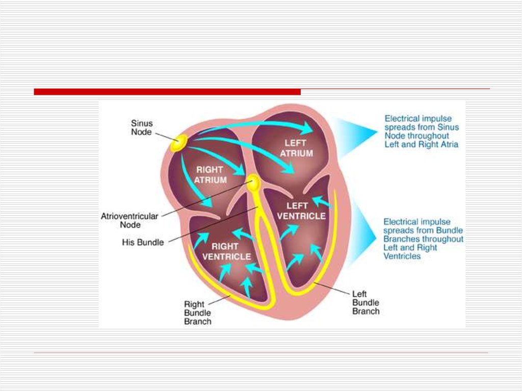
2.
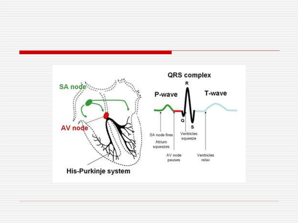
3.
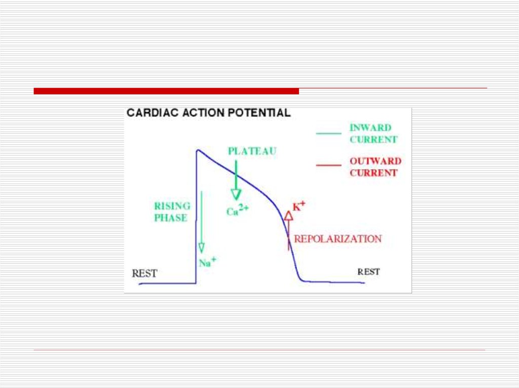
4.
5.
6.
7.
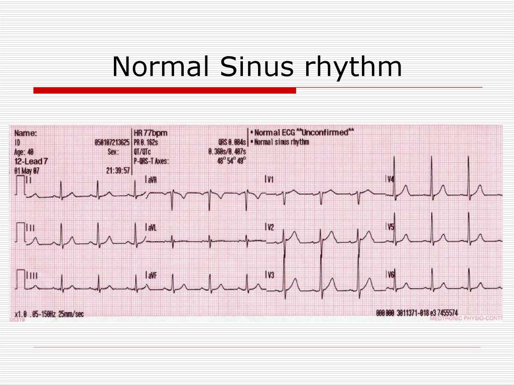
Normal Sinus rhythm8.
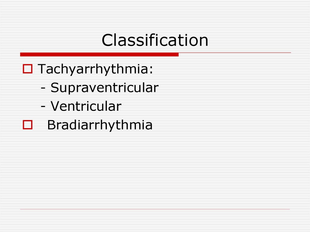
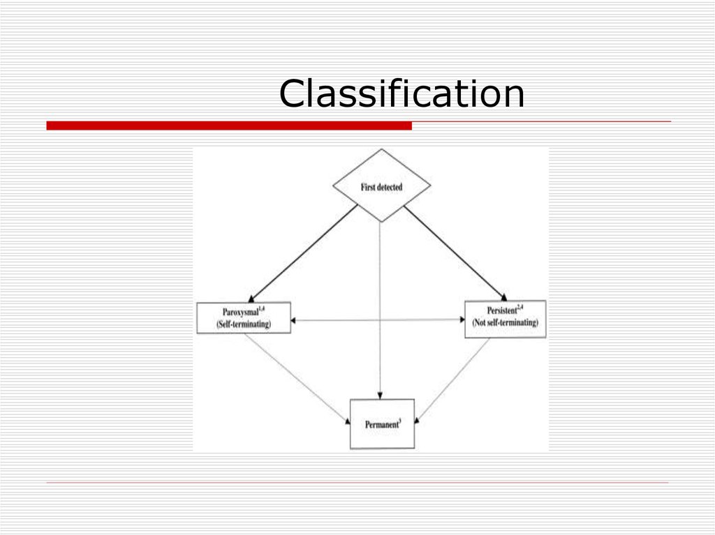
ClassificationTachyarrhythmia:
- Supraventricular
- Ventricular
Bradiarrhythmia
9.
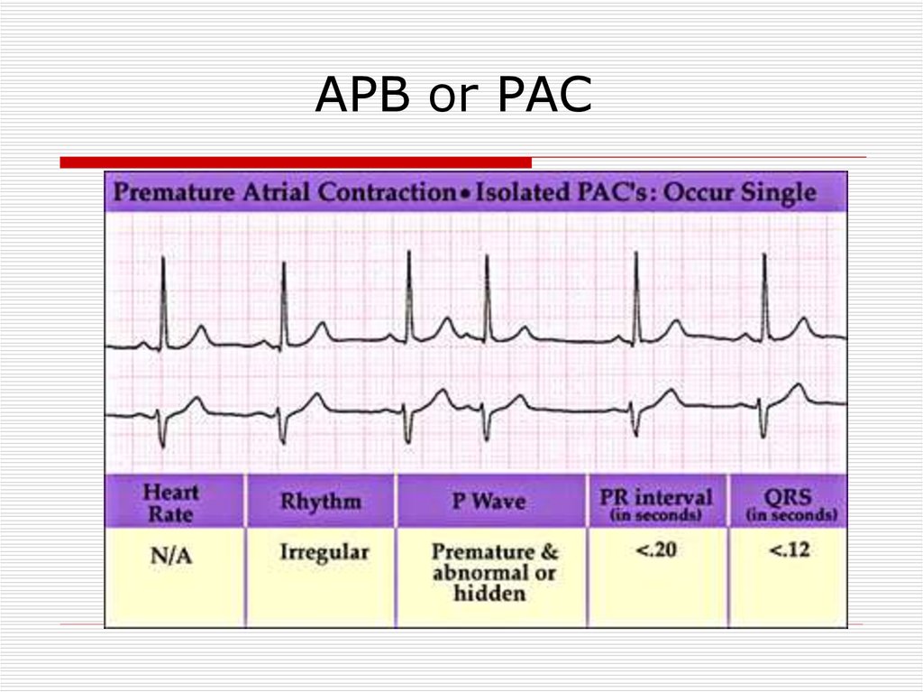
APB or PAC10.
Atrial FibrillationThe most common arrhythmia in
clinical practice
Frequency increases with age
11.
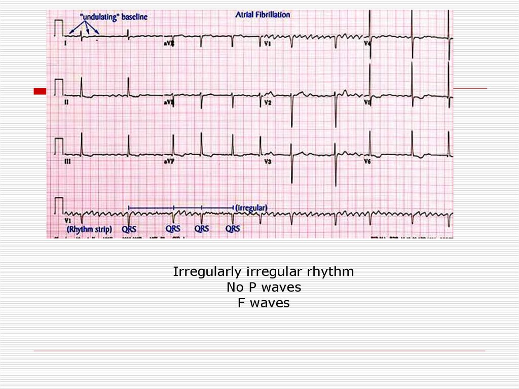
Irregularly irregular rhythmNo P waves
F waves
12.
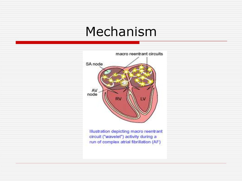
Mechanism13.
Most common causesValvular heart disease: (MS,MR)
LV hypertrophy (HTN, other cause)
Cardiomyopathy
Thyrotoxicosis
Alcohol (“holiday heart”)
Atrial septal defect
Lone AF (structurally normal heart)
14.
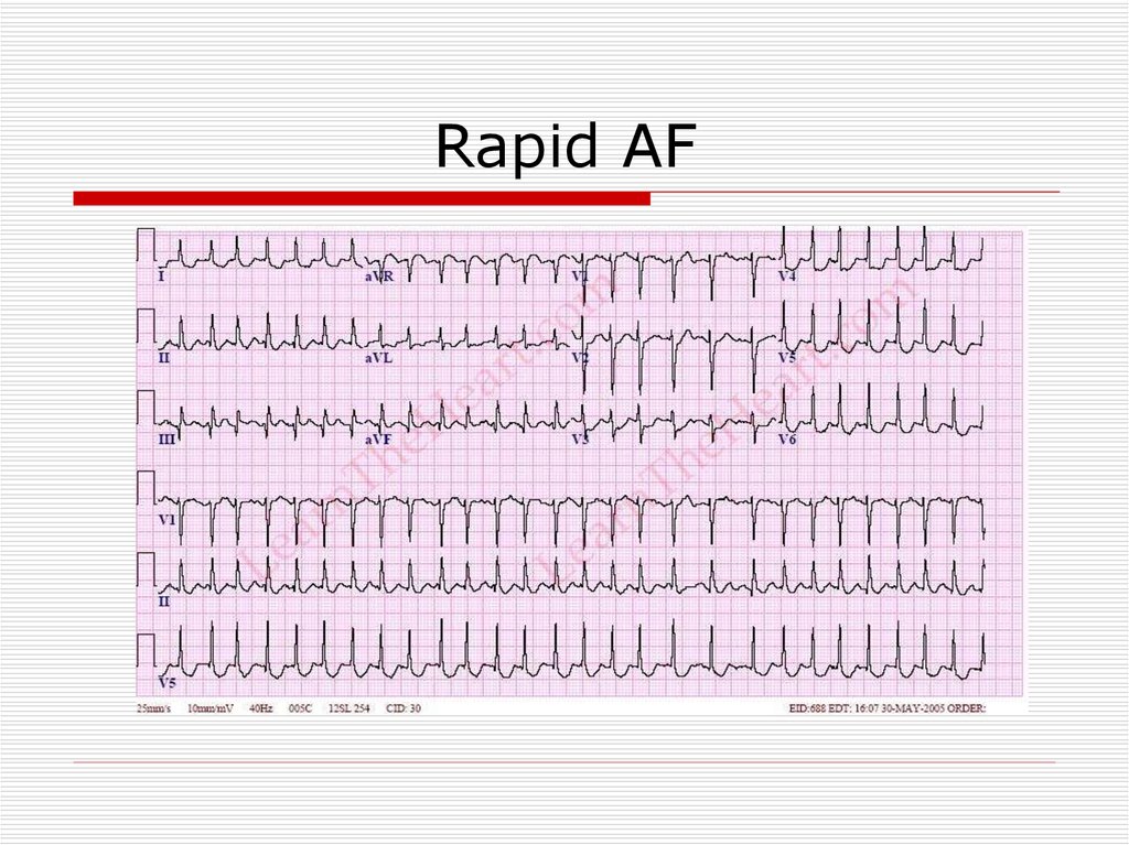
Rapid AF15.
Consequences of AtrialFibrillation
Hemodynamic
loss of synchronous atrial mechanical activity
irregularity of ventricular response
inappropriately rapid heart rate
Myocardial – persistently rapid rate can lead to:
atrial cardiomyopathy
dilated ventricular cardiomyopathy
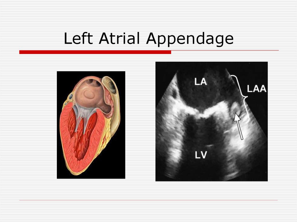
Thromboembolism
ischemic stroke and systemic arterial occlusion
attributed to LA and LAA thrombus
16.
Classification17.
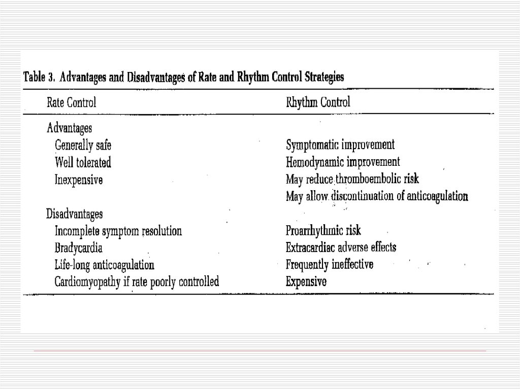
Treatment options1. Rhythm control – restoration and
maintenance of sinus rhythm
2. Rate control
Prevention of Thromboembolysm !
18.
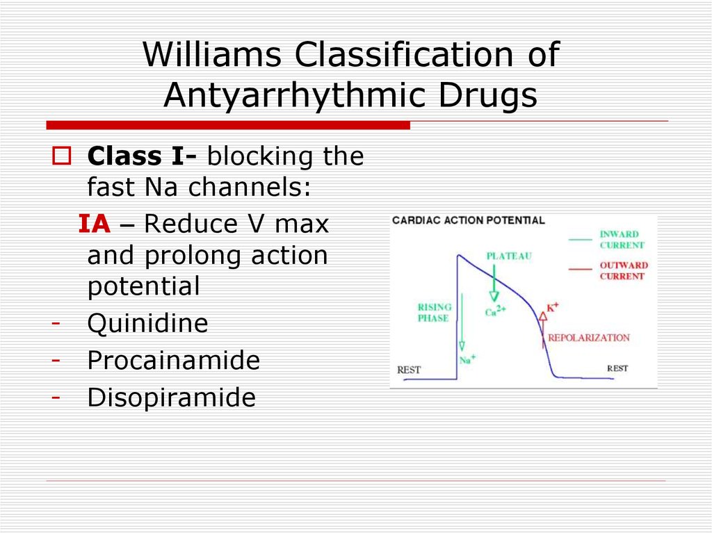
Williams Classification ofAntyarrhythmic Drugs
Class I- blocking the
fast Na channels:
IA – Reduce V max
and prolong action
potential
- Quinidine
- Procainamide
- Disopiramide
19.
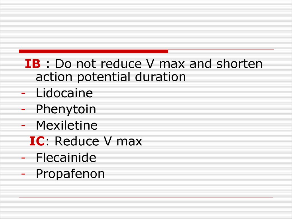
IB : Do not reduce V max and shortenaction potential duration
- Lidocaine
- Phenytoin
- Mexiletine
IC: Reduce V max
- Flecainide
- Propafenon
20.
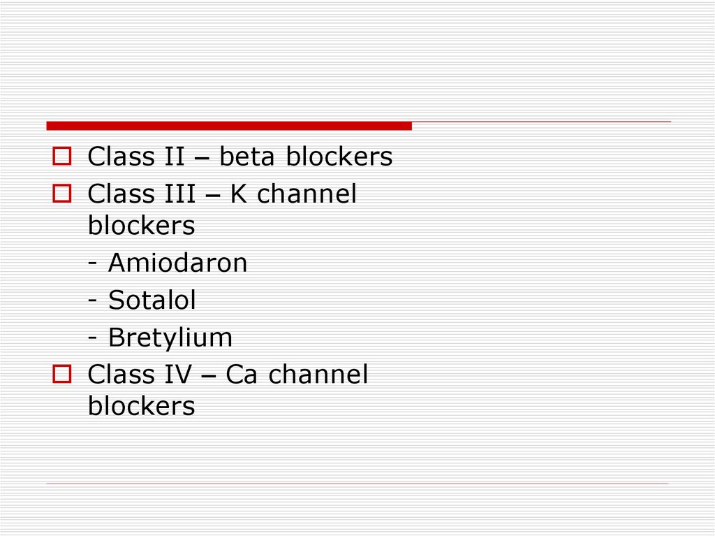
Class II – beta blockersClass III – K channel
blockers
- Amiodaron
- Sotalol
- Bretylium
Class IV – Ca channel
blockers
21.
CardioversionPharmacological
Propafenon
Amiodaron
Flecainide
22.
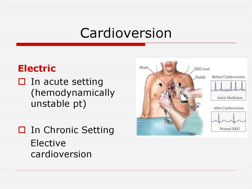
CardioversionElectric
In acute setting
(hemodynamically
unstable pt)
In Chronic Setting
Elective
cardioversion
23.
Predictors of successfulcardioverson
Short AF duration
Young age
Normal atrial size
No organic heart pathology
24.
Maintenance of sinus rhythmPropafenon
Amiodaron
Dronedaron
Sotalol
Flecainide
25.
26.
27.
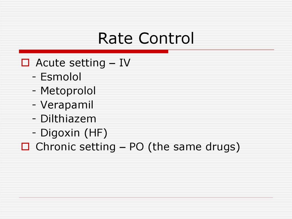
Rate ControlAcute setting – IV
- Esmolol
- Metoprolol
- Verapamil
- Dilthiazem
- Digoxin (HF)
Chronic setting – PO (the same drugs)
28.
29.
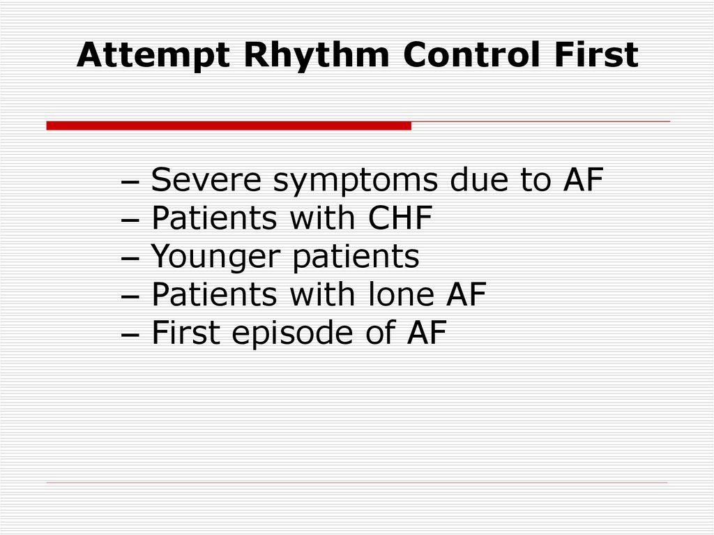
Attempt Rhythm Control First–
–
–
–
–
Severe symptoms due to AF
Patients with CHF
Younger patients
Patients with lone AF
First episode of AF
30.
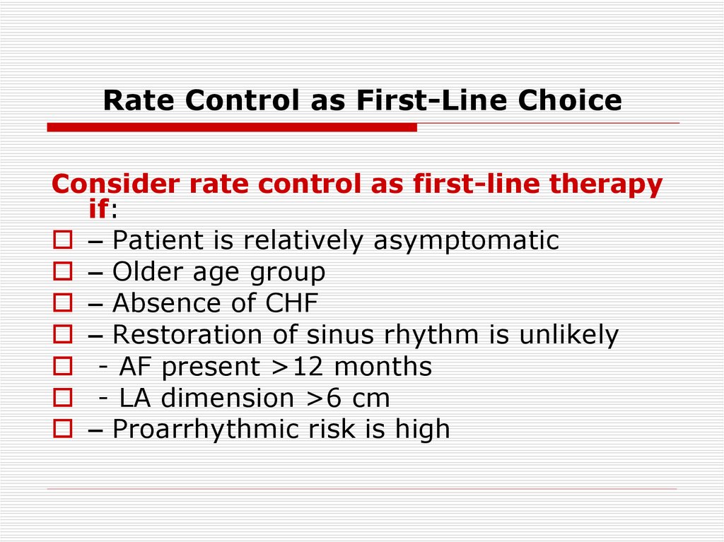
Rate Control as First-Line ChoiceConsider rate control as first-line therapy
if:
– Patient is relatively asymptomatic
– Older age group
– Absence of CHF
– Restoration of sinus rhythm is unlikely
- AF present >12 months
- LA dimension >6 cm
– Proarrhythmic risk is high
31.
Left Atrial Appendage32.
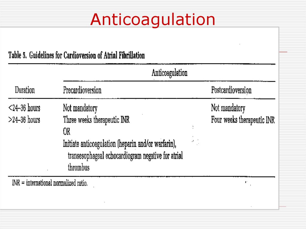
Anticoagulation33.
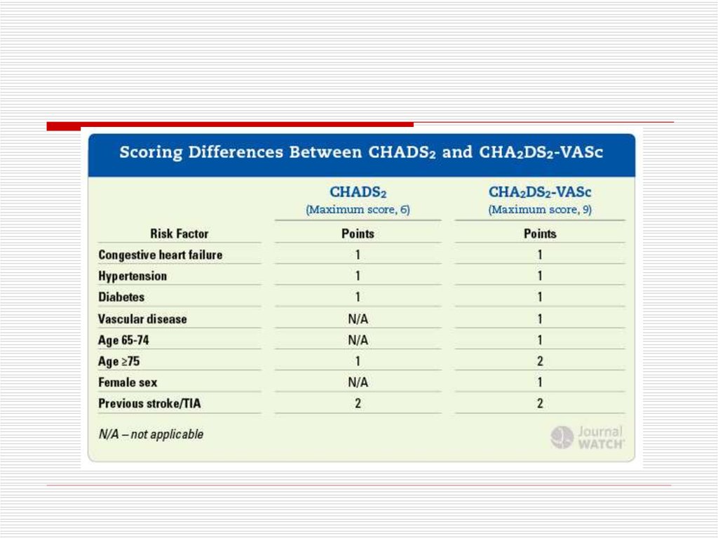
CHADS2 score34.
35.
Novel Oral AnticoagulantsDabigatran (Pradaxa)- direct oral
thrombin inhibitor
Rivaroxaban (Xarelto)– direct oral
factor Xa inhibitor
Apixaban (Eliquis) - direct oral factor
Xa inhibitor
36.
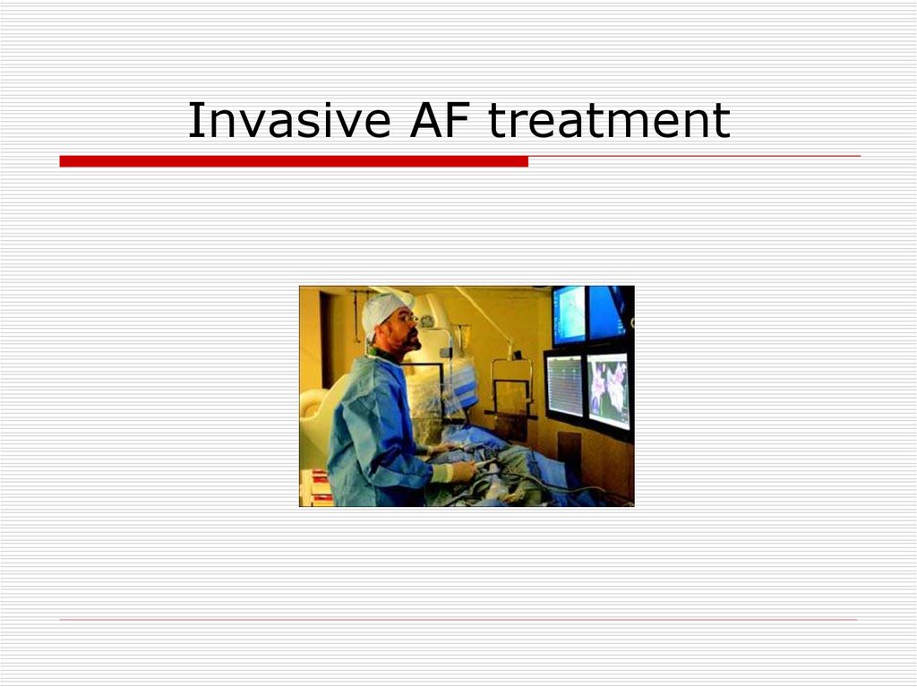
Invasive AF treatment37.
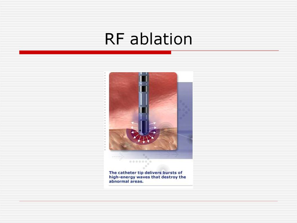
RF ablation38.
Invasive AF managementRate control
“Ablate and pace” –
A-v nodal ablation
& Permanent
pacemaker
39.
Pulmonary Venous IsolationFor recurrent paroxysmal AF
40.
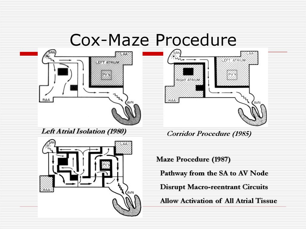
Cox-Maze ProcedureLeft Atrial Isolation (1980)
Corridor Procedure (1985)
Maze Procedure (1987)
Pathway from the SA to AV Node
Disrupt Macro-reentrant Circuits
Allow Activation of All Atrial Tissue
41.
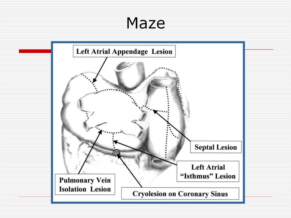
Maze42.
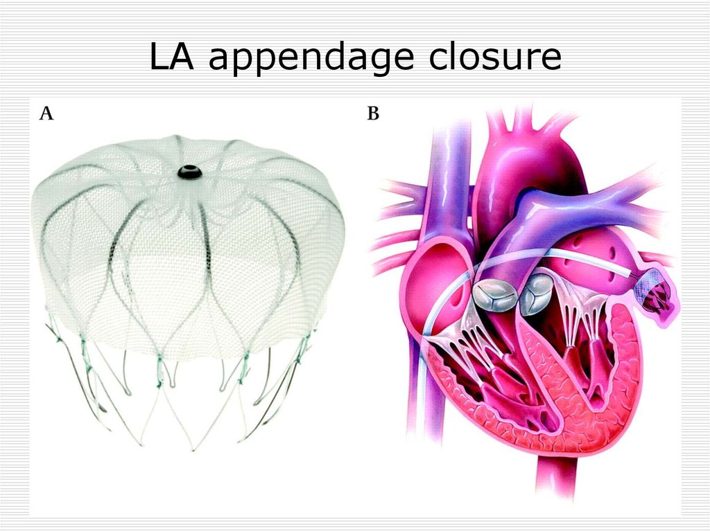
LA appendage closure43.
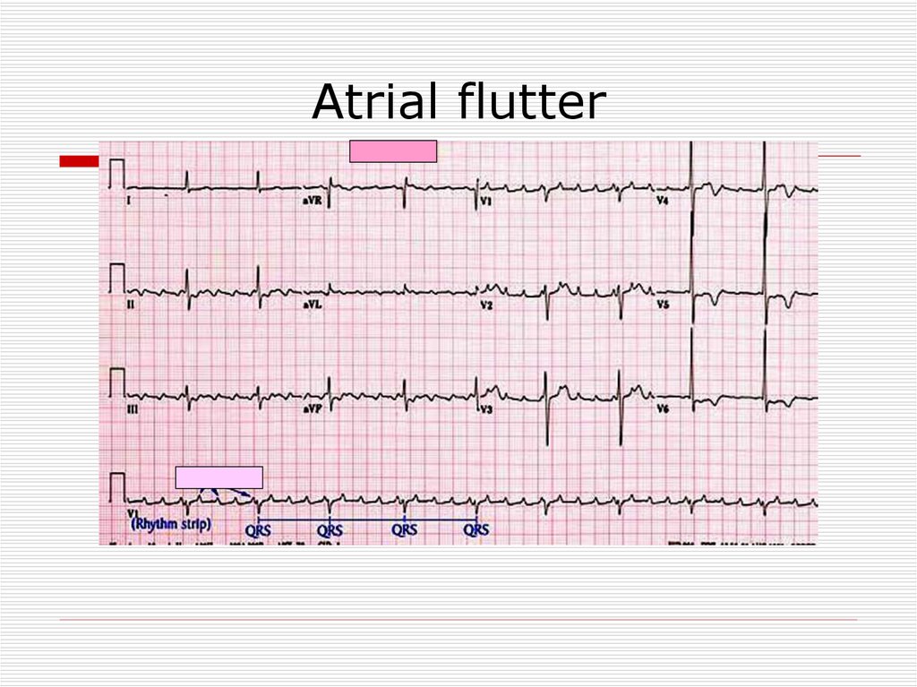
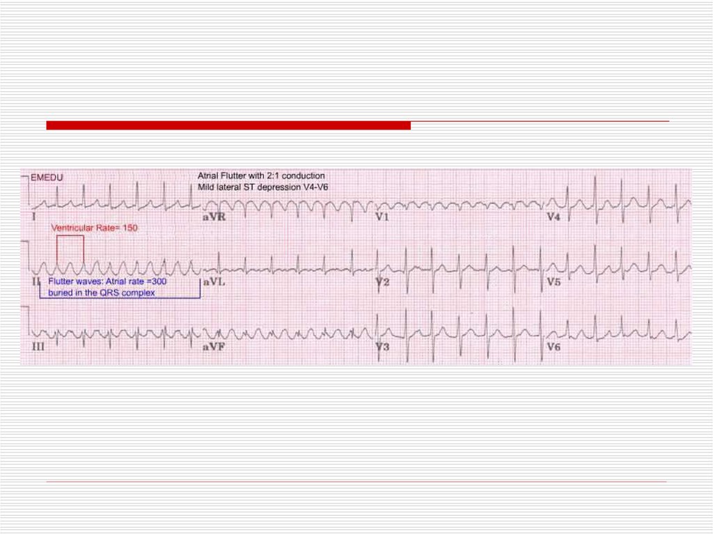
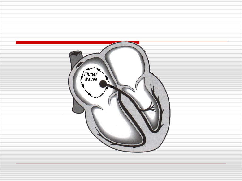
Atrial flutter44.
45.
46.
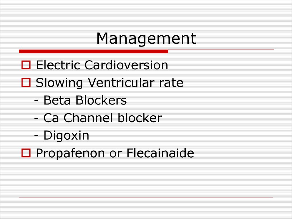
ManagementElectric Cardioversion
Slowing Ventricular rate
- Beta Blockers
- Ca Channel blocker
- Digoxin
Propafenon or Flecainaide
47.
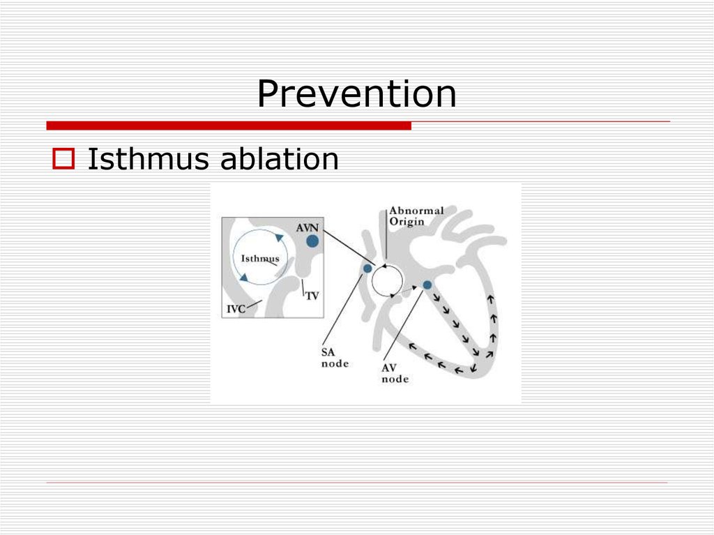
PreventionIsthmus ablation
48.
Preexitation – WPW syndrome(accessory pathway(
49.
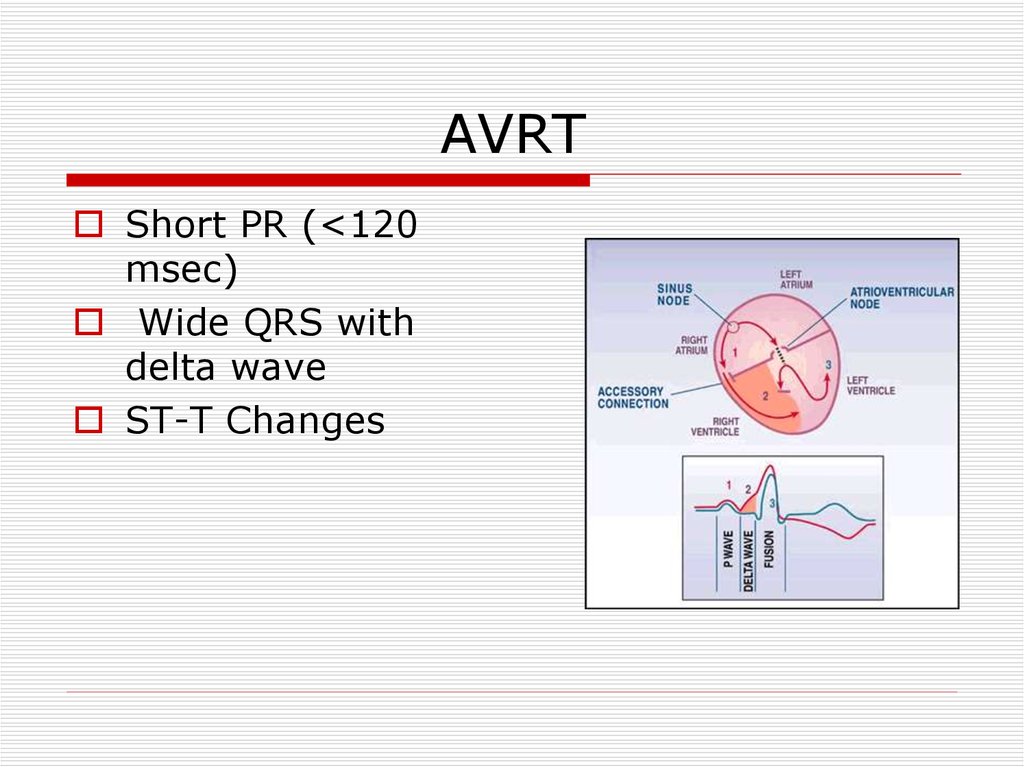
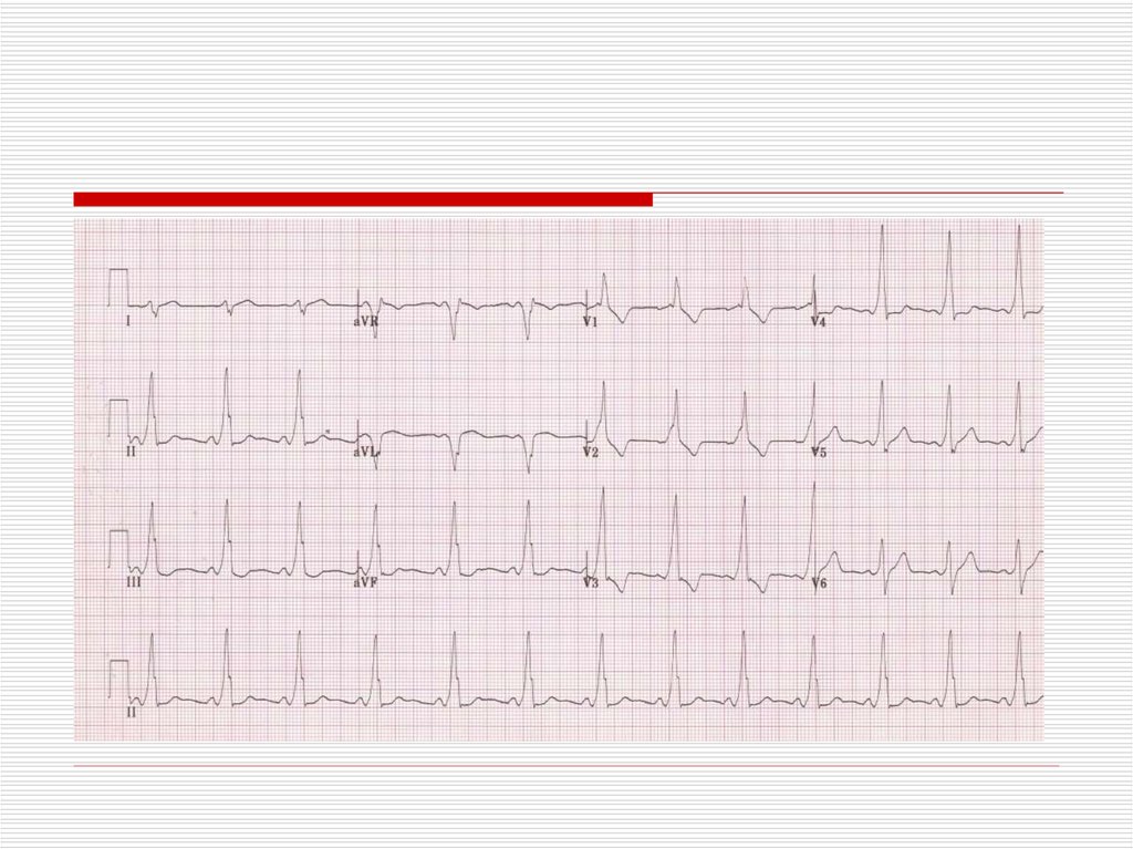
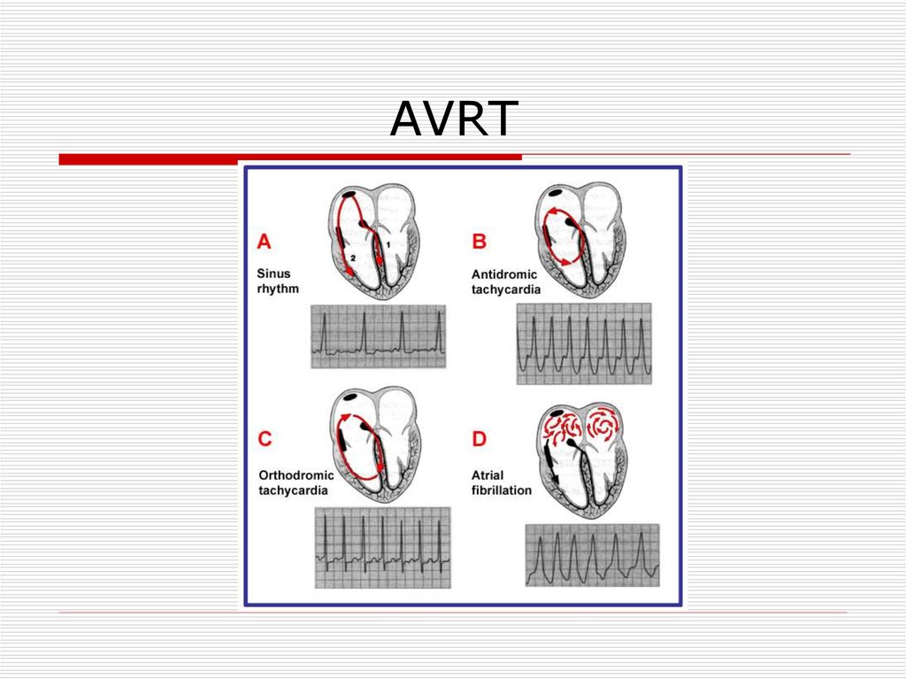
AVRTShort PR (<120
msec)
Wide QRS with
delta wave
ST-T Changes
50.
51.
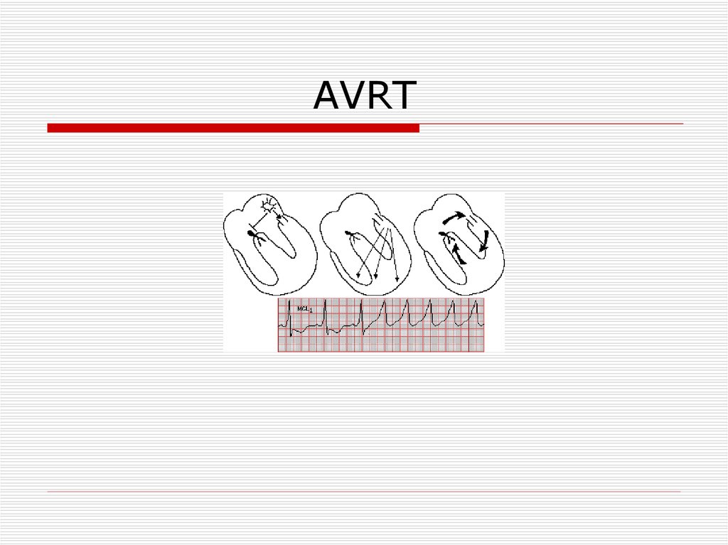
AVRT52.
AVRT53.
TreatmentAcute treatment:
Wide complex – Procainamide
DC Shock
Narrow complex – Verapamil,
Beta Blockers
Preventive treatment : accessory
pathway ablation
54.
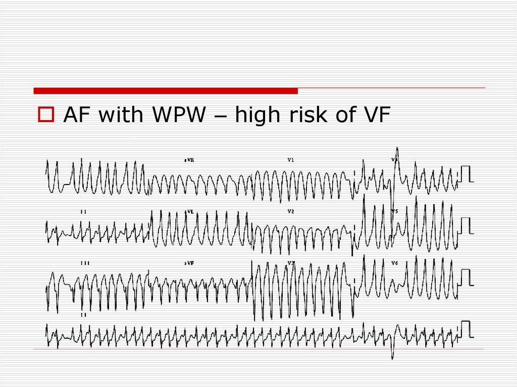
AF with WPW – high risk of VF55.
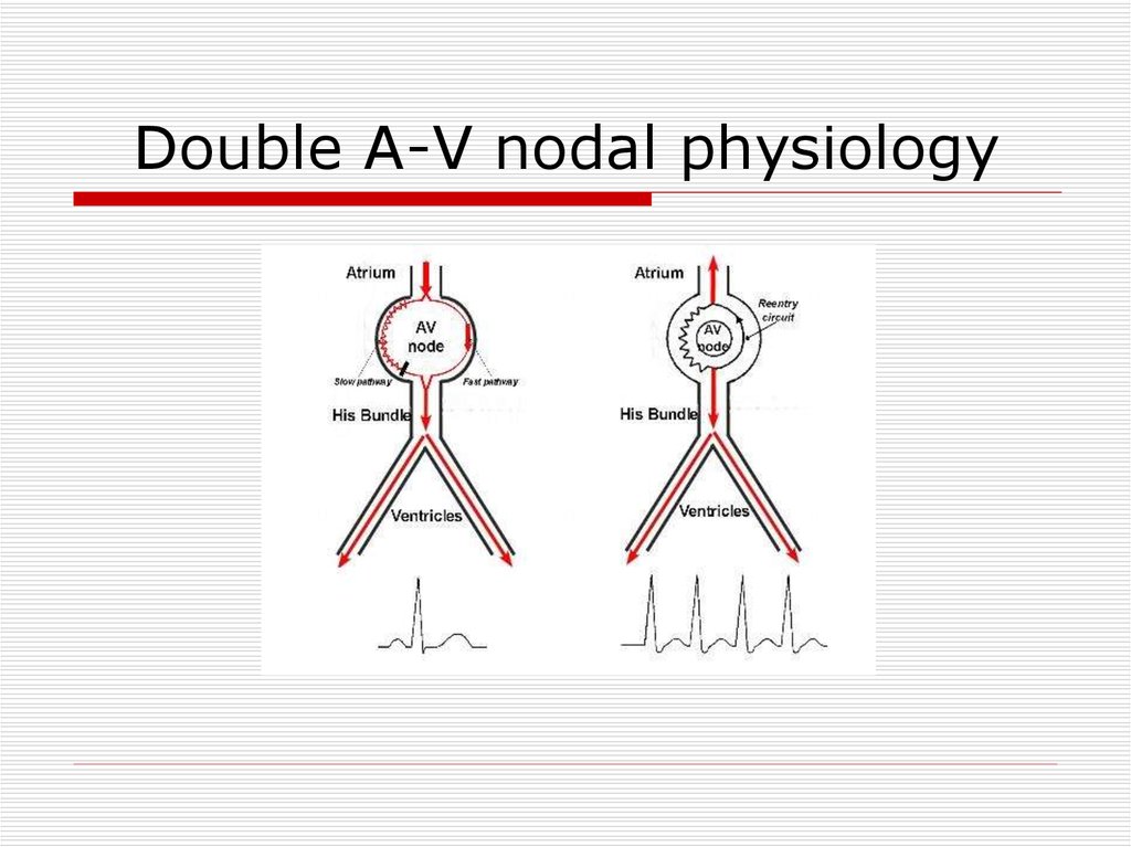
Double A-V nodal physiology56.
57.
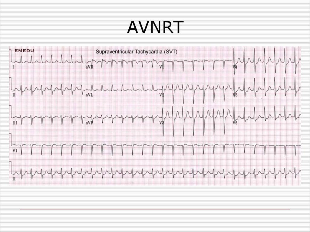
AVNRT58.
Management of narrow complexSVT
If unstable – DC shock
If Stable :
1. Vagal maneuvers
2. Adenosin
3. Verapamil
59.
Preventive treatmentDrugs
EPS
60.
Ventricular Arrhythmias61.
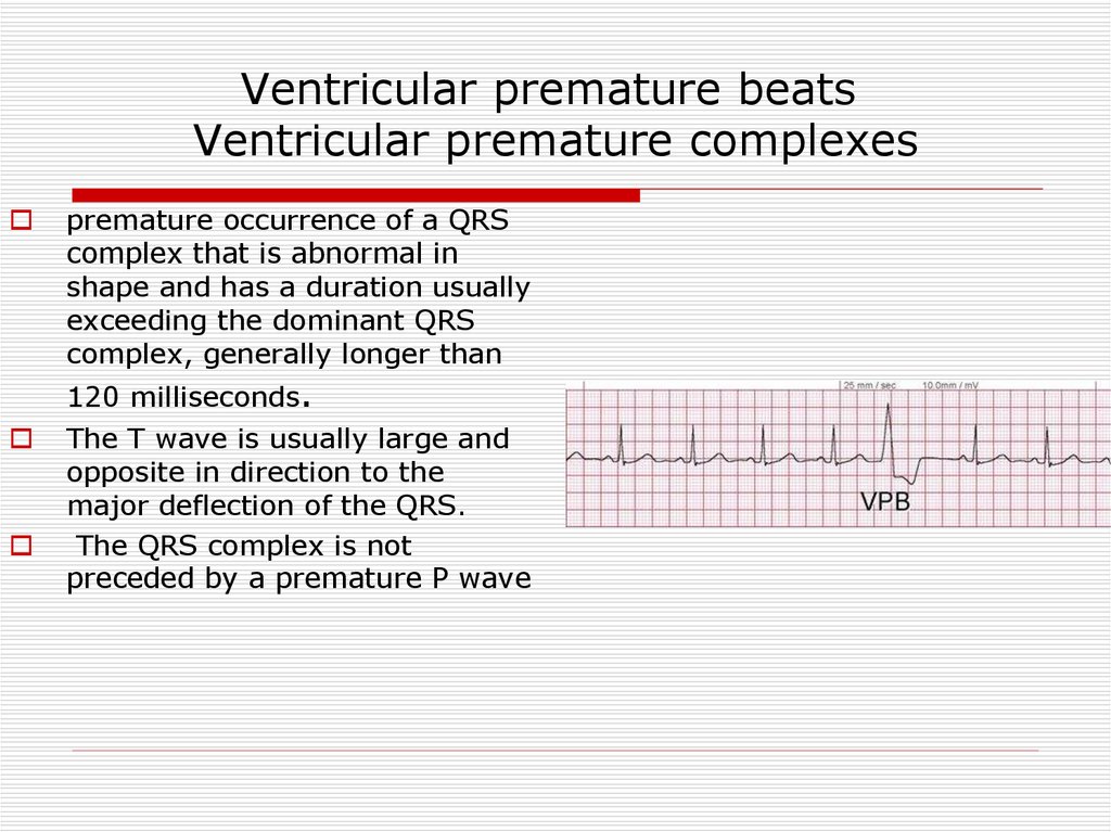
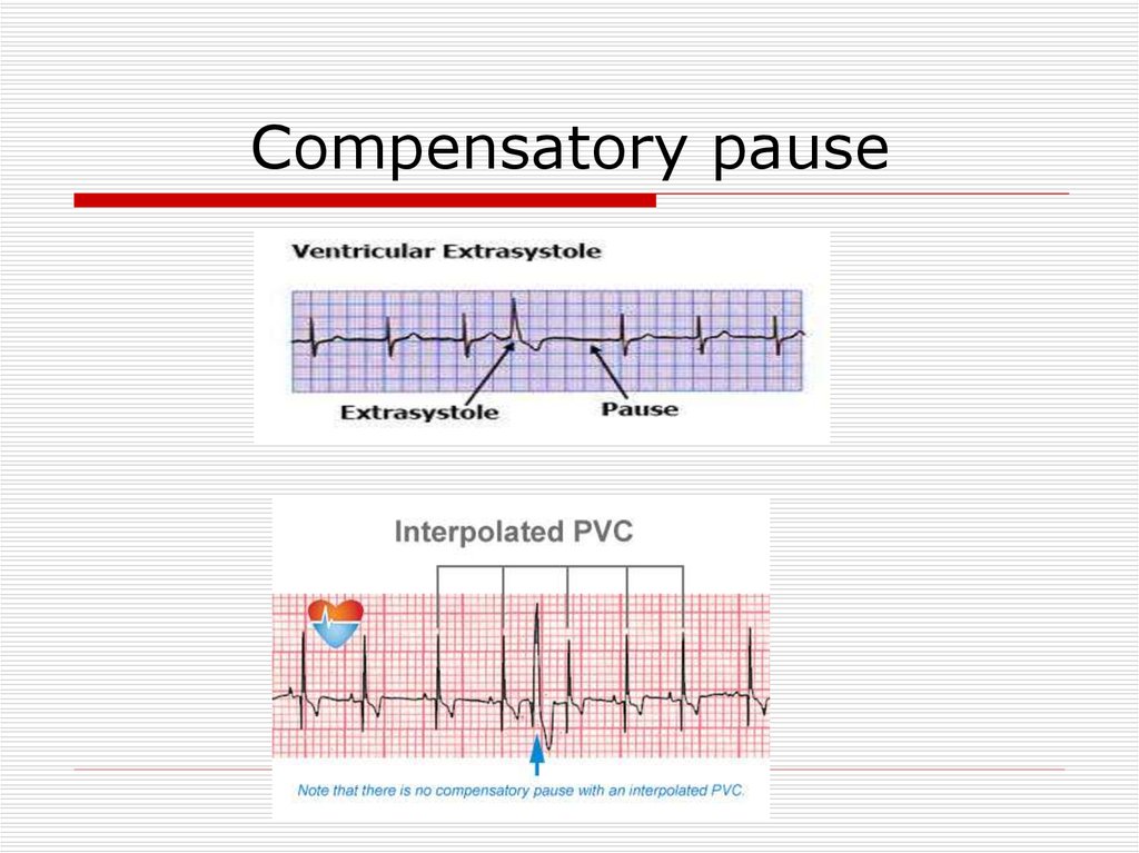
Ventricular premature beatsVentricular premature complexes
premature occurrence of a QRS
complex that is abnormal in
shape and has a duration usually
exceeding the dominant QRS
complex, generally longer than
120 milliseconds.
The T wave is usually large and
opposite in direction to the
major deflection of the QRS.
The QRS complex is not
preceded by a premature P wave
62.
Compensatory pause63.
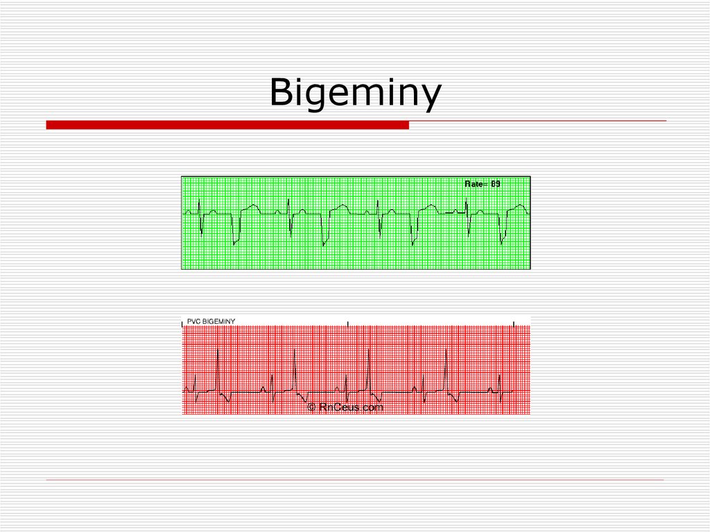
Bigeminy64.
Trigeminy65.
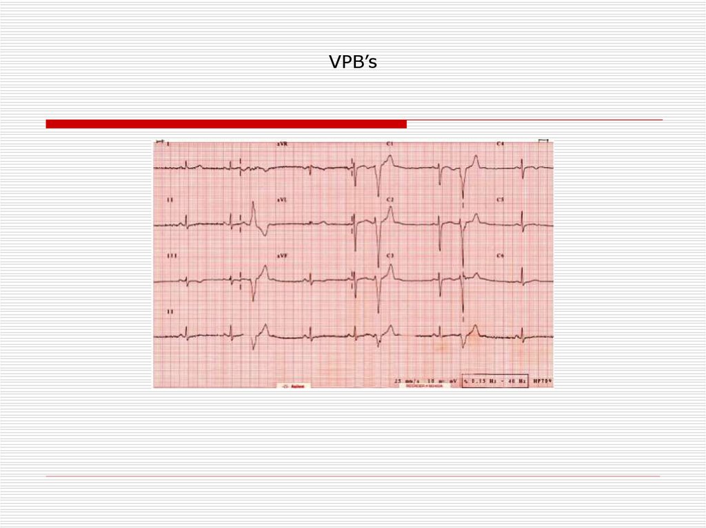
VPB’s66.
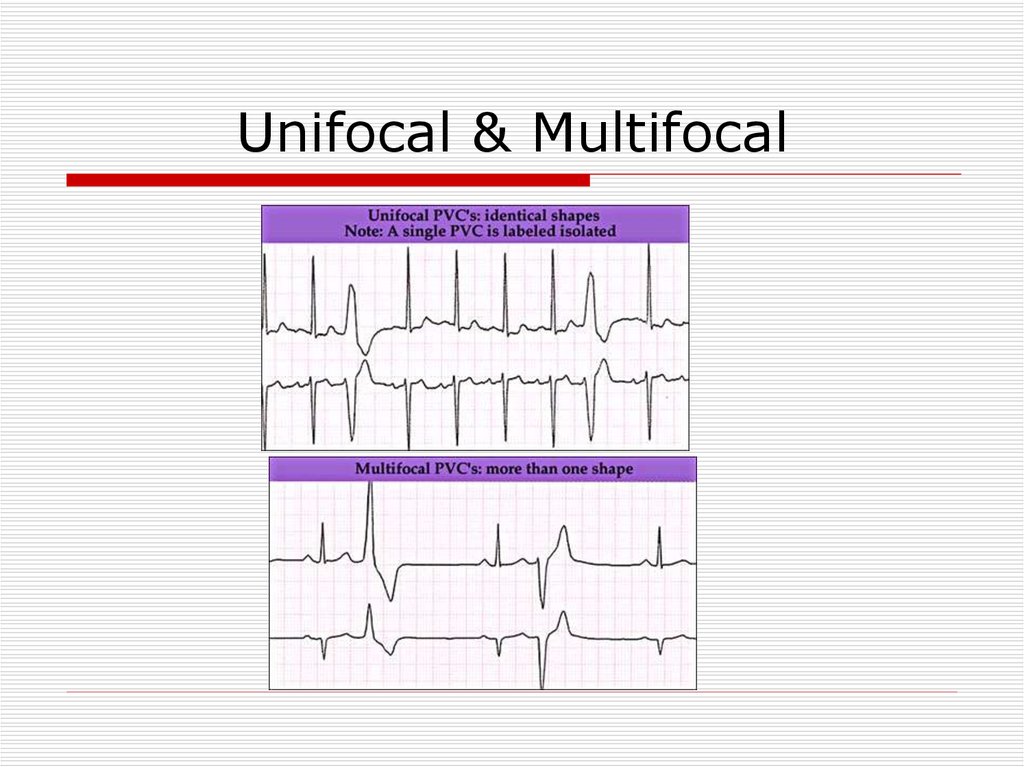
Unifocal & Multifocal67.
Couplet & Triplet68.
CausesLV false tendons,
infection
in ischemic or inflamed myocardium,
hypoxia,
Anesthesiaor
surgery.
Medications
electrolyte imbalance,
tension states,
myocardial stretch,
excessive use of tobacco, caffeine, or alcohol.
69.
Complex Ventricular Arrhythmia•Nonsustained ventricular tachycardia (VT)
♥ Monomorphic
♥ Polymorphic
•Sustained VT
♥ Monomorphic
♥ Polymorphic
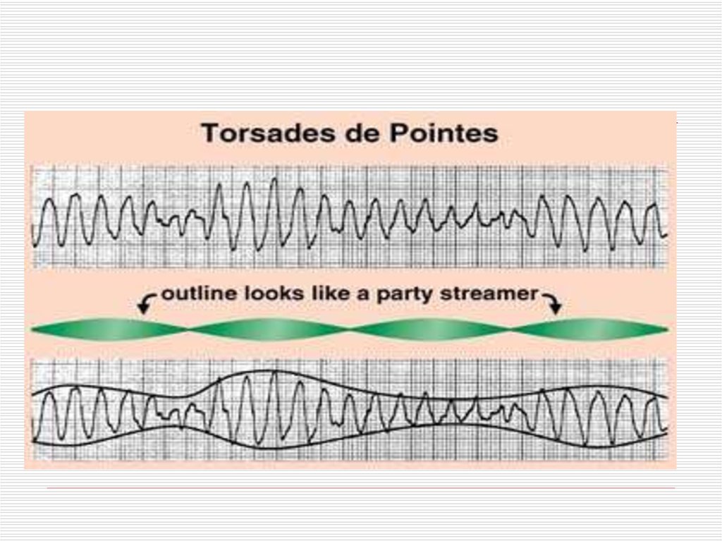
•Torsades de pointes
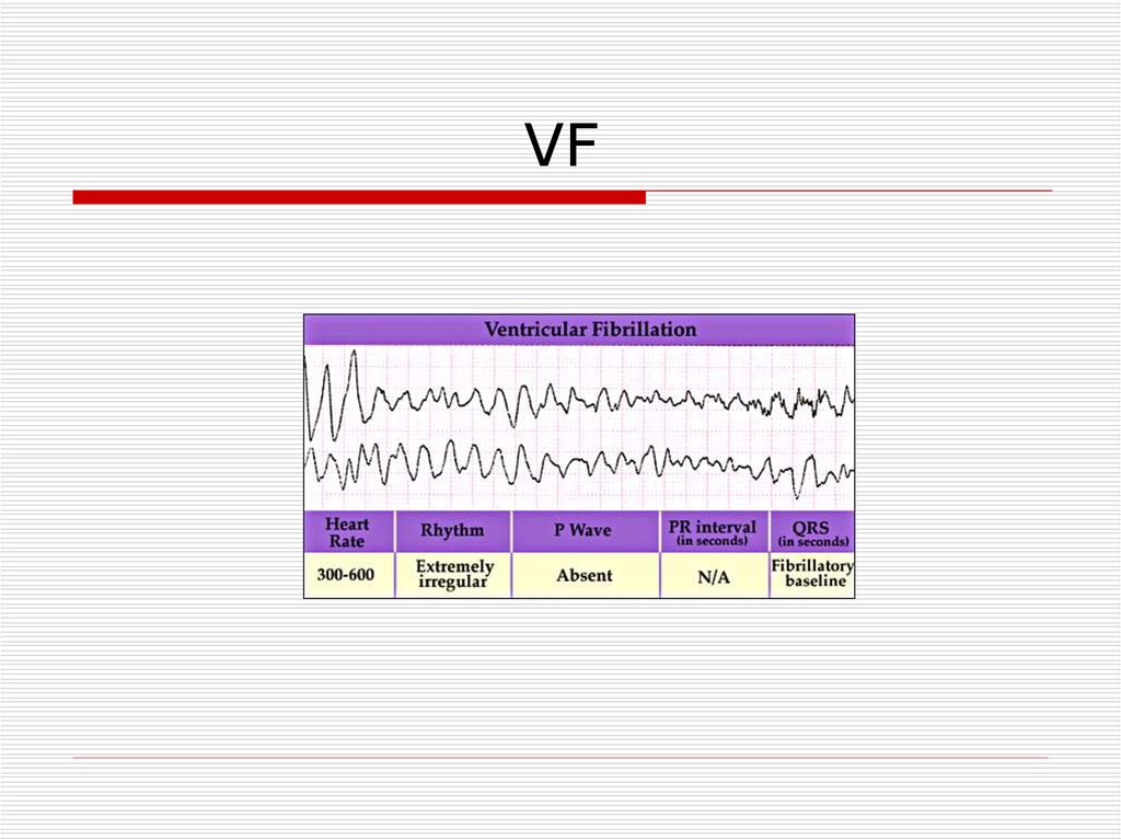
•Ventricular fibrillation
70.
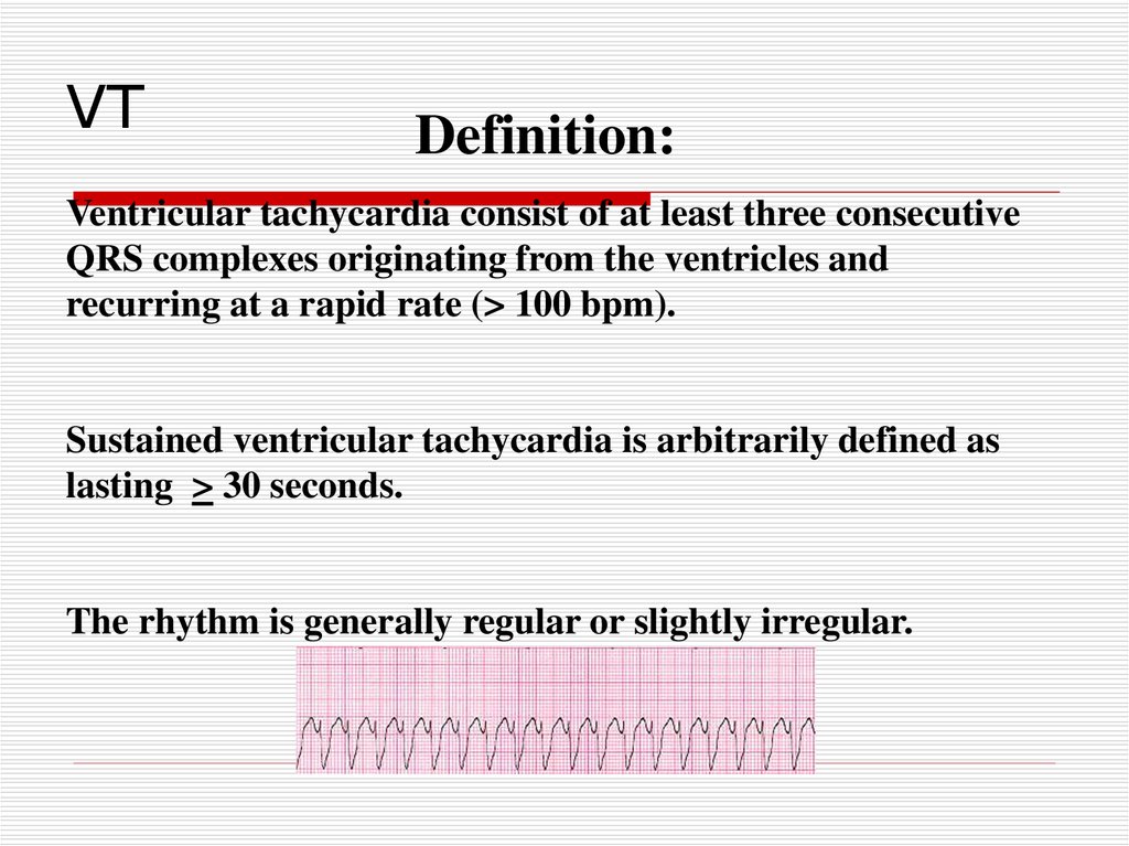
VTDefinition:
Ventricular tachycardia consist of at least three consecutive
QRS complexes originating from the ventricles and
recurring at a rapid rate (> 100 bpm).
Sustained ventricular tachycardia is arbitrarily defined as
lasting > 30 seconds.
The rhythm is generally regular or slightly irregular.
71.
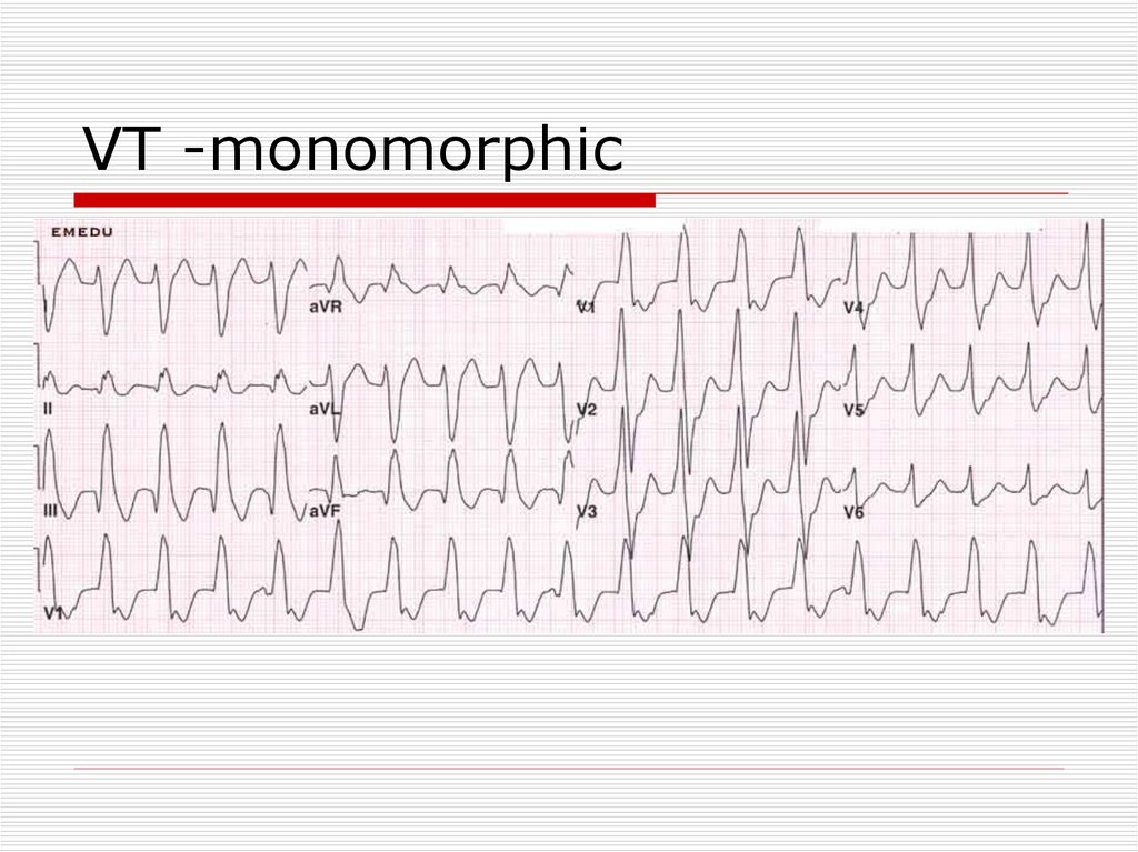
VT -monomorphic72.
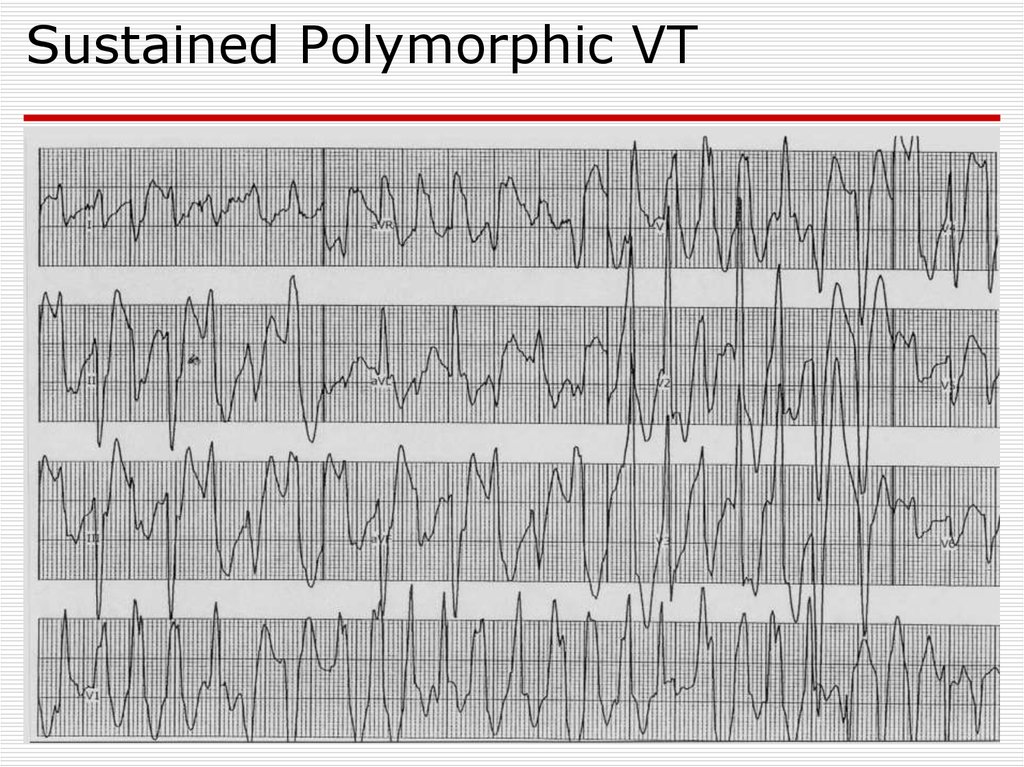
Sustained Polymorphic VT73.
VF74.
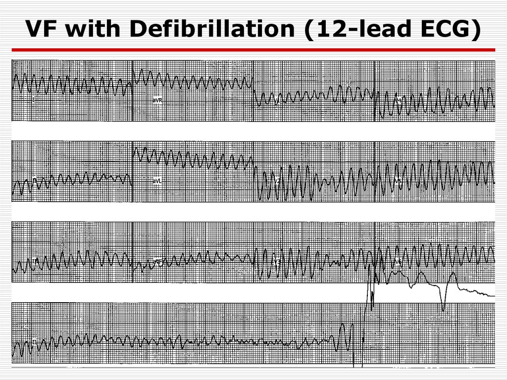
VF with Defibrillation (12-lead ECG)75.
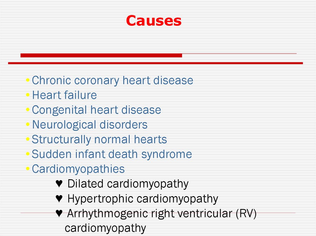
Causes•Chronic coronary heart disease
•Heart failure
•Congenital heart disease
•Neurological disorders
•Structurally normal hearts
•Sudden infant death syndrome
•Cardiomyopathies
♥ Dilated cardiomyopathy
♥ Hypertrophic cardiomyopathy
♥ Arrhythmogenic right ventricular (RV)
cardiomyopathy
76.
Mechanisms of Sudden Cardiac Death• Ventricular fibrillation - 62.4%
• Bradyarrhythmias (including advanced AV block and
asystole) - 16.5%
• Torsades de pointes - 12.7%
• Primary VT - 8.3%
Bayes de Luna et al. Am Heart J 1989;117:151–9.
77.
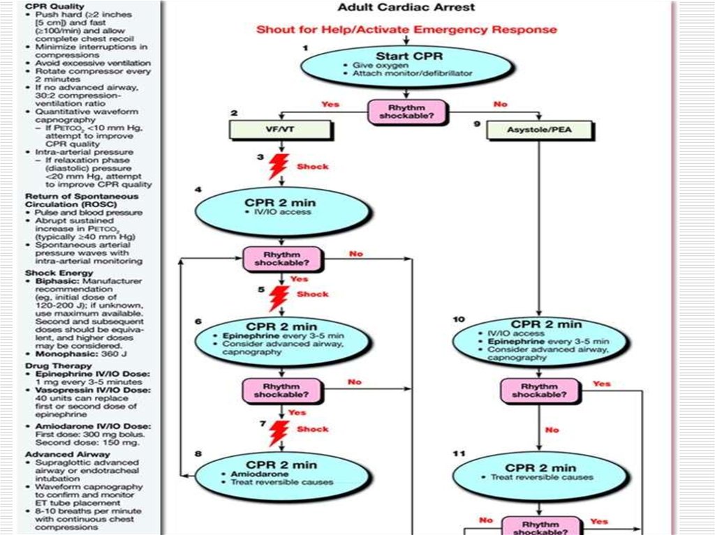
VA managementAcute
Chronic (secondary prevention)
78.
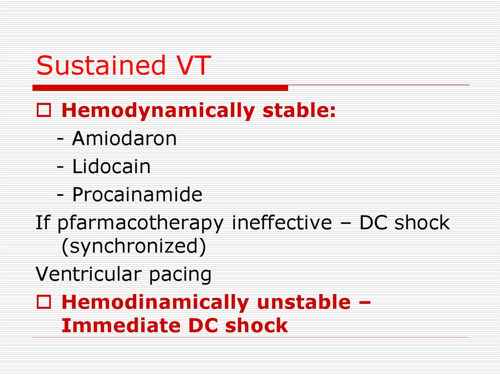
Sustained VTHemodynamically stable:
- Amiodaron
- Lidocain
- Procainamide
If pfarmacotherapy ineffective – DC shock
(synchronized)
Ventricular pacing
Hemodinamically unstable –
Immediate DC shock
79.
Polymorphic VTPolymorphic VT with long QT –
Torsades de pointes
Treatment – Mg , Pacing
Polymorphic VT w/o long QT
Antyarrhytmic drugs
80.
81.
Chronic Management (secondaryprevention)
Evaluation
- Rest ECG
- Exersise test
- Ambulatory ECG
- Imaging (LV function, CMP,
Valves etc…
- EPS
82.
Treatment of the underlyingdisease
Revascularisation
Valve surgery
CHD repair
83.
Non-antiarrhythmic Drugs♥ Electrolytes: Mg & K
♥ ACE inhibitors,
♥ Antithrombotic and antiplatelet agents
♥ Statins
84.
Antiarrhytmic drugsAntiarrhythmic drugs (except for
BB) should not be used as
primary preventive therapy of
VA and the prevention of SCD
85.
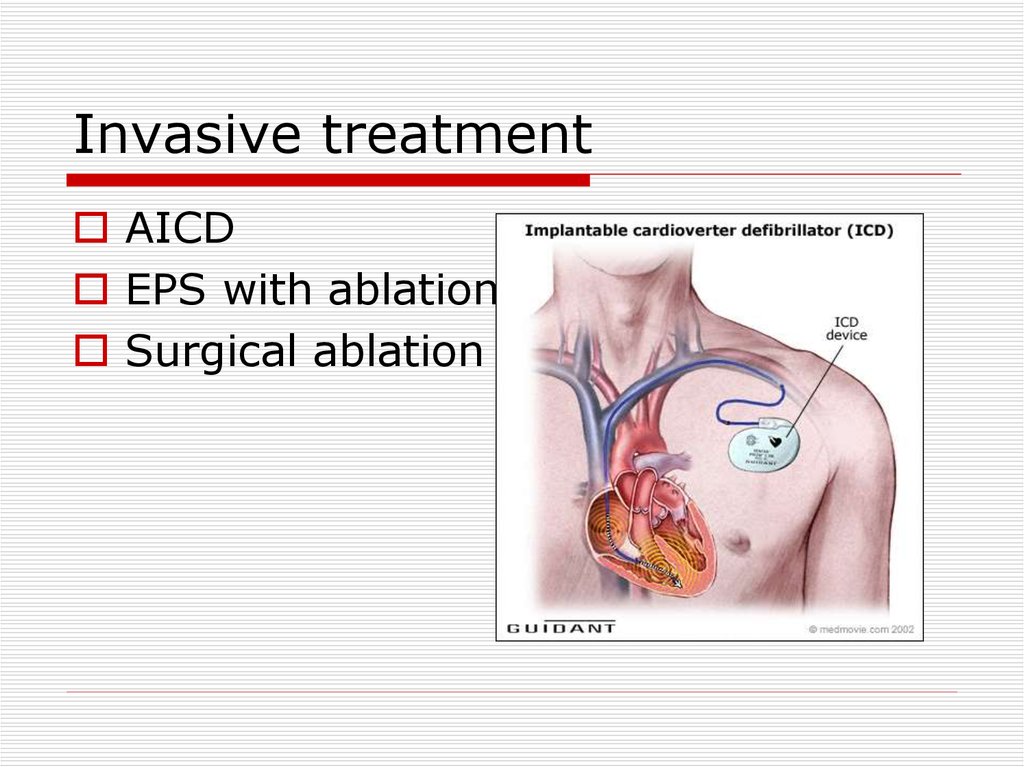
Invasive treatmentAICD
EPS with ablation
Surgical ablation
86.
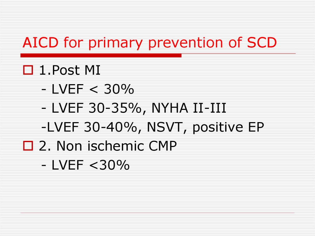
AICD for primary prevention of SCD1.Post MI
- LVEF < 30%
- LVEF 30-35%, NYHA II-III
-LVEF 30-40%, NSVT, positive EP
2. Non ischemic CMP
- LVEF <30%
87.
Long QT syndrome1. Congenital (family)
2. Acquired:
Electrolyte
anomalies – K, Mg
Drug induced
-Antiarrhytmics
- Tricyclic
antydepressants
- Antihistamines
CNS lesions
88.
89.
Long QT syndrome treatmentAcute
1.Remove the precipitating factor
2. Mg IV
3. Pacing
4. Isoproterenol
5. IB antiarrhythmic
90.
Long QT syndrome treatmentChronic – for congenital long QT
1.Beta blockers
2. AICD
91.
92.
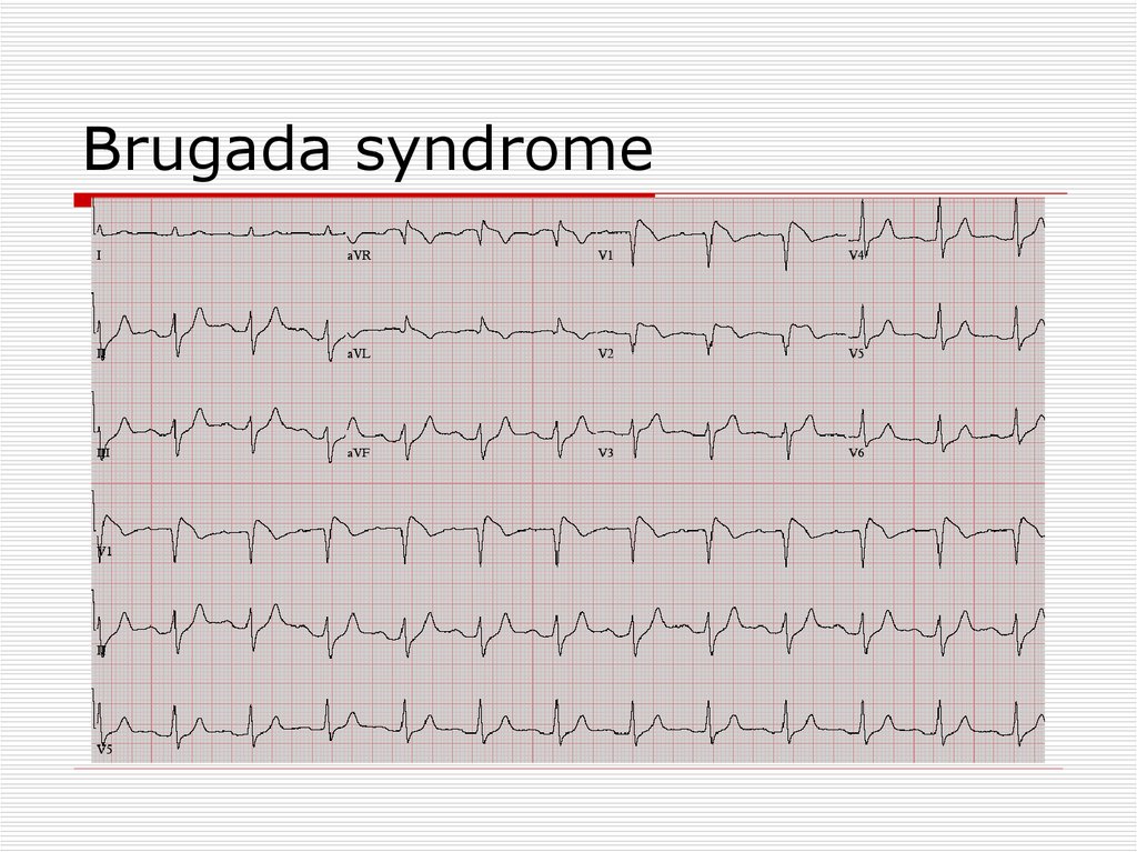
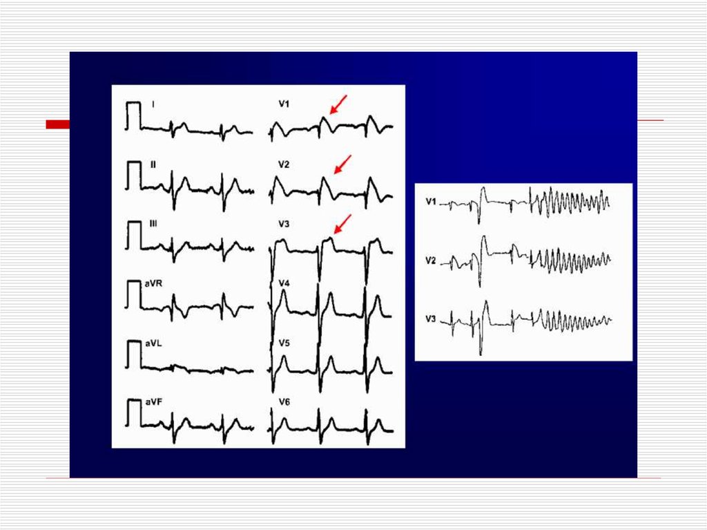
Brugada syndrome93.
94.
95.
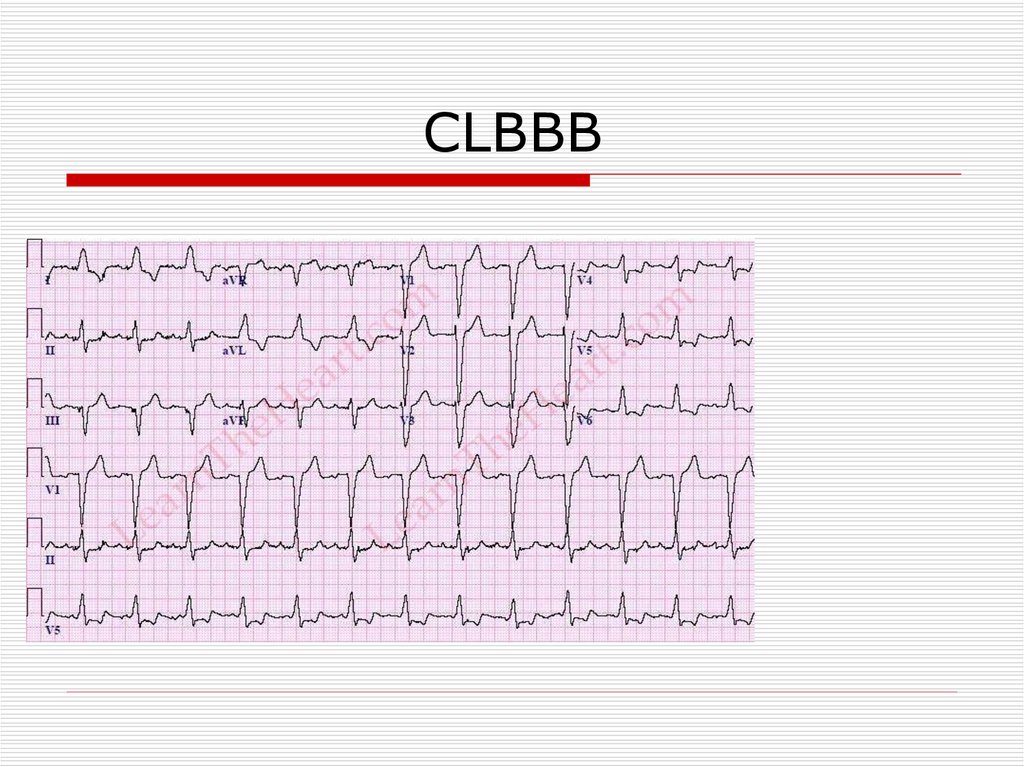
CLBBB96.
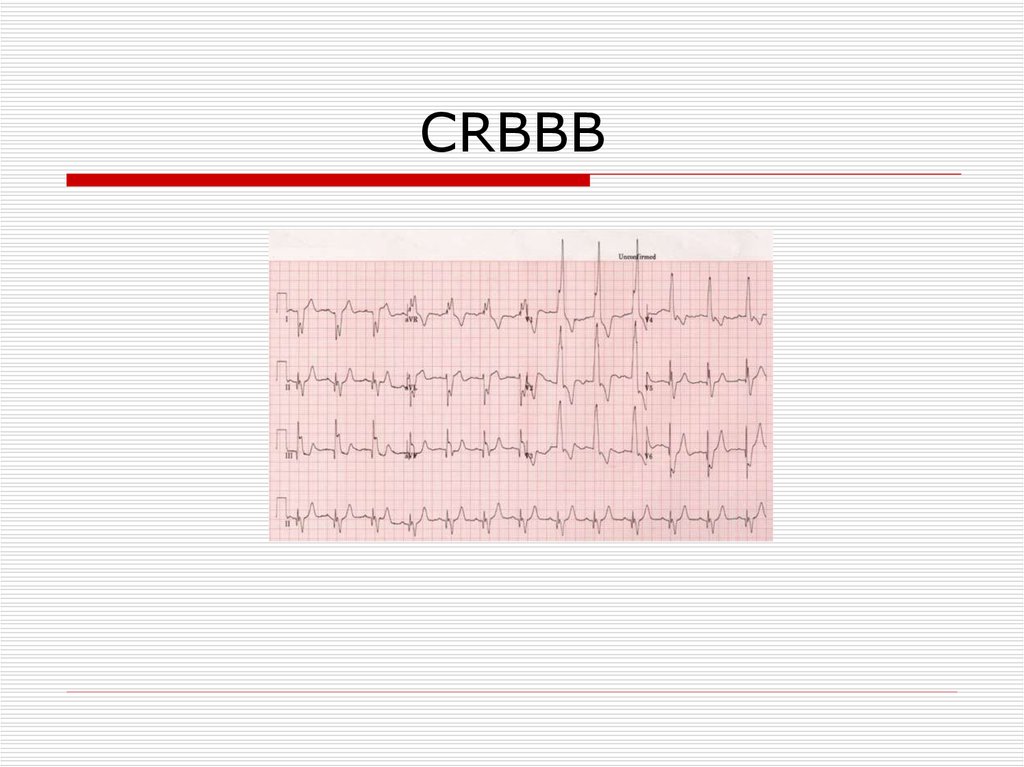
CRBBB97.
98.
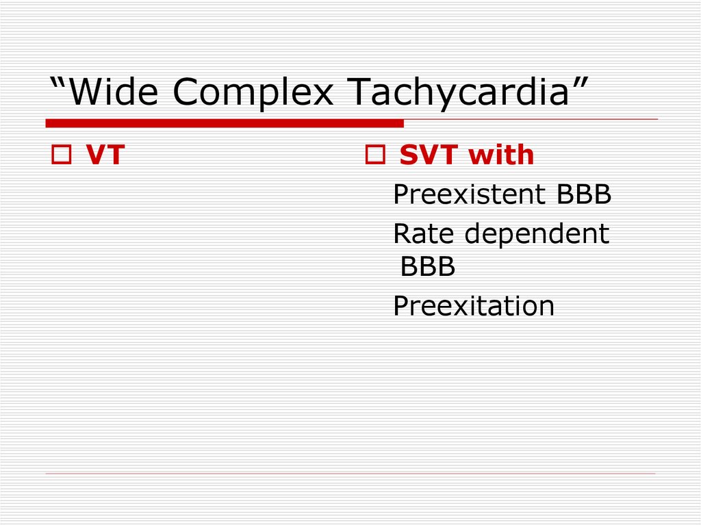
“Wide Complex Tachycardia”VT
SVT with
Preexistent BBB
Rate dependent
BBB
Preexitation
99.
100.
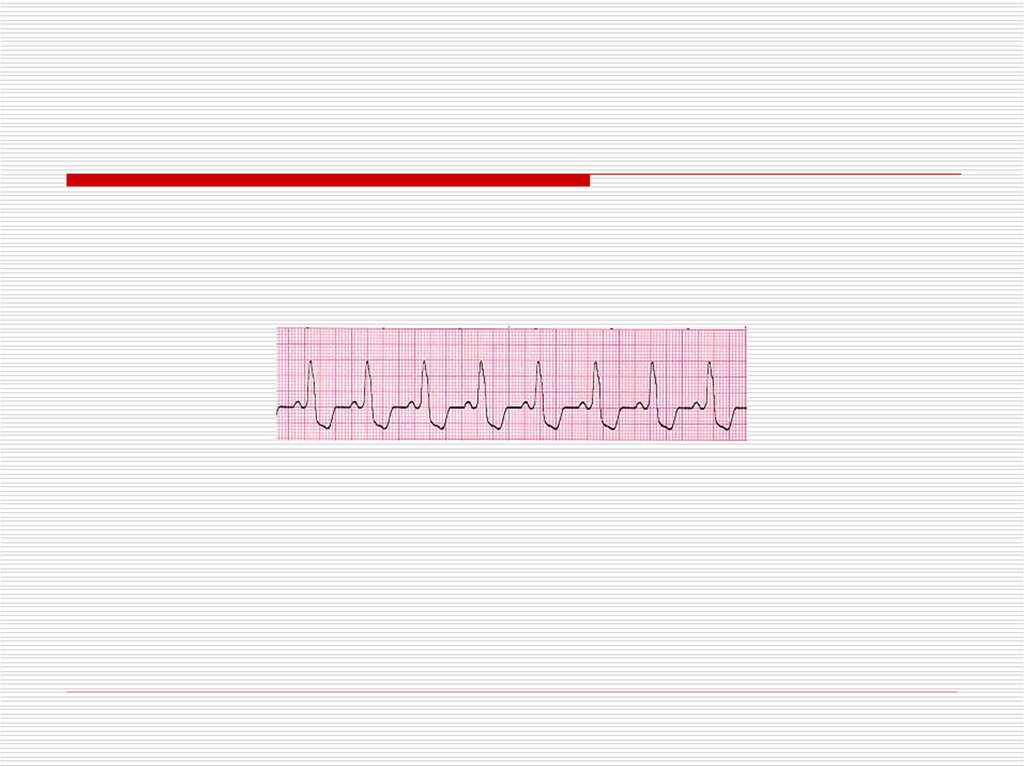
Wide QRS Irregular Tachycardia:Atrial Fibrillation with antidromic conduction in patient
with accessory pathway – Not VT
101.
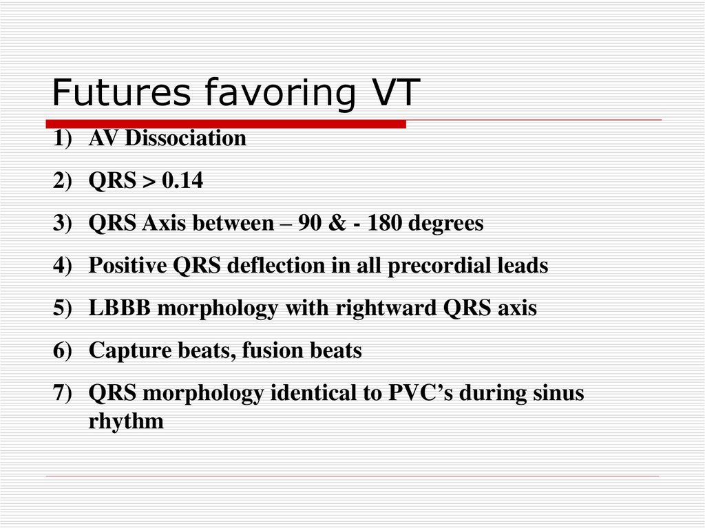
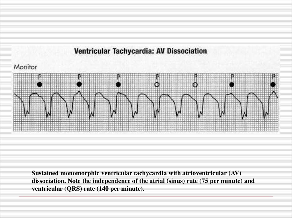
Futures favoring VT1) AV Dissociation
2) QRS > 0.14
3) QRS Axis between – 90 & - 180 degrees
4) Positive QRS deflection in all precordial leads
5) LBBB morphology with rightward QRS axis
6) Capture beats, fusion beats
7) QRS morphology identical to PVC’s during sinus
rhythm
102.
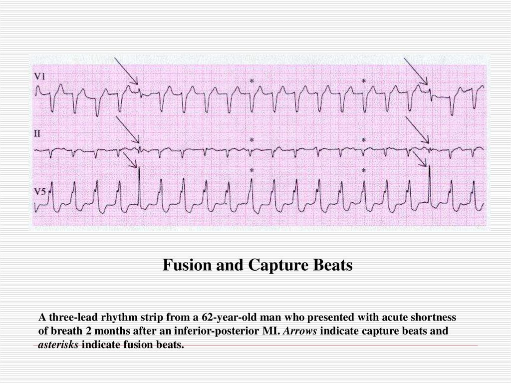
Fusion and Capture BeatsA three-lead rhythm strip from a 62-year-old man who presented with acute shortness
of breath 2 months after an inferior-posterior MI. Arrows indicate capture beats and
asterisks indicate fusion beats.
103.
Sustained monomorphic ventricular tachycardia with atrioventricular (AV)dissociation. Note the independence of the atrial (sinus) rate (75 per minute) and
ventricular (QRS) rate (140 per minute).
104.
?105.
106.
Atrioventricular ConductionDisturbances and
Bradyarrhythmias
107.
Sites of Disturbances in Impulse Formationor Conduction Leading to Bradyarrhythmias
SA Node
AV Node
His-Purkinje
System
108.
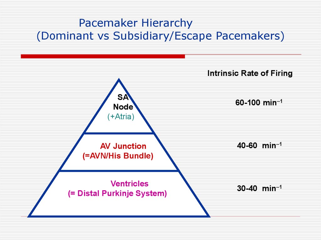
Pacemaker Hierarchy(Dominant vs Subsidiary/Escape Pacemakers)
Intrinsic Rate of Firing
SA
Node
(+Atria)
60-100 min 1
AV Junction
(=AVN/His Bundle)
40-60 min 1
Ventricles
(= Distal Purkinje System)
30-40 min 1
109.
AV Block110.
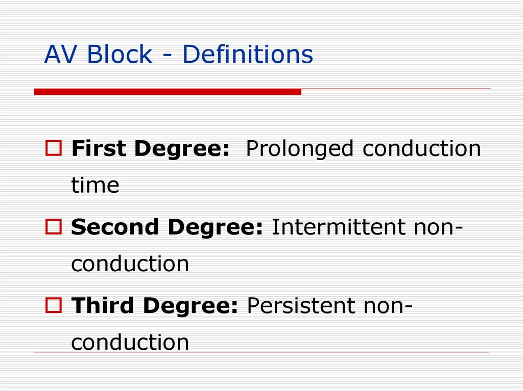
AV Block - DefinitionsFirst Degree: Prolonged conduction
time
Second Degree: Intermittent nonconduction
Third Degree: Persistent nonconduction
111.
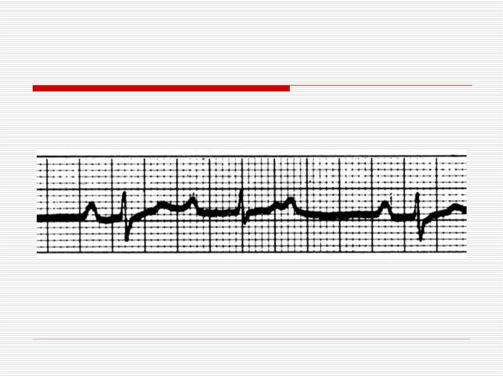
IIFirst Degree AV
Block
(PR > .20 sec [1 big
box])
P
P
P
.36
Site of delay most commonly the AV node,
but may be localized to the His-Purkinje system
112.
113.
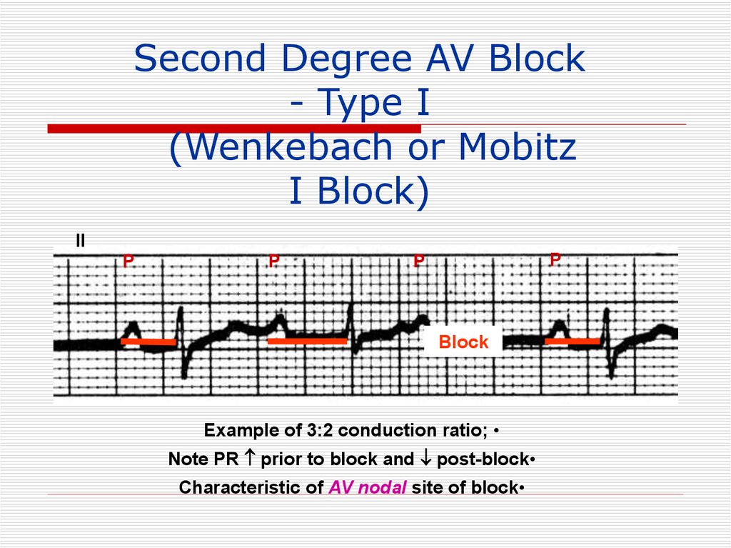
Second Degree AV Block- Type I
(Wenkebach or Mobitz
I Block)
II
P
P
P
P
Block
Example of 3:2 conduction ratio;
Note PR prior to block and post-block
Characteristic of AV nodal site of block
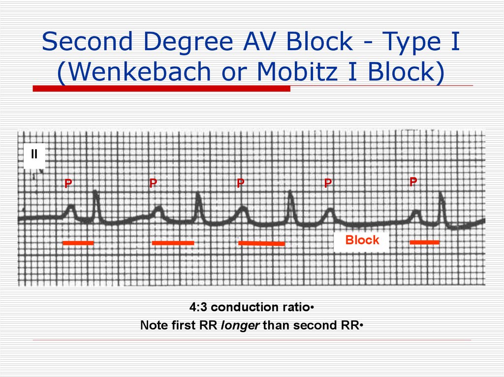
114.
Second Degree AV Block - Type I(Wenkebach or Mobitz I Block)
II
P
P
P
P
P
Block
4:3 conduction ratio
Note first RR longer than second RR
115.
II116.
Second Degree AV Block- Type II
(Mobitz II)
II
P
P
P
Block
P
P
P
Block
Example of 3:2 conduction ratio;
Note fixed PR for all conducted beats
Characteristic of His-Purkinje system site of block
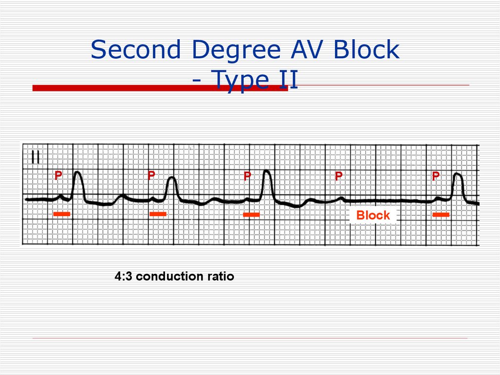
117.
Second Degree AV Block- Type II
P
P
P
P
P
Block
4:3 conduction ratio
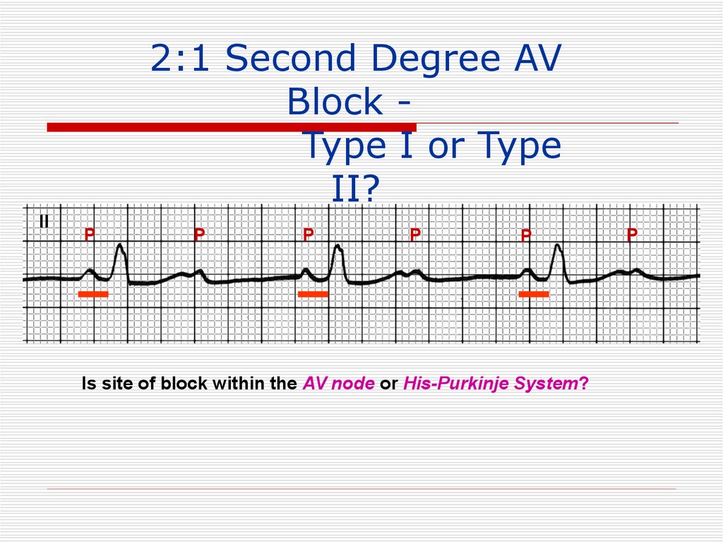
118.
2:1 Second Degree AVBlock Type I or Type
II?
II
P
P
P
P
P
Is site of block within the AV node or His-Purkinje System?
P
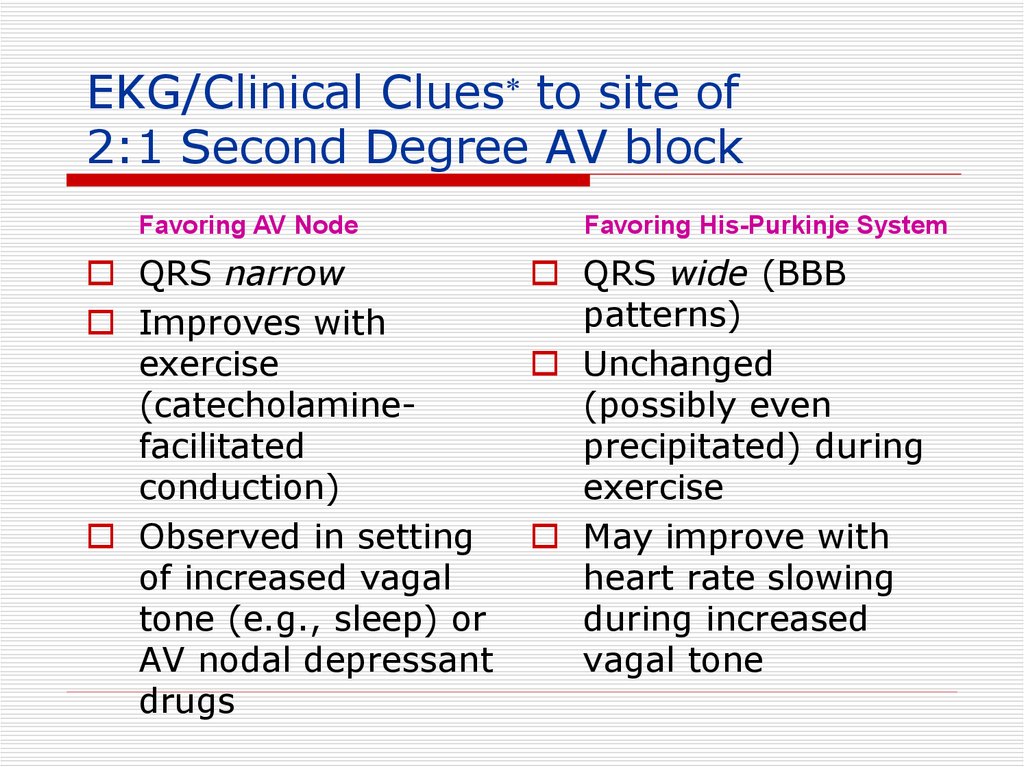
119.
EKG/Clinical Clues to site of2:1 Second Degree AV block
Favoring AV Node
QRS narrow
Improves with
exercise
(catecholaminefacilitated
conduction)
Observed in setting
of increased vagal
tone (e.g., sleep) or
AV nodal depressant
drugs
Favoring His-Purkinje System
QRS wide (BBB
patterns)
Unchanged
(possibly even
precipitated) during
exercise
May improve with
heart rate slowing
during increased
vagal tone
120.
Advanced Second DegreeAV Block
(Block of 2 Consecutive
P Waves)
II
P
P
P
P
P
P
P
P
3:1 conduction ratio, with ventricular rate in the 30’s
P
121.
Site of AV Block vs. EscapeRhythm
AV Node: Junctional or ventricular
His-Purkinje System: Ventricular
122.
123.
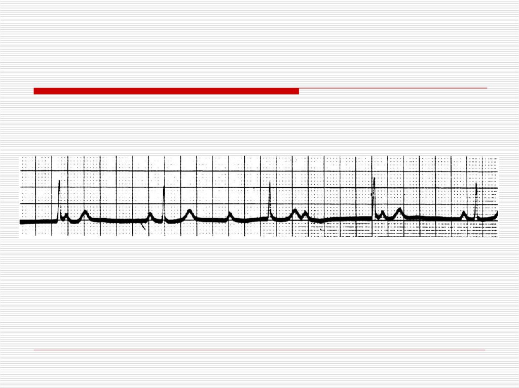
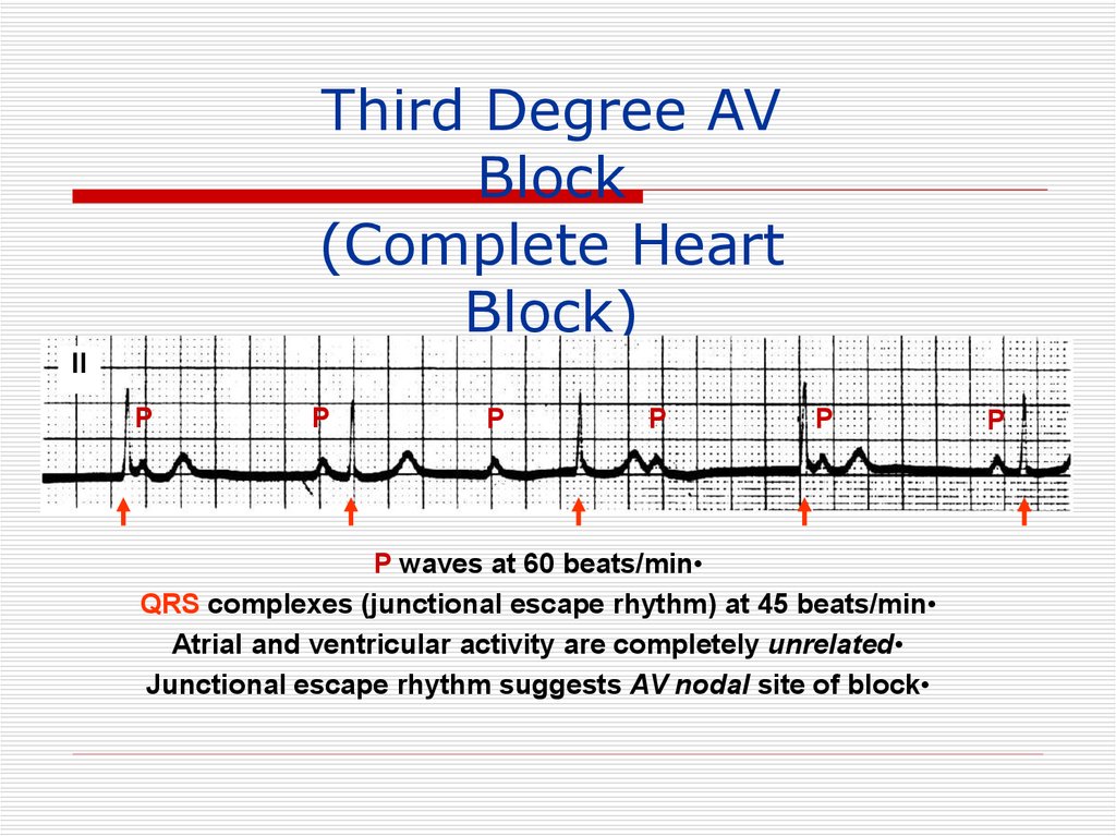
Third Degree AVBlock
(Complete Heart
Block)
II
P
P
P
P
P
P waves at 60 beats/min
QRS complexes (junctional escape rhythm) at 45 beats/min
Atrial and ventricular activity are completely unrelated
Junctional escape rhythm suggests AV nodal site of block
P
124.
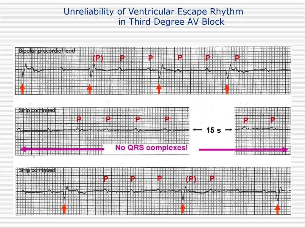
Unreliability of Ventricular Escape Rhythmin Third Degree AV Block
(P)
P
P
P
P
P
P
P
P
P
15 s
No QRS complexes!
P
P
P
P
(P)
P
P
125.
126.
127.
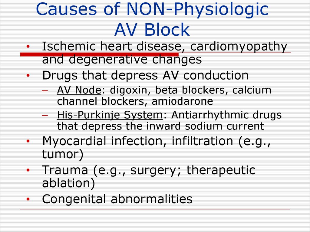
Causes of NON-PhysiologicAV Block
• Ischemic heart disease, cardiomyopathy
and degenerative changes
• Drugs that depress AV conduction
– AV Node: digoxin, beta blockers, calcium
channel blockers, amiodarone
– His-Purkinje System: Antiarrhythmic drugs
that depress the inward sodium current
• Myocardial infection, infiltration (e.g.,
tumor)
• Trauma (e.g., surgery; therapeutic
ablation)
• Congenital abnormalities
128.
Sinus Bradyarrhythmias129.
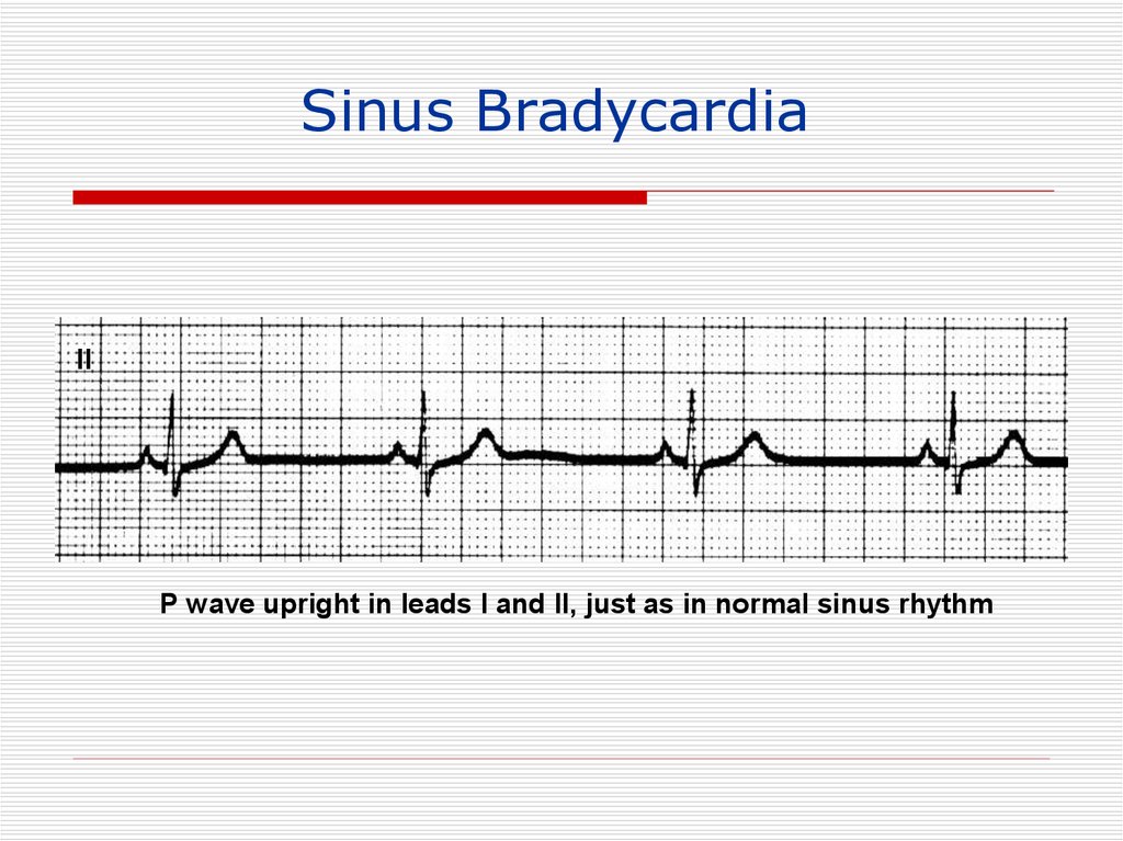
Sinus BradycardiaII
P wave upright in leads I and II, just as in normal sinus rhythm
130.
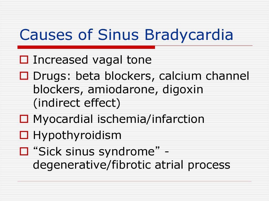
Causes of Sinus BradycardiaIncreased vagal tone
Drugs: beta blockers, calcium channel
blockers, amiodarone, digoxin
(indirect effect)
Myocardial ischemia/infarction
Hypothyroidism
“Sick sinus syndrome” degenerative/fibrotic atrial process
131.
Sequence of P Wave GenerationSinus
Node
SA
Junction
Non-visible process on the EKG
Atrium
(P wave)
132.
Sinus ArrhythmiaInspiration
SA nodal acceleration
Expiration
SA nodal deceleration
133.
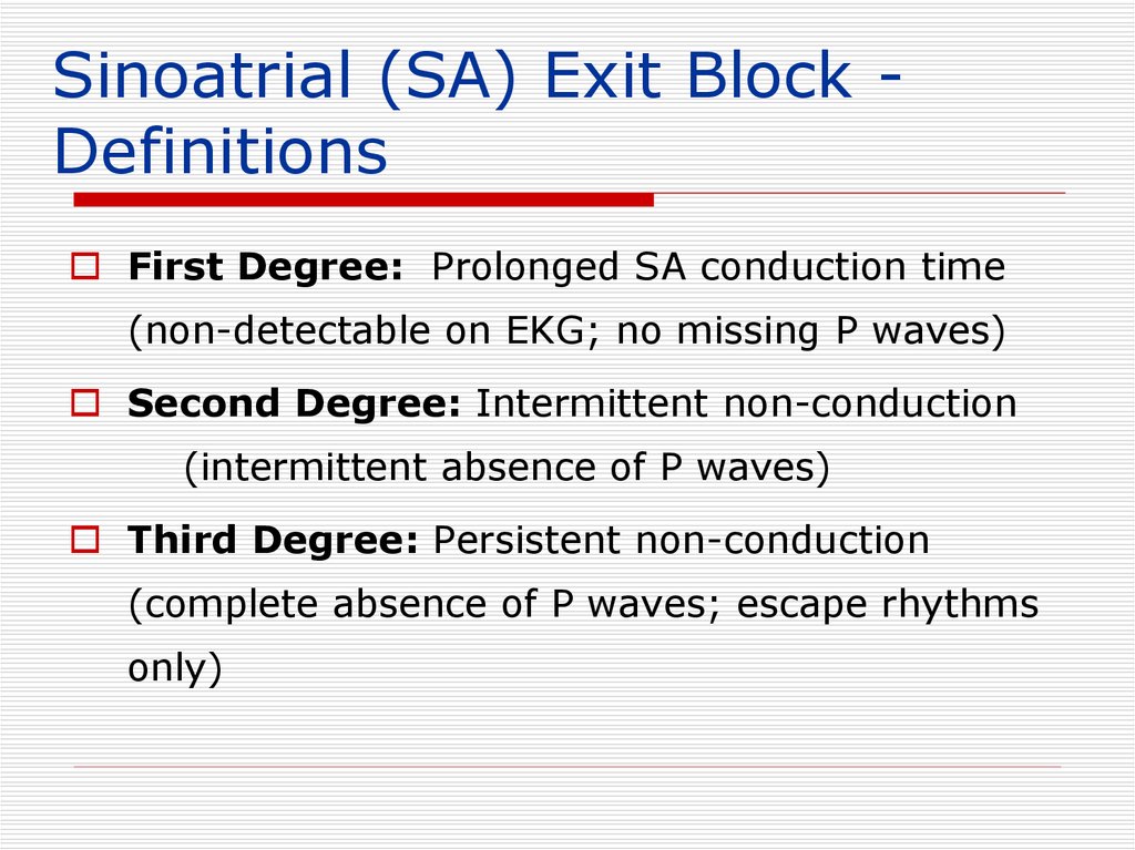
Sinoatrial (SA) Exit Block DefinitionsFirst Degree: Prolonged SA conduction time
(non-detectable on EKG; no missing P waves)
Second Degree: Intermittent non-conduction
(intermittent absence of P waves)
Third Degree: Persistent non-conduction
(complete absence of P waves; escape rhythms
only)
134.
Second Degree SA Exit Block - Type I(Wenkebach)
4:3 pattern
P
P
P
P
Missing
P wave
PP:
PP intervals shorten prior to block
Note unaffected, fixed PR intervals
135.
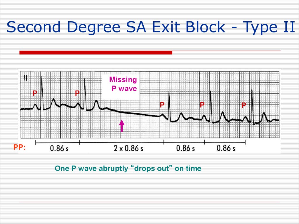
Second Degree SA Exit Block - Type IIP
P
Missing
P wave
P
P
PP:
One P wave abruptly “drops out” on time
P
136.
2:1 SA Exit Block(Every Other P wave is “Dropped”)
X
P
P
2X
P
P
P
Atrial rate is abruptly cut in half
2X
P
X
P
P
P
Resolution of block
137.
Sinus ArrestP
P
P’
P’
Sinus bradycardia Sinus arrest Slow junctional escape rhythm
(with retrograde p waves)
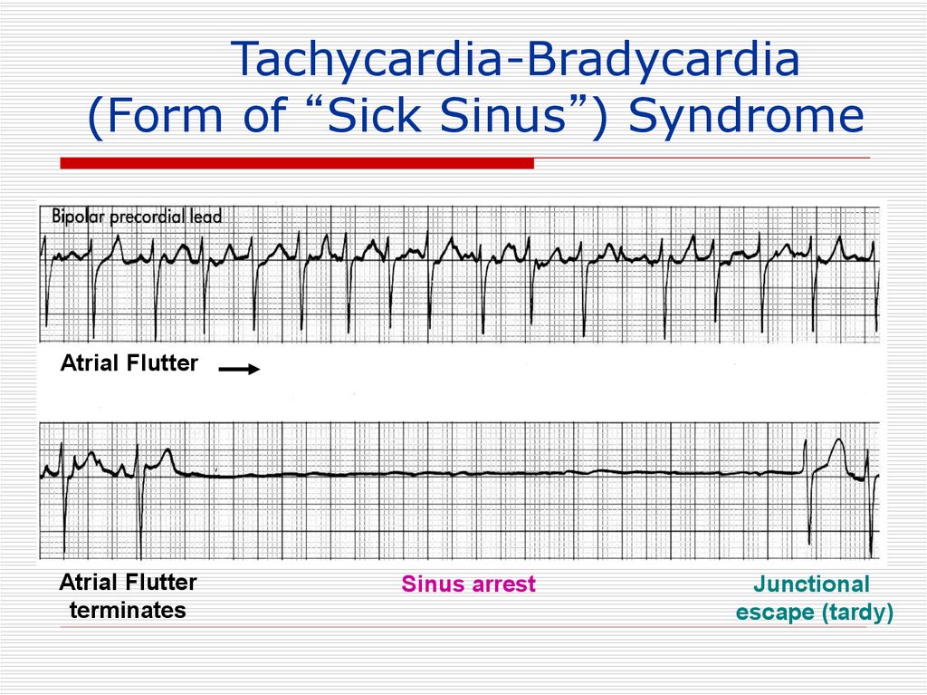
138.
Tachycardia-Bradycardia(Form of “Sick Sinus”) Syndrome
Atrial Flutter
Atrial Flutter
terminates
Sinus arrest
Junctional
escape (tardy)
139.
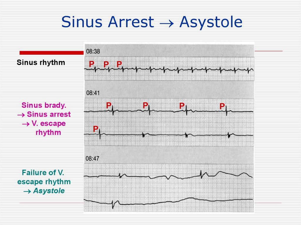
Sinus Arrest AsystoleSinus rhythm
Sinus brady.
Sinus arrest
V. escape
rhythm
Failure of V.
escape rhythm
Asystole
P
P P
P
P
P
P
P
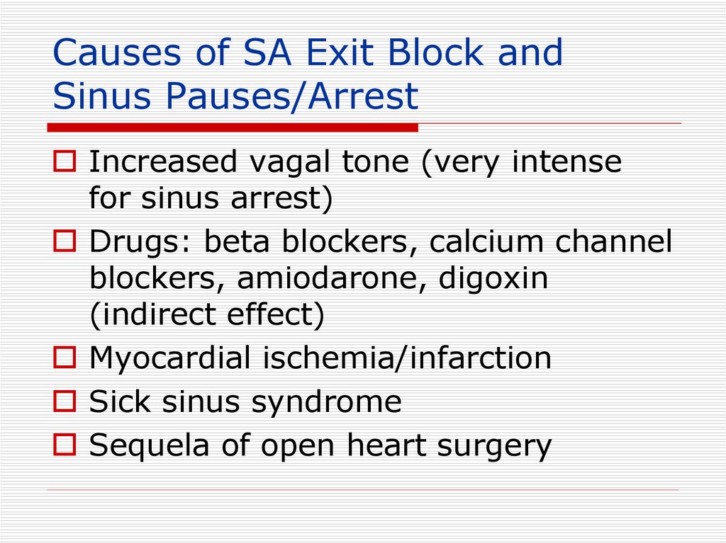
140.
Causes of SA Exit Block andSinus Pauses/Arrest
Increased vagal tone (very intense
for sinus arrest)
Drugs: beta blockers, calcium channel
blockers, amiodarone, digoxin
(indirect effect)
Myocardial ischemia/infarction
Sick sinus syndrome
Sequela of open heart surgery
141.
Sick Sinus Syndrome(1) persistent spontaneous sinus bradycardia
not caused by drugs and inappropriate for
the physiologic circumstance;
(2) sinus arrest or exit block
(3) combinations of SA and AV conduction
disturbances
(4) alternation of paroxysms of rapid regular
or irregular atrial tachyarrhythmias and
periods of slow atrial and ventricular rates
(bradycardia-tachycardia syndrome