


.")


")
 Keytruda (Pembrolizumab)")


 Checkmate 069")


 Медицина
Похожие презентации:


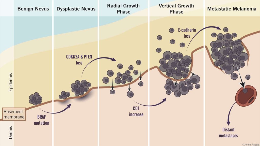
Malignant Melanoma
1. Malignant Melanoma
Dr Olga VornicovaResident in Clinical oncology
Rambam Medical Center
2. RISK FACTORS
Fair skinned.Hair color other than black.
Excessive sun exposure .
Melanoma in first-degree relative(s) .
Prior nonmelanoma skin cancer (basal cell and squamous cell
carcinoma).
Presence of xeroderma pigmentosum or familial atypical mole
melanoma syndrome.
3. Familial Atypical Mole Melanoma Syndrome
Autosomal dominantNeoplastic risk
"atypical melanocytic nevus“
25-40% with CDKN2A
mutation
4. Xeroderma Pigmentosum
Rare Autosomal recessive diseaseDNA repair enzyme defect
Photosensitivity
Photodamage
Cutaneous malignancies
Severe ophthalmological abnormalities
Early death from malignancy
5. Ultraviolet light
6.
UVC (< 290 nm)Completely absorbed by the atmosphere and is non-relevant for UV
induced skin carcinogenesis.
UVB (290-390 nm)
Absorbed by ozone, but 5-10% of it reaches the earth surface.
The exposure to the high penetrating UVB radiation leads to DNA damage
.
UVA (520-400 nm)
Genotoxicity seems to be induced by indirect mechanisms
mediated by reactive oxygen radicals and associated with
chronic sun damage changes.
7.
8. The ABCDEs of Melanoma Diagnosis
AsymmetryOne half of the lesion is shaped
differently than the other
Border
Color
The border of the
lesion is irregular,
blurred, or ragged
Inconsistent pigmentation, with
varying shades of brown and black
Evolution
Diameter
>6 mm, or a
progressive
change in size
History of change in the lesion
Photos courtesy of the American Cancer Society.
9.
TYPES OF MELANOMA10. NODULAR
– Commoner in males– Trunk is a common
site
– Poor prognosis
– Black/brown nodule
– Ulceration and
bleeding are common
11. SUPERFICIAL SPREADING
– The most common type of MMin the white-skinned population
– 70% of cases
– Commonest sites – lower leg in
females and back in males
– In early stages may be small,
then growth becomes irregular
12. ACRAL LENTIGINOUS MELANOMA
– Commonest MM in nonwhiteskinned nations– Usually comprises a flat lentiginous
area with an invasive nodular
component.
– Poorer prognosis.
13. SUBUNGAL MELANOMA
– Rare– Often diagnosed late –
confusion with benign subungal
naevus, paronychial infections,
trauma.
– Hutchinson’s sign – spillage of
pigment onto the surrounding
nailfold
14. LENTIGO MALIGNA MELANOMA
– Occurs as a late development ina lentigo maligna.
– Mainly on the face in elderly
patients .
– May be many years before an
invasive nodule develops.
15. AMELANOTIC MELANOMA
– Diagnosis is often missedclinically.
– The lack of pigmentation is due
to the rapid growth of the
tumour and the differentiation
of the malignant melanocytes.
16. Mucosal melanoma
– Muc M approximately 1 % of allmelanomas .
– Arise primarily in the head and neck,
anorectal, and vulvovaginal regions (55,
24, and 18 percent of cases,
respectively).
– Rarer sites of origin include the urinary
tract, gall bladder, and small intestine.
–
Worse prognosis
17. Ocular melanoma
– OM is the most common type of cancerto affect the eye, although it's still quite
rare.
– Incidence: 5.3 to 10.9 cases per million
– The incidence of ocular melanoma
increases with age, and most cases are
diagnosed in people in their 50s.
– OM may be more common in people
who have atypical mole syndrome .
18. Skin biopsy
Excisional Bx.Location
Breslow thickness
Ulceration
Peripheral and deep margins.
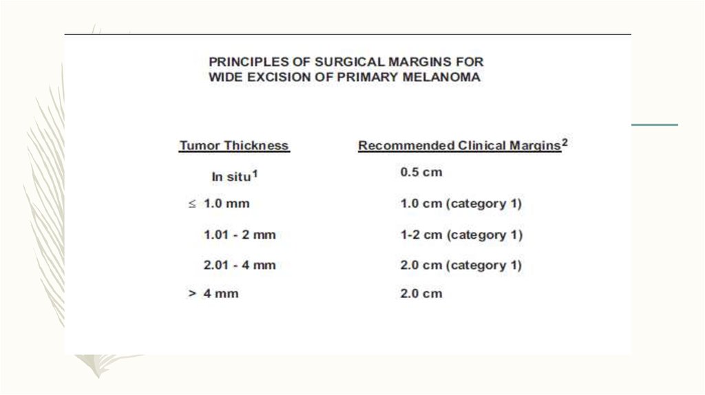
19. Breslow Thickness:
• < 1 mm (T1)thin
• 1-2 mm (T2)
• 2-4 mm (T3)
• > 4.0 mm (T4)
Intermediate
thick
20. Clark Level
21. Stage 0: (TisN0M0).
melanoma in situ22. Stage I: Local disease - superficial
23. Stage II: Local disease - deep invasion.
24. Stage III: Regional disease
25. Stage IV: Metastatic disease
26. Prognostic factors
Depth of InvasionUlceration
Lymph Node
Mitotic Rate (TNM staging system 2010)
LDH level
Patient Gender : women better than men
Anatomic site:
– head and neck- scalp worse
– extremity better than trunk
27.
28.
29.
30.
31. Sentinel lymph node biopsy
– SLN = First node(s) draining the area of primary lesion.– Sentinel node biopsy is generally recommended for patients with
melanomas at least 1 mm thick or more then 0.75 mm with 1 or more
mitoses
– Prognostic factor - data for patient.
– Applying adjuvant therapy.
– Survival benefit.
32. Sentinel lymph node mapping and biopsy
33.
34. Adjuvant therapy
– Potential candidates–
–
Stage IIB
Stage III
(+/-50% recurrence rate)
– Chemotherapy - not effective (DTIC).
– Immunotherapy - IFN and Ipillimumab
– Vaccination – not effective.
– Clinical trails ( anti BRAF , anti PD1, anti PD1+anti CTLA4- ongoing)
35.
36. IPILIMUMAB
YervoyAnti CTLA4 Antibody
37.
38.
39.
40.
41.
42. IFN - Side effects
IFN - Side effects– Acute toxicity :
(Due to PGE2 synthesis and/or other cytokines)
–
–
–
–
Flue like syndrome
malaise
Arthralgia
DLT - hepatotoxicity
– Chronic constitutional effects:
(Due to hypothalamic, endocrine and/or neurotransmitter dysfunction)
–
–
–
–
–
–
–
–
fatigue
anorexia
weight loss
depression
impaired cognitive function
diminished libido and potency
myelosuppression
Hepatic toxicity
43. Treatment Options for advanced Melanoma
44. BRAF\MEK Inhibitors
Dabrafenib (TAFINLAR) Trametinib( MEKINIST)
Vemurafenib ( ZELBORAF)
Cobimetinib (COTELIC)
45.
46.
47.
48.
49.
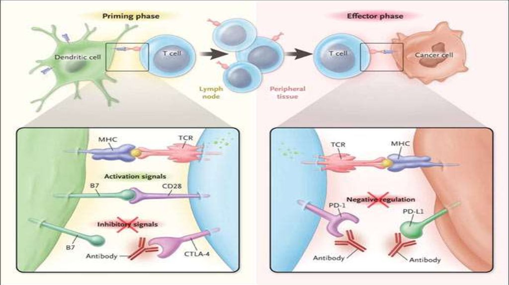
50. Imunotherapy
51.
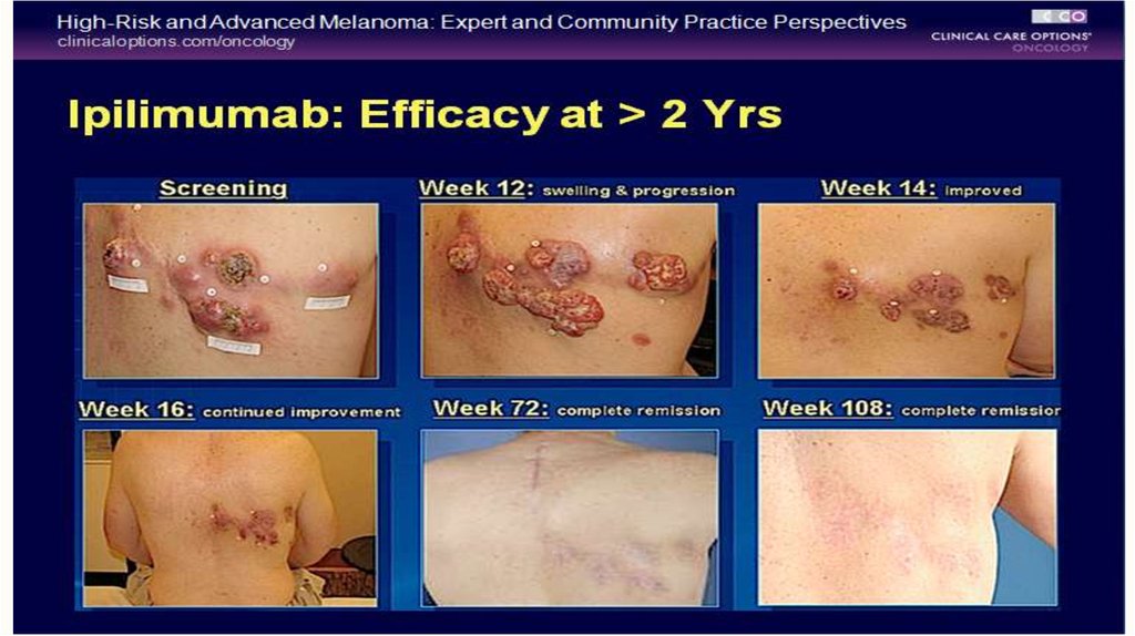
52. Ipillimumab (Yervoy)
In pooled analysis of 12 studies, a plateau in the survival curve begins atapproximately three years, with some patients followed for up to ten years
Three-year and five-year estimated survival rate of 22% and 18% respectively
observed in patients treated with Yervoy
53.
54. Anti PD1 therapy : Opdivo (Nivolumab) Keytruda (Pembrolizumab)
55. Opdivo Monotherapy Phase 3 Trial: Improved OS Versus Dacarbazine in BRAF Wild-type, Untreated Patients
Median OS,mo (95% CI)
HR (95% CI)
Phase III CheckMate 066
NIVO
DTIC
NR
(23.1, NR)
11.2
(9.6, 13.0)
0.43 (0.33, 0.57); P <0.001
1.0
Probability of Survival
0.9
0.8
NIVO 3 mg/kg Q2W (n=210)
1-yr OS=70.7%
0.7
2-yr OS=57.7%
0.6
1-yr OS=46.3%
0.5
0.4
2-yr OS=26.7%
0.3
0.2
Dacarbazine (n=208)
0.1
0.0
0
3
6
9
12
15
18
21
24
27
30
111
60
81
38
30
16
4
1
0
0
Overall Survival (Months)
Patients at Risk
Nivolumab
Dacarbazine
210
208
186
179
171
146
154
122
143
92
135
76
1. Atkinson V et al. Presented at SMR 2015. 2. Robert C, et al. N Engl J Med. 2015;372:320-323.
56.
57.
58.
59Updated Results From a Phase III Trial of Nivolumab
Combined With Ipilimumab in Treatment-naïve Patients
With Advanced Melanoma (Checkmate 067)
59. Updated Results From a Phase III Trial of Nivolumab Combined With Ipilimumab in Treatment-naïve Patients With Advanced Melanoma
OS at 2 Years of Follow-up(All Randomized Patients)
60
Checkmate 069
NR
NR (11.9‒NR)
HR (95% CI)
0.9
Probability of Overall Survival
IPI (N = 47)
Median OS, months (95% CI)
1.0
0.8
0.74 (0.43‒1.26)*
*Exploratory
endpoint
NR = not reached
73%
0.7
64%
0.6
65%
0.5
54%
0.4
0.3
0.2
NIVO + IPI
0.1
0.0
0
IPI
3
6
9
12
95
47
15
18
21
24
27
30
65
26
63
25
57
22
6
3
0
0
Months
Number of Patients at Risk
NIVO+ IPI
IPI
NIVO + IPI (N = 95)
82
41
77
36
74
33
69
29
67
27
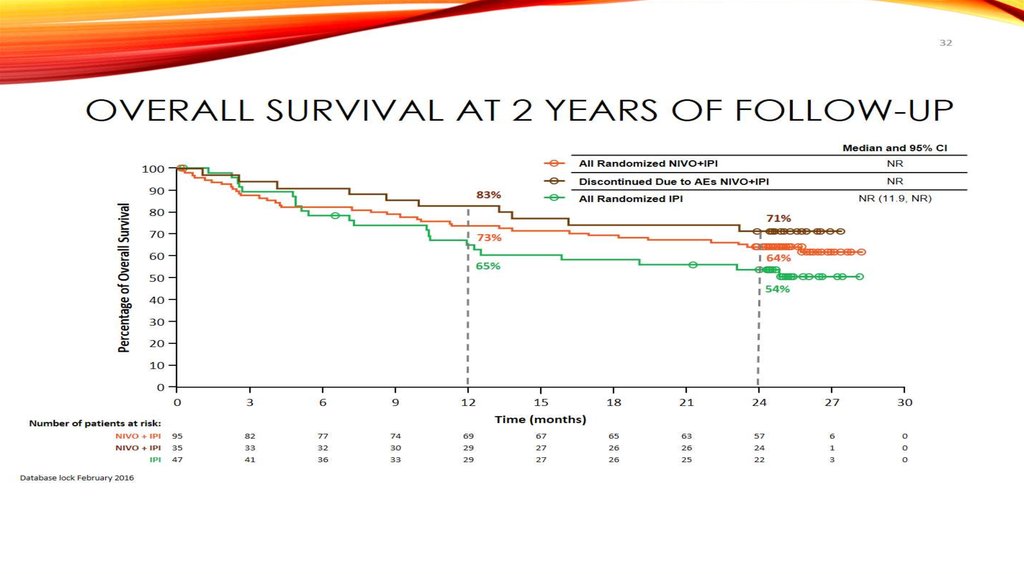
30/47 (64%) of patients randomized to IPI crossed over to receive any systemic therapy at progression
AACR 2016
60. OS at 2 Years of Follow-up (All Randomized Patients) Checkmate 069
Response To Treatment61
61. Response To Treatment
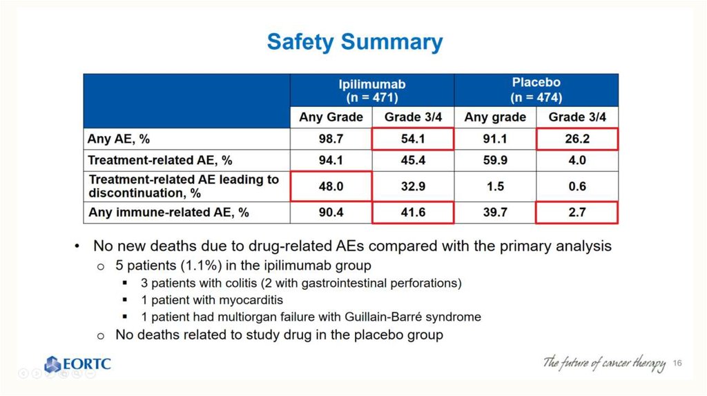
Safety Summary62
–
Updated safety information with 9 additional months of follow-up were consistent with the initial report
NIVO+IPI
(N=313)
Patients reporting
event, %
Treatment-related adverse
event (AE)
Treatment-related AE
leading to discontinuation
Treatment-related death*
–
NIVO
(N=313)
IPI
(N=311)
Any
Grade
Grade 3-4
Any
Grade
Grade 34
Any
Grade
Grade 34
95.8
56.5
84.0
19.8
85.9
27.0
38.7
30.7
10.5
7.3
15.4
13.5
0
0.3
0.3
68.8% of patients who discontinued NIVO+IPI due to treatment-related AEs achieved a response
*One reported in the NIVO group (neutropenia) and one in the IPI group (colon perforation)
Database lock Nov 2015
62. Safety Summary
63.
64.
Overall Survival at 2 Years of Follow-up65
Median and 95% CI
100
Percentage of Overall Survival
90
83%
All Randomized NIVO+IPI
NR
Discontinued Due to AEs NIVO+IPI
NR
NR (11.9, NR)
All Randomized IPI
80
71%
70
73%
60
64%
65%
50
54%
40
30
20
10
0
0
3
6
9
12
95
35
47
82
33
41
77
32
36
74
30
33
69
29
29
18
21
24
27
30
65
26
26
63
26
25
57
24
22
6
1
3
0
0
0
Time (months)
Number of patients at risk:
NIVO + IPI
NIVO + IPI
IPI
15
Database lock February 2016
67
27
27
65. Overall Survival at 2 Years of Follow-up
J.M. Michot et al. EuropeanJournal of Cancer 54 (2016)
66.
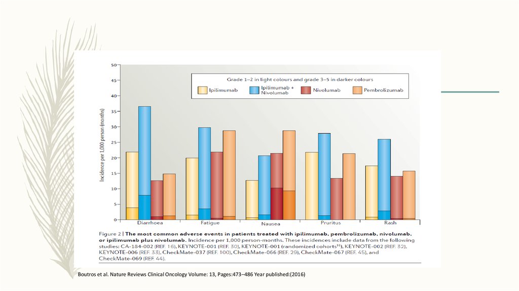
Boutros et al. Nature Reviews Clinical Oncology Volume: 13, Pages:473–486 Year published:(2016)67.
Boutros et al. Nature Reviews Clinical Oncology 13, 473–486 (2016)68.
Webber JS , Safety profile of nivolumab in patients with advanced melanoma, Pooled Analysis. ASCO 2016( Poster).