


 Медицина
Похожие презентации:


")
Myeloprolifirative disorders
1.
Dr. TzoranMYELOPROLIFERATIVE
DISORDERS
2.
IntroductionHematopoietic stem cell disorder
Clonal
Characterized by proliferation
Granulocytic
Erythroid
Megakaryocytic
Interrelationship between
Polycythaemia
Essential thrombocythaemia
myelofibrosis
3.
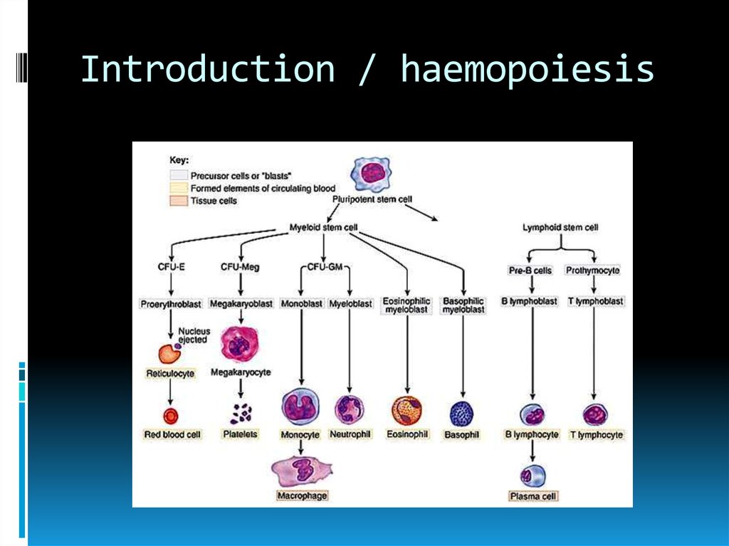
Introduction / haemopoiesis4.
IntroductionNormal maturation (effective)
Increased number of
Red cells
Granulocytes
Platelets
(Note: myeloproliferation in myelodysplastic syndrome is
ineffective)
Frequent overlap of the clinical, laboratory & morphologic
findings
Leucocytosis, thrombocytosis, increased
megakaeryocytes, fibrosis & organomegaly blurs the
boundaries
Hepatosplenomegaly
Sequestration of excess blood
Extramedullary haematopoiesis
Leukaemic infiltration
5.
Rationale for classificationClassification is based on the lineage of the
predominant proliferation
Level of marrow fibrosis
Clinical and laboratory data (FBP, BM,
cytogenetic & molecular genetic)
6.
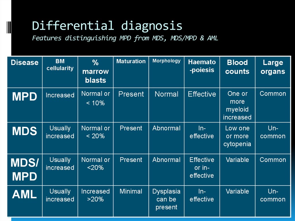
Differential diagnosisFeatures distinguishing MPD from MDS, MDS/MPD & AML
Disease
BM
cellularity
%
marrow
blasts
Maturation
Morphology
Haemato
-poiesis
Blood
counts
Large
organs
MPD
Increased
Normal or
< 10%
Present
Normal
Effective
One or
more
myeloid
increased
Common
MDS
Usually
increased
Normal or
< 20%
Present
Abnormal
Ineffective
Low one
or more
cytopenia
Uncommon
MDS/
MPD
Usually
increased
Normal or
<20%
Present
Abnormal
Effective
or ineffective
Variable
Common
AML
Usually
increased
Increased
>20%
Minimal
Dysplasia
can be
present
Ineffective
Variable
Uncommon
7.
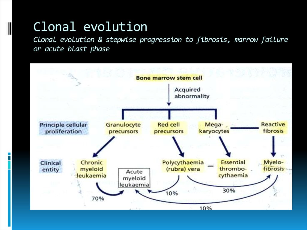
Clonal evolutionClonal evolution & stepwise progression to fibrosis, marrow failure
or acute blast phase
8.
Incidence and epidemiologyDisease of adult
Peak incidence in 7th decade
6-9/100,000
9.
PathogenesisDysregulated proliferation
No specific genetic abnormality
CML (Ph chromosome t(9;22) BCR/ABL)
Growth-factor independent proliferation
PV, hypersensitiviy to IGF-1
Bone marrow fibrosis in all MPD
Fibrosis is secondary phenomena
Fibroblasts are not from malignant clone
TGF-β & Platelet like growth factor
10.
Molecular basis ofPhiladelphia-negative
myeloproliferative neoplasms
Polycythemia Vera:~95% JAK2(V617F)
Essential thrombocythemia: 50-60% JAK2(V617F)
Primary myelofibrosis 50-60% JAK2(V617F)
11.
PrognosisDepends on the proper diagnosis and early
treatment
Role of
IFN
BMT
Tyrosine kinase inhibitors
12.
Polycythaemia vera(Polycythaemia rubra vera)
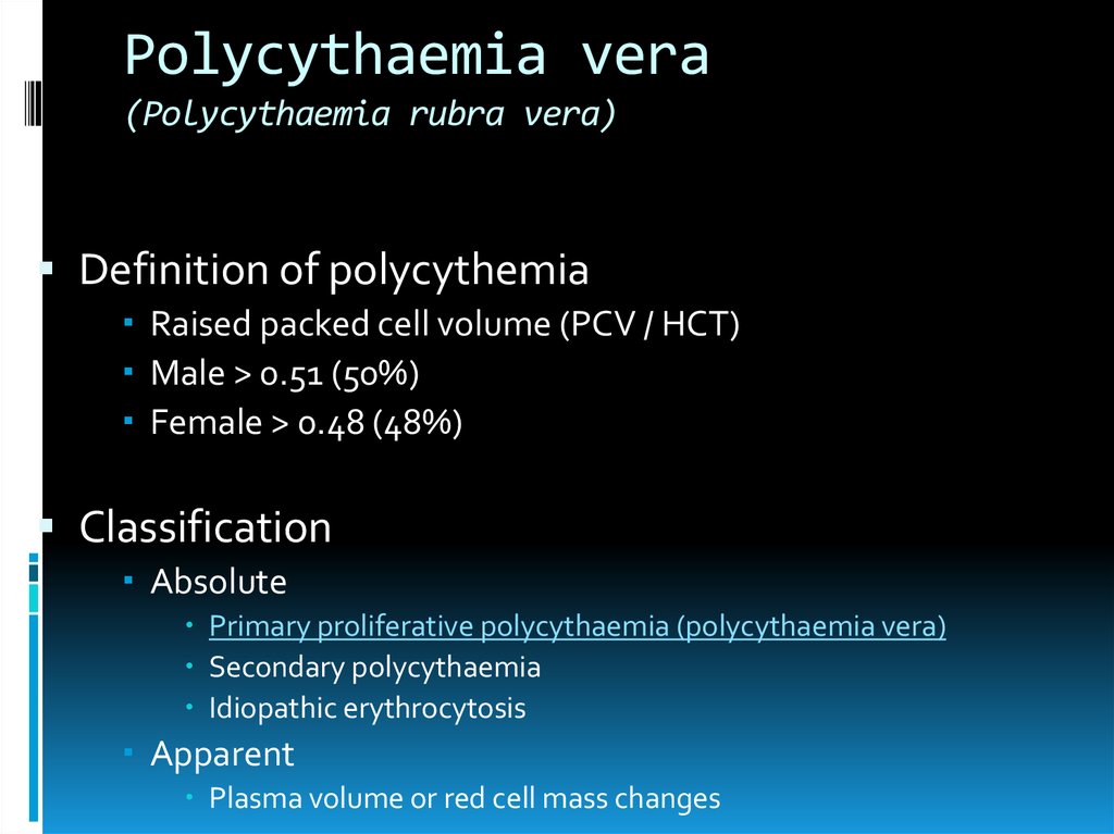
Definition of polycythemia
Raised packed cell volume (PCV / HCT)
Male > 0.51 (50%)
Female > 0.48 (48%)
Classification
Absolute
Primary proliferative polycythaemia (polycythaemia vera)
Secondary polycythaemia
Idiopathic erythrocytosis
Apparent
Plasma volume or red cell mass changes
13.
Polycythaemia vera(Polycythaemia rubra vera)
Polycythaemia vera is a clonal stem cell disorder
characterised by increased red cell production
Abnormal clones behave autonomous
Same abnormal stem cell give rise to granulocytes and platelets
Disease phase
Proliferative phase
“Spent” post-polycythaemic phase
Rarely transformed into acute leukemia
14.
Polycythaemia vera(Polycythaemia rubra vera)
Clinical features
Age
55-60 years
May occur in young adults and rare in childhood
Majority patients present due to vascular complications
Thrombosis (including portal and splenic vein)
DVT
Hypertension
Headache, poor vision and dizziness
Skin complications (pruritus, erythromelalgia)
Haemorrhage (GIT) due to platelet defect
15.
Polycythaemia vera(Polycythaemia rubra vera)
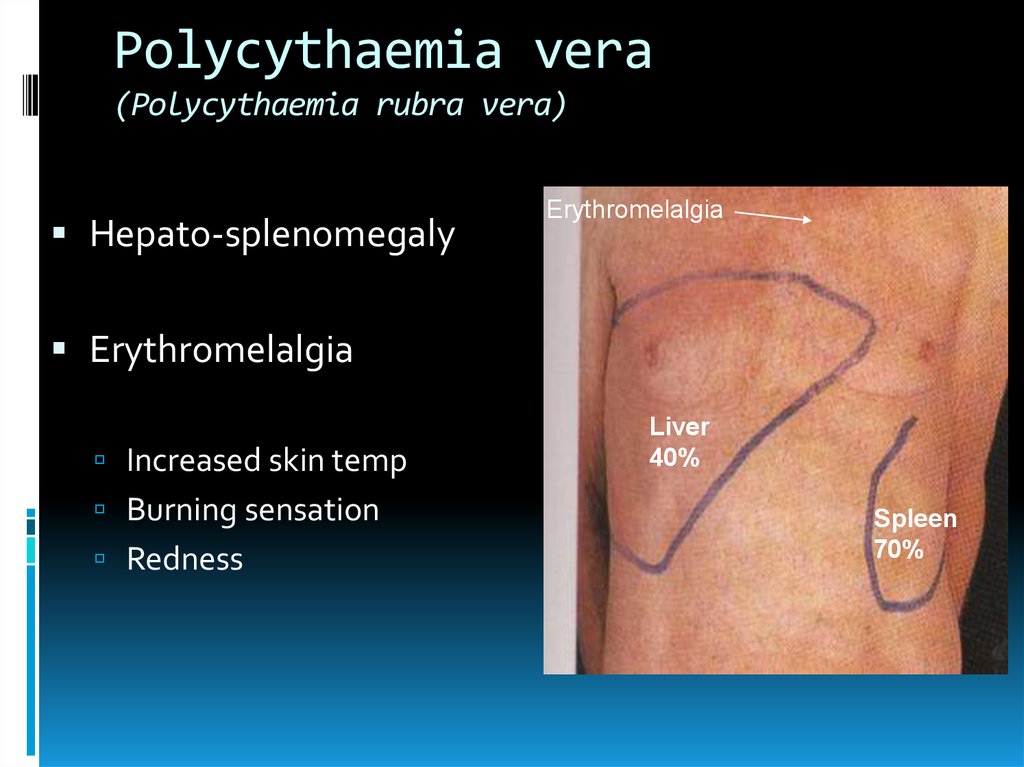
Hepato-splenomegaly
Erythromelalgia
Erythromelalgia
Increased skin temp
Burning sensation
Redness
Liver
40%
Spleen
70%
16.
Polycythaemia vera(Polycythaemia rubra vera)
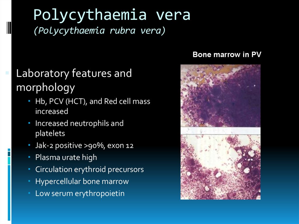
Bone marrow in PV
Laboratory features and
morphology
Hb, PCV (HCT), and Red cell mass
increased
Increased neutrophils and
platelets
Jak-2 positive >90%, exon 12
Plasma urate high
Circulation erythroid precursors
Hypercellular bone marrow
Low serum erythropoietin
17.
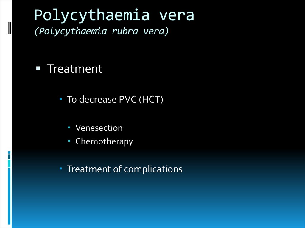
Polycythaemia vera(Polycythaemia rubra vera)
Treatment
To decrease PVC (HCT)
Venesection
Chemotherapy
Treatment of complications
18.
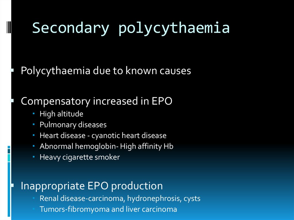
Secondary polycythaemiaPolycythaemia due to known causes
Compensatory increased in EPO
High altitude
Pulmonary diseases
Heart disease - cyanotic heart disease
Abnormal hemoglobin- High affinity Hb
Heavy cigarette smoker
Inappropriate EPO production
Renal disease-carcinoma, hydronephrosis, cysts
Tumors-fibromyoma and liver carcinoma
19.
Secondary polycythaemiaArterial blood gas
Hb electrophoresis
Oxygen dissociation curve
EPO level
Ultrasound abdomen
Chest X ray
Total red cell volume(51Cr)
Total plasma volume(125 I-albumin)
20.
Relative polycythaemiaApparent polycythaemia or
pseudopolycythaemia due to plasma volume
contraction
Causes
Stress
Cigarette smoker or alcohol intake
Dehydration
Plasma loss- burn injury
21.
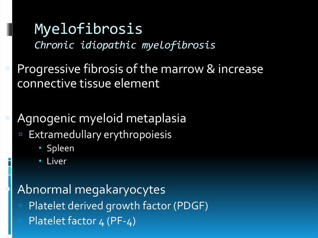
MyelofibrosisChronic idiopathic myelofibrosis
Progressive fibrosis of the marrow & increase
connective tissue element
Agnogenic myeloid metaplasia
Extramedullary erythropoiesis
Spleen
Liver
Abnormal megakaryocytes
Platelet derived growth factor (PDGF)
Platelet factor 4 (PF-4)
22.
MyelofibrosisChronic idiopathic myelofibrosis
Insidious onset in older people
Splenomegaly- massive
Hypermetabolic symptoms
Loss of weight, fever and night
sweats Myelofibrosis
Chronic idiopathic myelofibrosisc
Bleeding problems
Bone pain
Gout
Can transform to acute
leukaemia in 10-20% of cases
23.
MyelofibrosisChronic idiopathic myelofibrosis
Anaemia (bad prognosis)
High WBC at presentation
Later leucopenia and
thrombocytopenia
Leucoerythroblastic blood film
Tear drops red cells
Bone marrow aspirationFailed due to fibrosis
Trephine biopsy- fibrotic
hypercellular marrow
Increase in LAP score
24.
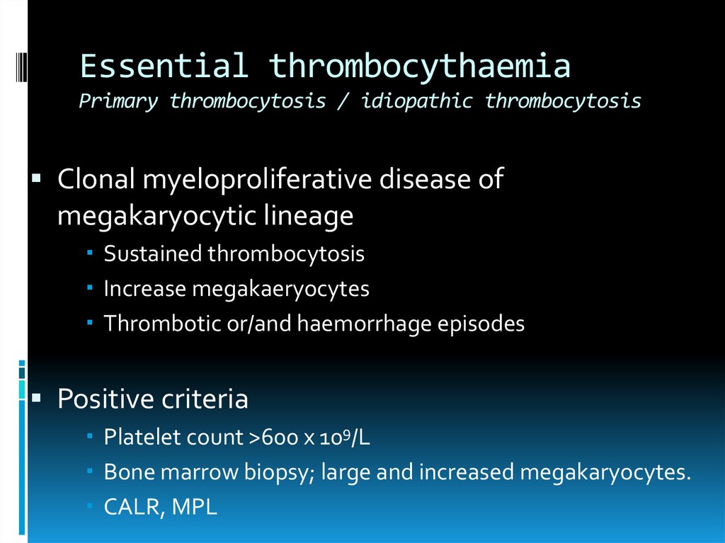
Essential thrombocythaemiaPrimary thrombocytosis / idiopathic thrombocytosis
Clonal myeloproliferative disease of
megakaryocytic lineage
Sustained thrombocytosis
Increase megakaeryocytes
Thrombotic or/and haemorrhage episodes
Positive criteria
Platelet count >600 x 109/L
Bone marrow biopsy; large and increased megakaryocytes.
CALR, MPL
25.
Essential thrombocythaemiaPrimary thrombocytosis / idiopathic thrombocytosis
Criteria of exclusion
No evidence of Polycythaemia vera
No evidence of CML
No evidence of myelofibrosis (CIMF)
No evidence of myelodysplastic syndrome
No evidence of reactive thrombocytosis
Bleeding
Trauma
Post operation
Chronic iron def
Malignancy
Chronic infection
Connective tissue disorders
Post splenectomy
26.
Essential thrombocythaemiaPrimary thrombocytosis / idiopathic
thrombocytosis
Clinical features
Haemorrhage
Microvascular occlusion
TIA, gangrene
Splenic or hepatic vein
thrombosis
Hepatosplenomegaly
27.
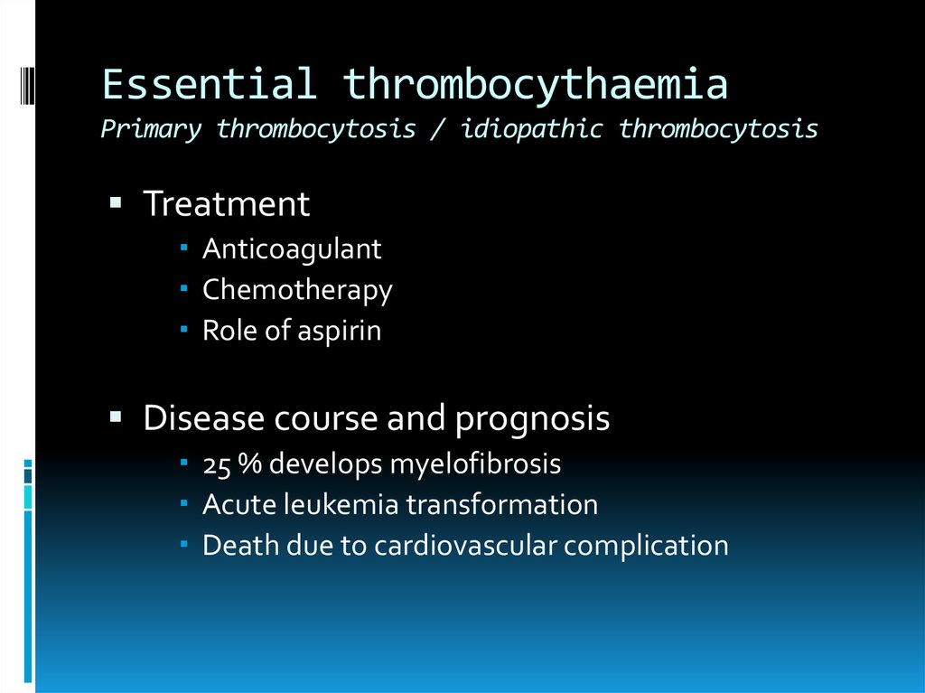
Essential thrombocythaemiaPrimary thrombocytosis / idiopathic thrombocytosis
Treatment
Anticoagulant
Chemotherapy
Role of aspirin
Disease course and prognosis
25 % develops myelofibrosis
Acute leukemia transformation
Death due to cardiovascular complication