


 Медицина
Похожие презентации:


Anatomy of orbit
1. ..
ANATOMY OF ORBITRajvin Samuel Ponraj
2. Development of orbit
Develops from mesenchymeossification
by
6 th to 7 th week laying down of bones
starting with maxilla bone around the
Optic vesicle
During this time optic vesicle 170 degree
apart rotates anteriorly
3. Developmental Anomalies :
Craniosynosotosis:Brachycephaly
Oxycephaly
Scophocephaly
Trigonocephaly
4.
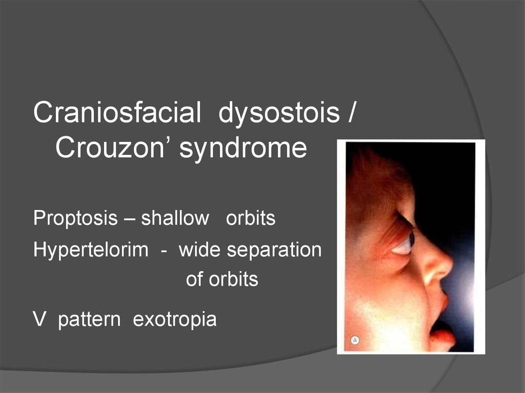
Craniosfacial dysostois /Crouzon’ syndrome
Proptosis – shallow orbits
Hypertelorim - wide separation
of orbits
V pattern exotropia
5.
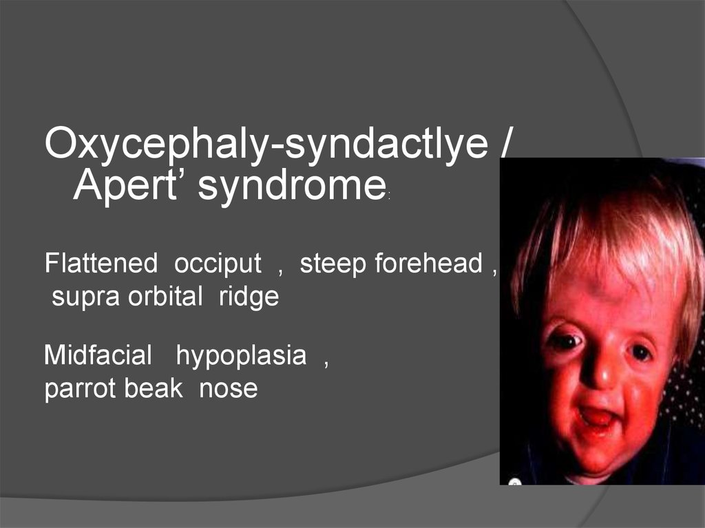
Oxycephaly-syndactlye /Apert’ syndrome
:
Flattened occiput , steep forehead ,
supra orbital ridge
Midfacial hypoplasia ,
parrot beak nose
6. Bones of Orbit
FrontalLacrimal
Maxillary
Ethmoid
Palatine
Zygomatic
Sphenoid
7. Dimensions - orbit
30 ml –volume35 mm vertically ,
40 mm horizontally
45 degree between lateral wall
and sagital plane
23 degree between visual
and orbital axis
8. Boundaries of Orbit
RoofFloor
Side walls
Orbital apex
9.
Roofof orbit
Frontal bone [Orbital plate] & lesser wing of sphenoid
Separated from frontal sinus and anterior
cranial fossa above
Lacrimal gland fossa and trochlear fossa
behind orbital rim
10.
Orbital roof anomaly / fractureCSF pulsation
pulsatile
exophthalmos
Orbital meningocele / encephalocele
11.
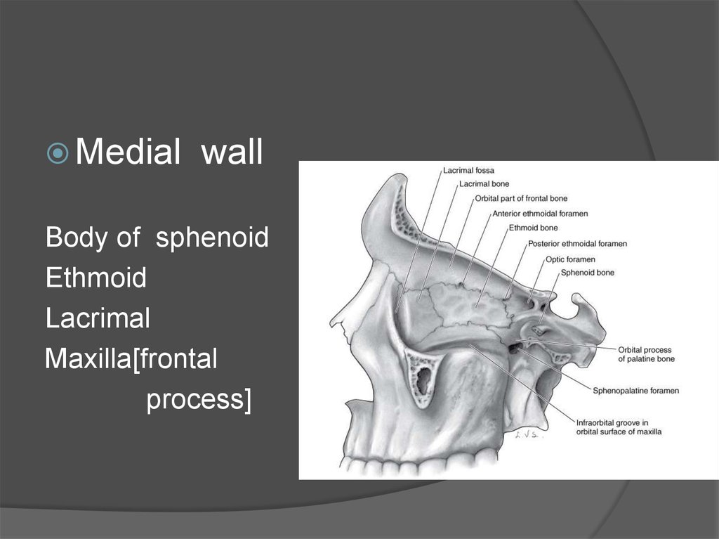
Medialwall
Body of sphenoid
Ethmoid
Lacrimal
Maxilla[frontal
process]
12.
Orbital cellulitisExtremely thin wall
Prone for damage & sinusitis spread
Infection across
Orbital cellulitis
13.
Floorof orbit
Maxilla
Zygomatic
Palatine
Triangular segment
-- thinnest
Inferior orbital groove
14.
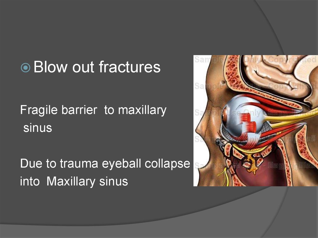
Blowout fractures
Fragile barrier to maxillary
sinus
Due to trauma eyeball collapse
into Maxillary sinus
15. Le fort’s fracture
Type 2 - PyramidalType 3 - Craniofacial
dissociation
16.
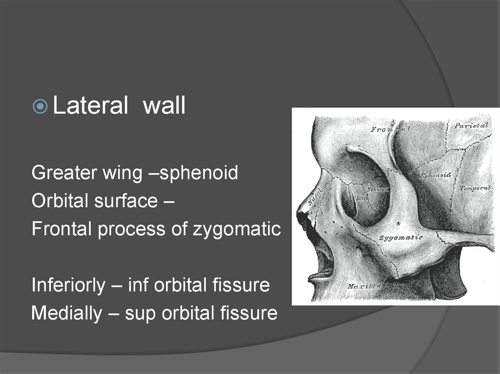
Lateralwall
Greater wing –sphenoid
Orbital surface –
Frontal process of zygomatic
Inferiorly – inf orbital fissure
Medially – sup orbital fissure
17.
Behind Zygomatic sphenoidal suturelateral orbitotomy of greater wing
( thin wall )
cancellous bone
middle cranial fossa
dura matter
18.
At frontal sphenoidal suture-- meningeal foramen
Site of anastomosis of Lacrimal artery and
meningeal artery collaterals
Periosteal elevation at this site
bleeding
Brisk
19.
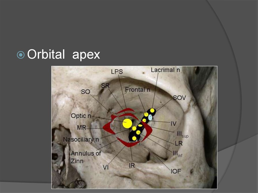
Orbitalapex
20.
Orbital apex syndrome/ Tolosa - hunt syndrome :
Damage to structures at apex 2 nd, 3 rd, 4
th ,6 th nerves
Symptoms : visual loss, ophthalmoplegia
periorbital & facial pain
21.
a.b.
c.
d.
e.
Other causes:
Inflammatory
Infectious
Neoplastic
Iatrogenic / traumatic
Vascular
22.
Superior orbital fissure syndrome/ Rochon – Duvigneaud syndrome :
Lesion anterior to orbital apex excluding
optic nerve pathology
23. Contents of orbit
Eye ballOrbital fat
Connective tissue system
Blood vessels
Nerves
Extraocular muscles
24. Eyeball - Applied anatomy:
Proptosis:
Dystopia
Enophthalmosis
Ophthalmoplegia
25. Connective tissue system
PeriorbitaOrbital septum
Tenon’s capsule
26.
Periorbita:Loosely attached to orbital bone
Attached firmly to
a.
b.
c.
d.
e.
f.
Arcus marginalis
Trochlea
Lateral orbital tubercle
Optic foramen
Orbital fissures
Dura and optic canal margins
27.
Orbitalseptum:
Interconnecting / circumferential radial
webs of fascial system
support and transmit forces in trauma
Compressive optic neuropathy following
trauma
28.
Anterior fascial systemFormed by condensation of fibrous septa
Lockwood lig,
whitnall sup susp lig
Lacrimal lig
Intermuscular septum
Posterior Fascial system
Incompletely
formed
29.
Tenon’scapsule
Dense elastic , vascular
Extent : from perilimbal sclera to optic
nerve meninges with bursa within
Sleeve like extensions for
extra ocular muscles continues as
fibrous capsule along its length
30.
31. Surgical spaces in orbit :
Sub periosteal spacePeripheral space
Central space
Tenon’s space
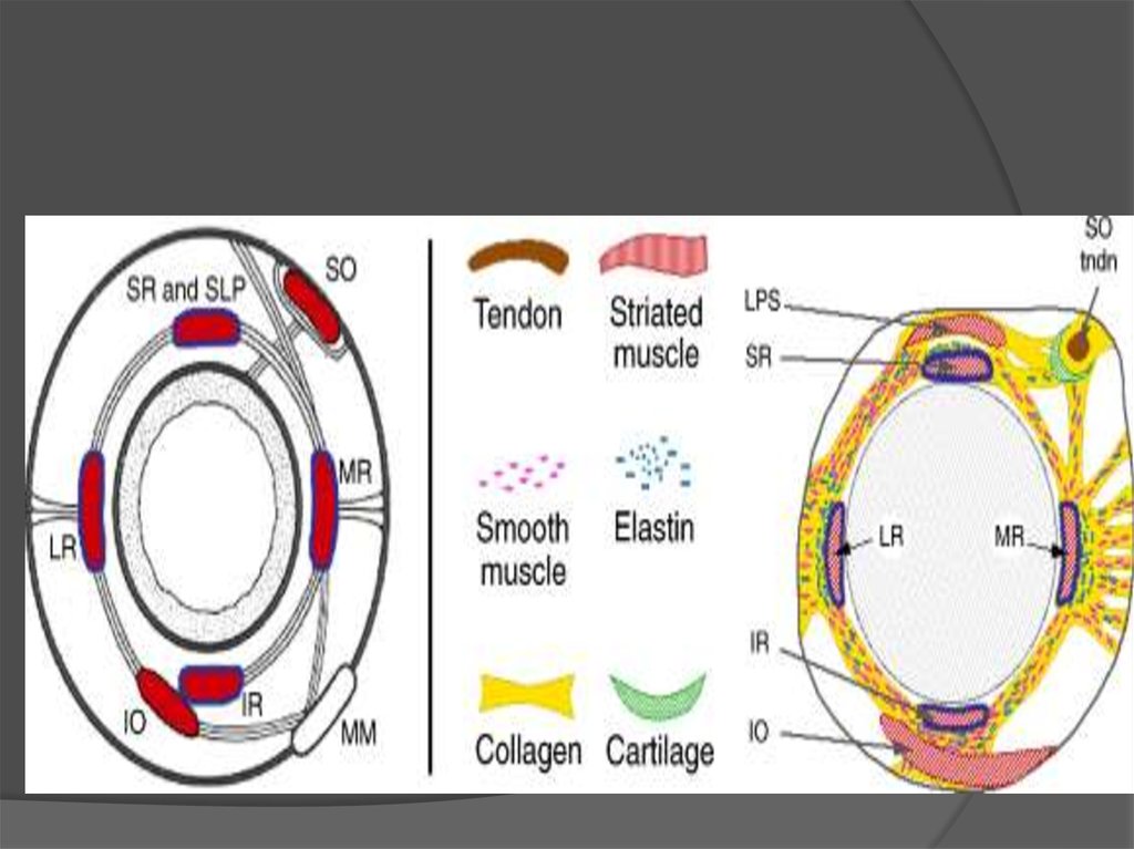
32. Extra ocular muscles
4 rectal muscles2 oblique muscles
Two lid retractors
To serve in eyeball movements in the
orbital cavity
33. Arterial supply
34. Venous drainage
35. Optic nerve
Intra orbital part = 25 mm outof 4 cm
Enclosed in three meningeal
sheaths
At apex surrounded by recti muscles ,
Central retinal artery and vein pierces optic nerve
1.25 cm behind optic nerve
Relations: superiorly
inferiorly
ophthalmic artery
sup ophthal vein
nasociliary nerve
nerve to medial rectus
36. Oculomotor nerve
Divides at anterior part ofcavernous sinus before
Entering sup orbital fissure
Sup division
Inf division
Sup rectus
LPS
Medial rectus
Inf rectus
Inf oblique
And motor root relay at ciliary ganglion
sphincter pupillae , ciliary muscle
37. Trochlear nerve
Runs medially from lateral wallof cavernous sinus
Above Levator palpebral sup
Then supplies orbital surface of
Superior oblique
38. Abducent nerve
Running inferior lateral to 3 rdnerve then supplies ocular
surface of lateral Rectus
39. Trigeminal nerve
Three terminal branches of ophthalmic division:I.
Frontal nerve
I.
Lacrimal nerve
supratrochlear
supraorbital
Sensory and secretomotor
fibres to lacrimal gland tru
zygomaticotemporal nerve
40.
1.2.
3.
4.
Nasociliary nerve:
Communicating branch to sensory root of ciliary
ganglion
Long ciliary nerves - dilator pupillae
Posterior and anterior ethmoidal branches
Infratrochlear nerve