


 Медицина
Похожие презентации:


Neurology
1.
NEUROLOGYHEADACHE , SYNCOPE, CRANIAL NERVE DISORDERS,
DISORDERS OF THE VISUAL PATHWAY
2.
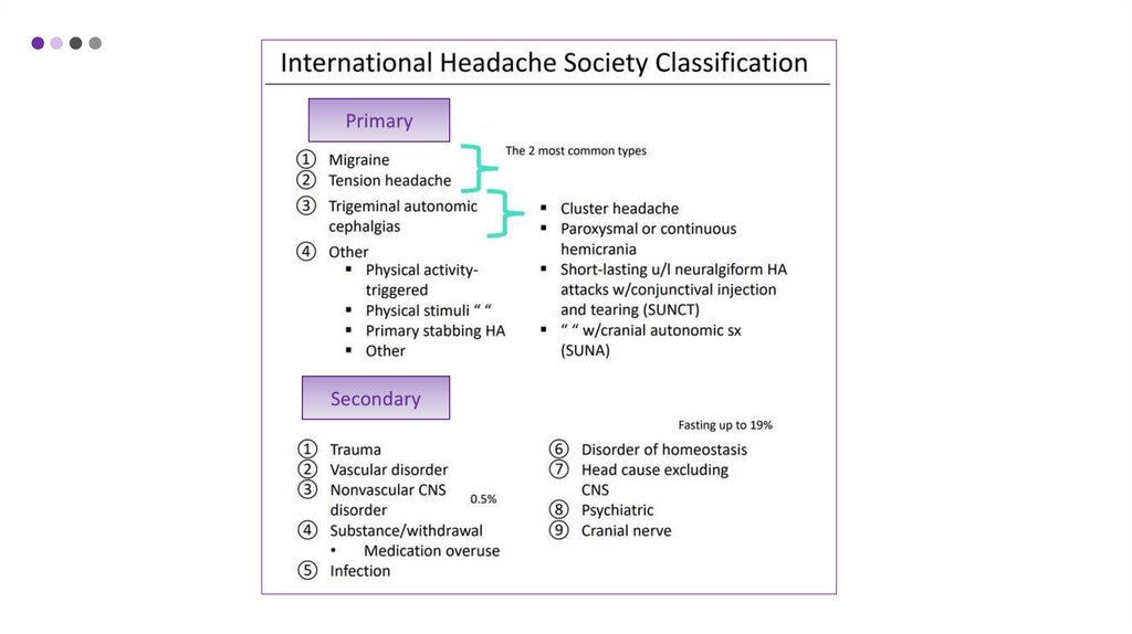
PrimarySecondary
3.
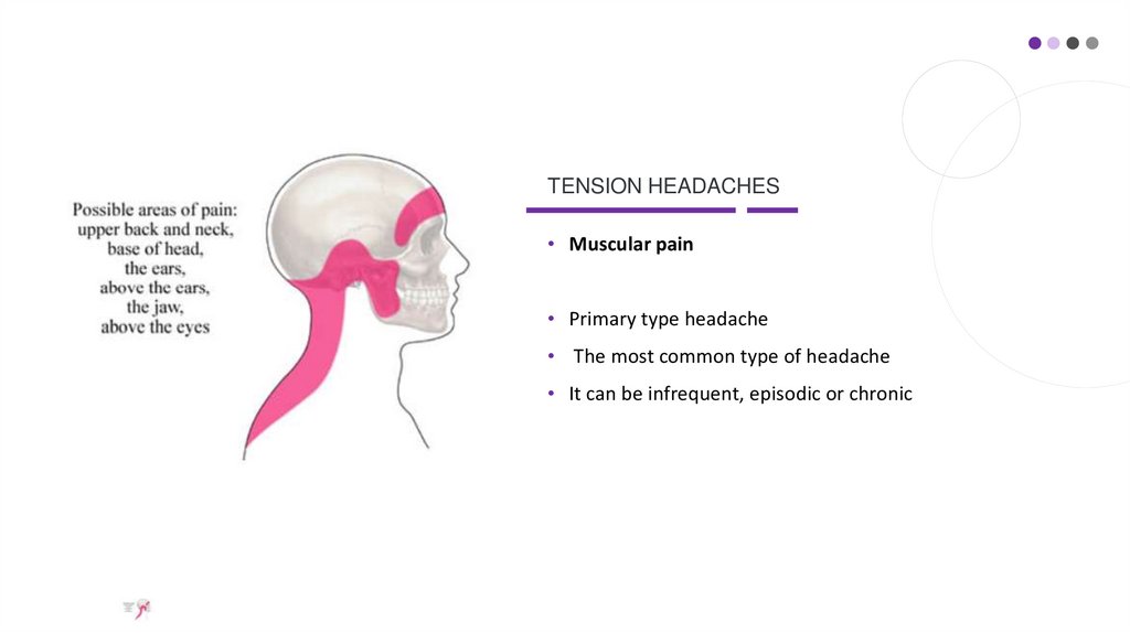
TENSION HEADACHES• Muscular pain
• Primary type headache
• The most common type of headache
• It can be infrequent, episodic or chronic
4.
Risk factorsSimptoms
• Stress
• dull ache, like a ‘tight pressure feeling’, ‘heavy
weight
• Hunger
• History of teeth grinding or jaw clenching
• Anxiety
• almost daily
• Depression
• hours (can last days)
• Sleep apnea or sleep disruption
• Onset: after rising, gets worse during day
• Eyestrain
• Poor posture
• Injuries or arthritis of the neck area
• Temporomandibular joint disease (TMJ)
• Medications
• Physical examination: muscle tension (e.g.
frowning), scalp often tender to touch, ‘invisible
pillow’ sign may be positive
• Low physical activity
• Obesity
• Smoking
Treatment: NSAIDs or acetaminophen
5.
CLUSTER HEADACHE• Site: over or about one eye
Diagnosis
• Horner’s syndrome
• retro-orbital headache + rhinorrhoea + lacrimation
→ cluster headache
• Radiation: frontal and temporal regions
• Frequency: one every other day and 8 per day for
more than half the time
• Duration: 15–180 minutes (average 30 minutes);
the clusters last 4–6 weeks (can last months)
• Onset: suddenly during night (usually), same time
about 2–3 hours after falling asleep; the ‘alarm
clock’ headache
• Offset: spontaneous
Treatment
• O2
• Triptants
• Prophylaxis: verapamil
6.
HORNER’S SYNDROME• Miosis
• a persistently small pupil. Denervation of
dilatator pupillae m.
• Ptosis
• dropping of the upper eyelid.
Denervation of smooth mm. serving
palpebra
• Pseudo-enophtalmos
• sunken globe. Appearance based on
ptosis.
• Hyperemia
• flushed skin. Denervation of vasomotor
fibers
• Anhydrosis
• lack of sweating. Denervation of
sudomotor fibers
7.
MIGRANE• Site: temporofrontal region (unilateral) can be
bilateral
• Radiation: retro-orbital and occipital
Diagnosis
• headache + vomiting + visual aura → migraine
with aura (classic)
• Quality: intense and throbbing
• Frequency: 1 or 2 per month
Treatment
• Duration: 4–72 hours (average 6–8 hours)
• Mild: NSAIDs
• Onset: paroxysmal, often wakes with it
• Severe or refractory: Triptans, ergots
• Offset: spontaneous (often after sleep)
• Prophylaxis:
• Aggravating factors: tension, activity
• Relieving factors: sleep, vomiting
• Associated factors: nausea, vomiting (90%)
irritability aura
• Beta blocker – propranolol
• Valproic acid or topiramate
8.
IDIOPATHIC INTRACRANIAL HYPERTENSIONClinical features
Diagnosis
• Change in LOC
• Lumbar puncture
• Pupillary changes
• OP>25 cm H2O
• Headache
• ↑ BP + widening pulse pressure
Treatment
• Bradycardia
• Acetazolelamide
• Fever
• Serial lumber
• Focal neurologic deficit
• VP shunt
• Nausea
• Vomiting
Usually woman
9.
CRANIAL NERVE PALSIESI
Olfactory nerve
II
Optic nerve
III
IV
Oculomotor nerve
Trochlear nerve
V
Trigeminal nerve
VI
Abducens nerve
VII
Facial nerve
VIII
Vestibulocochlear nerve
IX
Glossopharyngeal nerve
X
XI
Vagus nerve
Accessory spinal nerve
XII
Hypoglossal nerve
10.
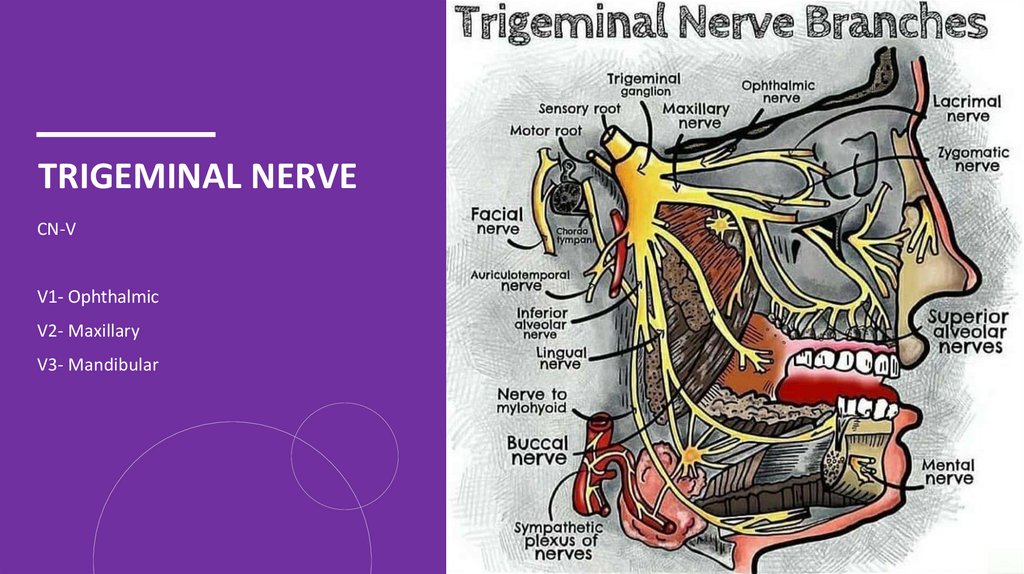
TRIGEMINAL NERVECN-V
V1- Ophthalmic
V2- Maxillary
V3- Mandibular
11.
TRIGEMINAL NEURALGIA• unilateral
Causes
• excruciating, searing jabs of pain like a burning knife
or electric shock
• Idiopathic or compression of the TN
• Duration of pain is variable
• Local pressure on the nerve root entry zone by vessels
(probably up to 75%)
• Seconds to 1–2 minutes (up to 15 minutes)
• Multiple sclerosis
• Onset: spontaneous or trigger point stimulus
• Neurosyphilis
• Offset: spontaneous
• Tumours of the posterior fossa
• Madibular affected most often
• Precipitating factors: talking, chewing, touching trigger
areas on face
12.
TRIGEMINAL NEURALGIADiagnosis
• Clx
• MRI to rule out secondary causes
Treatment
• Carbamazepine
• Oxcarbazepine
• Baclofen
• Lamotrigine
• Surgical
13.
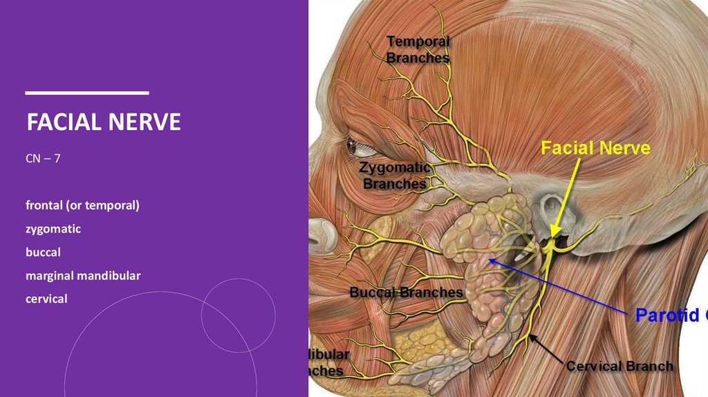
FACIAL NERVECN – 7
frontal (or temporal)
zygomatic
buccal
marginal mandibular
cervical
14.
BELL’S PALSY• Abrupt onset (can worsen over 2–5 days)
Causes
• Weakness in the face (complete or incomplete)
• infection or inflammation of the facial nerve
• Preceding pain in or behind the ear
• head trauma
• Impaired blinking
• head or neck tumor
• Bell phenomenon—when closing the eye it turns up
under the half-closed lid
• stroke
• Associations:
• herpes simplex virus (postulated)
• diabetes mellitus
• hypertension
• thyroid disorder
15.
BELL’S PALSYDiagnosis
• Clx
Treatment
• Supportive:
• Artificial tears if eye is dry and at bedtime
• Massage and facial exercises during recovery
• Prednisone taper
• Virus infection: acyclovir, valacyclovir
16.
LUMBAR PUNCTUREBleeding in the brain (intracranial hemorrhage).
Dementia.
Leukemia or other cancers.
Meningitis and encephalitis (brain and spinal cord
infections).
• Multiple sclerosis or other autoimmune disorders.
• Myelitis (spinal cord inflammation).
• Excess cerebrospinal fluid.
• Administer regional anesthesia, such as an epidural to
block pain in the lower part of the body.
• Inject dye for an X-ray diagnostic test (myelogram).
• Inject cancer medications or muscle relaxers.
• Relieve intracranial (head) pressure.
Preparing
• Stop taking blood-thinning medications, such as
aspirin and warfarin.
• Tell your doctor if you’re allergic to povidone-iodine
(an antiseptic) or procaine (an anesthetic).
17.
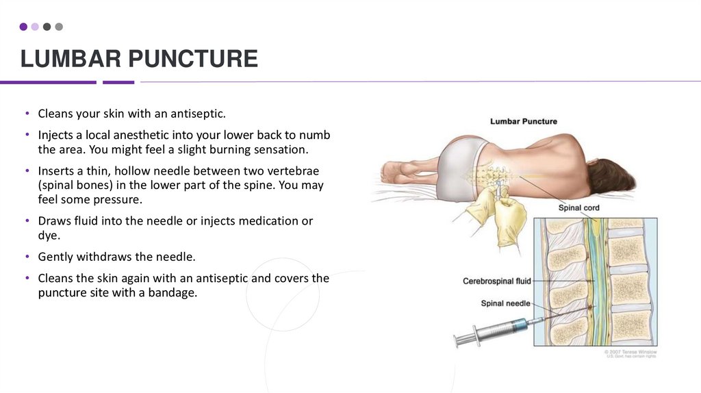
LUMBAR PUNCTURE• Cleans your skin with an antiseptic.
• Injects a local anesthetic into your lower back to numb
the area. You might feel a slight burning sensation.
• Inserts a thin, hollow needle between two vertebrae
(spinal bones) in the lower part of the spine. You may
feel some pressure.
• Draws fluid into the needle or injects medication or
dye.
• Gently withdraws the needle.
• Cleans the skin again with an antiseptic and covers the
puncture site with a bandage.
18.
19.
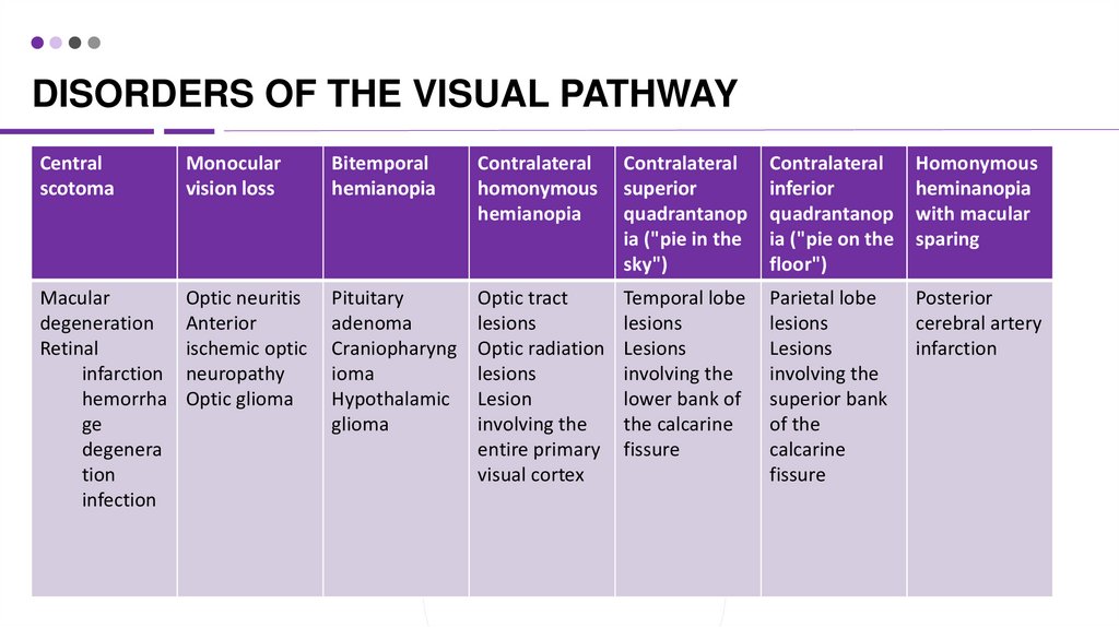
DISORDERS OF THE VISUAL PATHWAYCentral
scotoma
Monocular
vision loss
Bitemporal
hemianopia
Contralateral
homonymous
hemianopia
Contralateral
superior
quadrantanop
ia ("pie in the
sky")
Contralateral
inferior
quadrantanop
ia ("pie on the
floor")
Homonymous
heminanopia
with macular
sparing
Macular
degeneration
Retinal
infarction
hemorrha
ge
degenera
tion
infection
Optic neuritis
Anterior
ischemic optic
neuropathy
Optic glioma
Pituitary
adenoma
Craniopharyng
ioma
Hypothalamic
glioma
Optic tract
lesions
Optic radiation
lesions
Lesion
involving the
entire primary
visual cortex
Temporal lobe
lesions
Lesions
involving the
lower bank of
the calcarine
fissure
Parietal lobe
lesions
Lesions
involving the
superior bank
of the
calcarine
fissure
Posterior
cerebral artery
infarction
20.
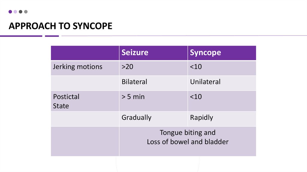
APPROACH TO SYNCOPEJerking motions
Postictal
State
Seizure
Syncope
>20
<10
Bilateral
Unilateral
> 5 min
<10
Gradually
Rapidly
Tongue biting and
Loss of bowel and bladder
21.
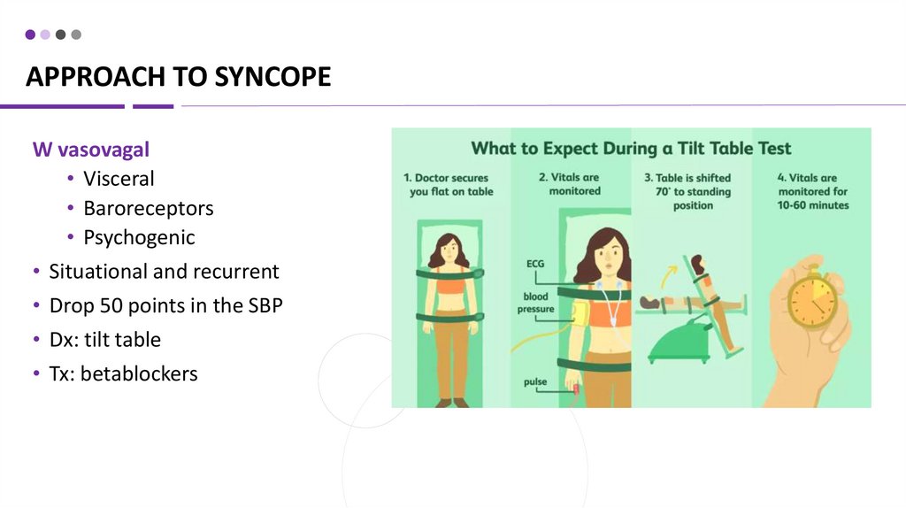
APPROACH TO SYNCOPEW vasovagal
• Visceral
• Baroreceptors
• Psychogenic
• Situational and recurrent
• Drop 50 points in the SBP
• Dx: tilt table
• Tx: betablockers
22.
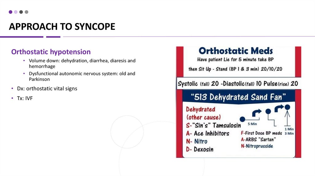
APPROACH TO SYNCOPEOrthostatic hypotension
• Volume down: dehydration, diarrhea, diaresis and
hemorrhage
• Dysfunctional autonomic nervous system: old and
Parkinson
• Dx: orthostatic vital signs
• Tx: IVF
23.
APPROACH TO SYNCOPEMechanical cardiac – a structural heart disease
Psych
• Exertional syncope, a murmur
• Dx: face-palm maneuver
• Dx: Echo
• Tx: surgery the valves
Arrhythmia
• Sudden onset
• Dx: Holter monitor
Neurogenic (rare)
• Sudden onset and focal neurologic deficit
• Dx: U/S
Electrolytes
• Dx: BMP
• Na, Ca – mental status
• K, Mg - weakness