


:")


 Медицина
Похожие презентации:


")


Disorders of the Cornea, Sclera and Orbit
1. Disorders of the Cornea, Sclera and Orbit
Departmentof ophthalmology
Head of department: Prof. N.V. Ivanova
Disorders of the Cornea,
Sclera and Orbit
2. Cornea is anterior part of fibrous cover. Is a part of dioptric apparatus of the eye.
3.
Anatomy of the corneaIn normal:
avascular
sensitive
transparent
smooth
glassy
spherical
resplendent
Tear film
Surface cells
Wing cells
Basal cells
Basement membrane
Epithelium
Basement membrane
Bowman’s layer
Stroma
Descemet’s
membrane
Endothelium
4.
Diseasesof the Cornea
Inflammations
Congenital
anomalies
Tumors
Dystrophies
Keratitis
Megalocornea
microcornea,
keratoglobus
Benign
Malignant
Primary
Secondary
5. Investigation of Corneal Disease
AnamnesisClinical examination
Biomicroscopy
Pachometry
Keratometry
Keratoscopy
Laboratory
investigations
6. Keratitis – inflammation of the cornea
Keratitis (objective signs)=
Corneal oedema
+
Cellular infiltration
+
Ciliary congestion
keratitis (adenovirus)
7. Corneal syndrome
photophobialacrimation
blepharospasm
a sensation of a foreign body present
behind the eyelids
pain
8. Classification
1. Exogenous keratitisCorneal erosions
Traumatic keratitis
Bacterial keratitis
Keratitis, caused by disease of conjunctiva, eyelids,
meibomite glands.
Fungal keratitis
Corneal ulcer
2. Endogenous keratitis
Infectious (tuberculous, viral, rheumatic,
toxicoallergic and others)
Neuroparalytic keratitis
Keratitis because of avitaminosis
3. Keratitis of unclear etiology (recurrent erosion,
rosacea keratitis and other)
9. Bacterial keratitis – often develop in chronic inflammatory diseases of conjunctiva and lacrimal ducts, as well as in the
trauma of the cornea.Predisposing Condition to Bacterial Keratitis:
1/ Contact Lens Wear
2/ Nonsurgical Trauma
Corneal abrasion or wound
Corneal foreign body
Toxic medications (e.g., anesthetic abuse, idoxuridine)
3/ Surgical Trauma
4/ Corneal Injury Caused by Lid Dysfunction
Trichiasis
Lagophthalmus
5/ Corneal, Conjunctival, or Lacrimal Dysfunction
Bullous keratopathy
Neurotrophic cornea
Stevens-Johnson syndrome
Tear insufficiency
Mucin insufficiency
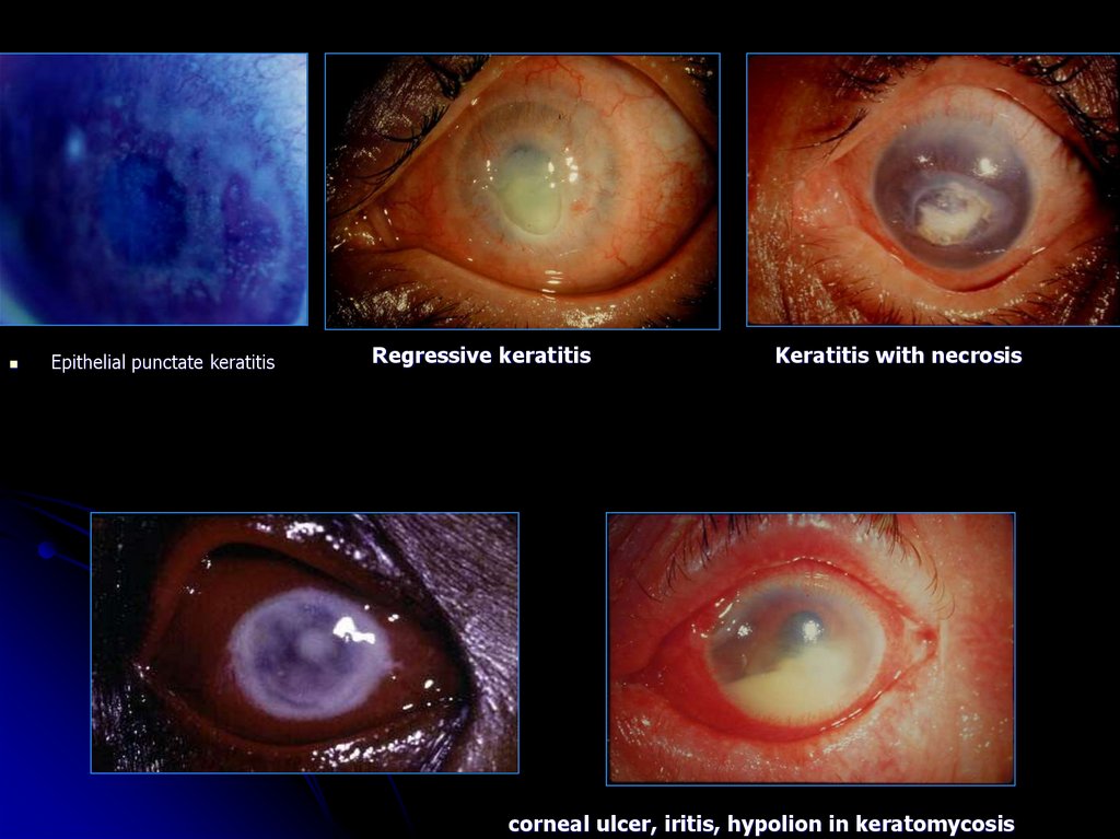
10.
Epithelial punctate keratitisRegressive keratitis
Keratitis with necrosis
corneal ulcer, iritis, hypolion in keratomycosis
11. Herpes Simplex Keratitis
Primary ocularinfection typically
occurs in children
between the ages of 6
months and 5 years.
Is more frequently
accompanied by
anterior uveitis and
keratic precipitates,
early vascularization,
hypoaesthesia,
photophobia,
blepharospasm,
lacrimation.
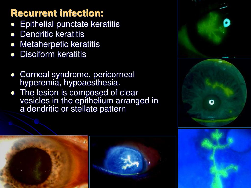
12.
Recurrent infection:Epithelial punctate keratitis
Dendritic keratitis
Metaherpetic keratitis
Disciform keratitis
Corneal syndrome, pericorneal
hyperemia, hypoaesthesia.
The lesion is composed of clear
vesicles in the epithelium arranged in
a dendritic or stellate pattern
13. additional investigations of patients with keratitis
review roentgenography of the additional nasal sinus andorgans of the chest
consultations of the otorhinolaryngologist and stomatologist
(sometimes of phthisiatrician and dermatovenerologist)
results of additional methods of investigation: clinical and
laboratory (detailed blood count, Wassermann reaction,
Mantoux reactions, focal test with tuberculin, toxoplasmin
and others).
14. Laboratory diagnosis
HerpeviralBacterial
Express-diagnosis-method of
Conjunctical smear in deep
fluorescining antibodies –
ulcers of the cornea revealing of virus in the scrub of
smear-print from the ulcer.
conjunctiva
Scrub from ulcerous
Monoclonal immunofermental
surface and margins of
test-system
ulcer
Finding of antiallergic antibodies
Staining – with methylene
in lacrimal fluid in the reaction of
blue and Gram’s staining
passive hemagglutination
inoclulation of media with
Finding of lymphocytes to herpes
investigation of antibiotic
in the reaction of
sensitivity
blasttansformation and inhibition
of leucocytes migration
Finding of IgA, IgG, IgM in
lacrimal fluid and blood serum
15. The differential diagnosis of primary ocular infection includes:
1. Keratitis with lid lesions: zoster, chickenpox,molluscum contagiosum, and ulcerative
blepharitis with keratitis due to
staphylococcal infection
2. Keratitis without lid lesions: vaccinia,
adenoviral infections (types 3, 7, and 8),
chlamydial infections, and zoster
16. Parasitic Keratitis - Acanthamoeba
Slit lamp photograph of a 42 year oldfemale patient with unilateral, red,
painful eye with epithelial defect;
Corneal sensitivity decreased.
Stromal ring infiltrate (1), fluorescein
staining (2).
Corneal scrapings and PCR for VZV
negative.
confocal microscopy in vitro
17. Keratomycoses
Risk factors: long treatment withantibiotics or coricosterois,
microtraumas of the cornea,
fungus skin diseases.
Greyish-white infiltrate with
crumb-like friable surface and
yellowish border appears on the
place of corneal erosion,
presence of hypopion is typical.
Vascularization is insignificant.
18. Ulcer of cornea – inflammation of corneal membrane, accompanying with necrosis with formation of its tissue defect
Infectious –bacterial, herpeviral, fungal and parasitic infectionof the eyes.
Noninfectious – ulcer of immune genesis, corneal xerosis, in
absolute glaucoma
Risk factors:
conjunctivites,
dacriocystites,
Corneal xerosis,
traumas of the cornea,
foreign bodies of the cornea,
injuries by chemical, thermal, radiation, laser affection.
long wearing of contact lenses,
various operations on the cornea,
bullous keratopathia.
19. Organisms Commonly Isolated From Corneal Ulcers
Healthy CorneaStaphylococcus
Streptococcus
Pseudomonas
Enterobacteriaceae
Moraxella
Klebsiella
Corneal creeping ulcer
with the thread of
perforation
Compromised Cornea*
Staphylococcus aureus
Staphylococcus epidermidis
a-Hemolytic Streptococcus
b-Hemolytic Streptococcus
Pseudomonas
Proteus
Pediatric
Pseudomonas
Staphylococcus
Fungi
20. Stages of ulcerous process:
Stage of infiltrationStage of infiltrate decay and
formation of ulcer
Stage of facet – regression of
ulcer, is characterized by
clearance from necrotic residues
of the fundus and margins of the
ulcer and rapid growth of
epithelium, that covers the fundus
and margins of the cornea defects
Stage of the scar formation
Stage of outcome – maturation
and thickness of connectivetissue scar
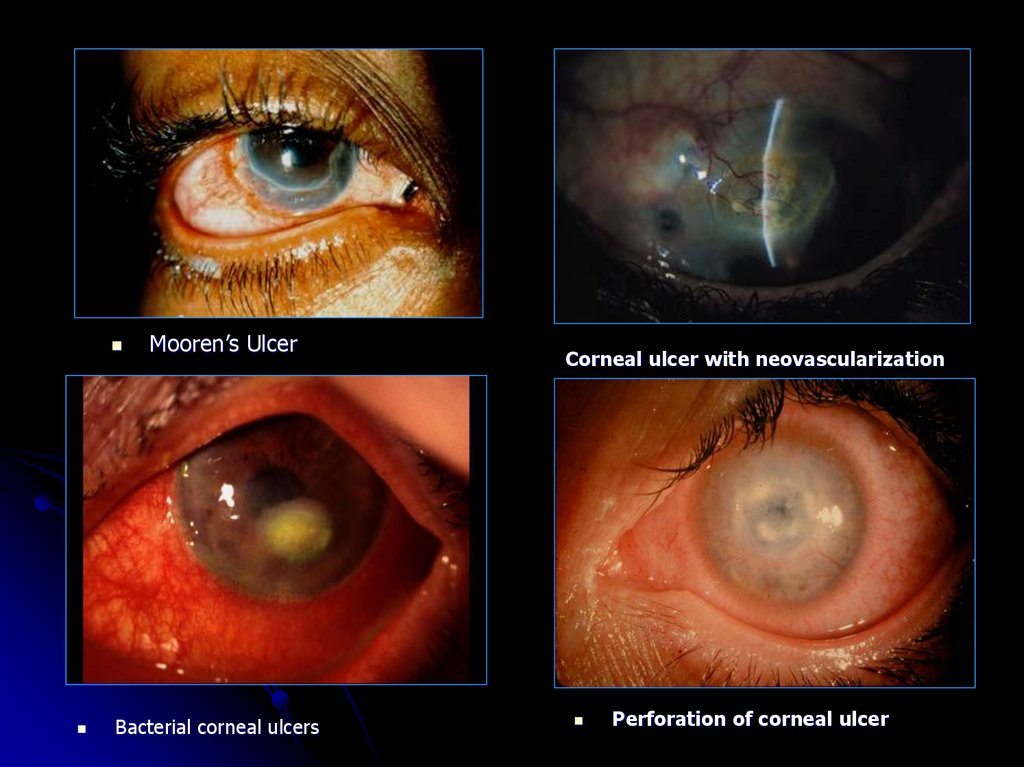
21.
Mooren’s UlcerBacterial corneal ulcers
Corneal ulcer with neovascularization
Perforation of corneal ulcer
22. Principles of Keratitis and Corneal Ulcers treatment
1. Specific therapy:a) Antiviral (Zovirax, Aceclovir ointment 3%
- 5 times a day or
Lokferon 8000 МЕ/ml instillation 8 times a day for 5-7 days + systemic
therapy)
b) Antibacterial
Local – moxifloxacin, Cefazolin, Tobramycin, Gentamicin 4-8 times a day,
Erythromycin, Tetracyclin ointment 1% 3-4 times a day
Parabular – Tobramycin, Gentamicin, Cefazolin
Systemically - Maxavin, Erythromycin, Doxicyclini, Ceftriaxone
c) Antifungal (local suspension of ketoconazole, miconazole ointment 4
times a day + Nizoral 200mg 2 times daily)
d) Antiparasitic (topical propamidine, neomysin, clotrimazole)
23.
2. Pathogenetic therapyAntiinflammatory (Eye drops – only
non-steroidal
antiinflammatory: Naclof. Parabular or intravenous injection of
dexamethazon (in severe course)
Antiallergic
Metabolic - taufon, actovegin, solcoseril, corneregel, vitamins
Hypotensive - in a case of the eye hypertesion or secondary
glaucoma.
Mydriatic-cycloplegic drugs - instillation of 1.0% mydriacyl,
tropicamid.
Physiotherapy, Criotherapy
24.
Following arresting of inflammatory process acourse of resolving therapy (fibrinolysin,
lidase)
Penetrating keratoplasty indicated for visual
rehabilitation in patients with sever corneal
scarring. Lamellar keratoplasty has
advantage over penetrating keratoplasty of
reduced potential for corneal graft rejection.
25. Complications of keratitis:
limbal and scleral extensioncorneal perforation
iridocyclitis
endophthalmitis
Panophthalmitis
Secondary glaucoma
Corneal scarring: nebula, macula,
leucoma, kerectasia, anterior staphyloma
26. Sclera – is a part of fibrous coat of the eyeball
The sclera is composed of three layers:the episclera
the sclera proper
the lamina fusca
The sclera is relatively poorly vascularized. Its blood supply is
derived from the anterior and posterior ciliary arteries.
27.
Diseasesof the sclera
Inflammations
Congenital
anomalies
Tumors
Episcleritis
scleritis
Blue discoloration
melanocytosis
Coloboma
Benign
Malignant
28. Episcleritis is a inflammatory disorder of the superficial layer of the sclera. Is a common, benign, self-limiting and
Episcleritis is a inflammatorydisorder of the superficial layer
of the sclera.
Is a common, benign, selflimiting and frequently recurrent
disorder which typically affects
young adults.
the episcleritis
may be nodular
or diffuse
29. Systemic diseases with episcleritis
30. Nodular Episcleritis
• Sudden onset of FB sensation,discomfort, tearing ± photophobia. It
may be recurrent.
• Red nodule arising from the
episclera; can be moved separately
from the sclera and conjunctiva;
blanches with topical
vasoconstrictor (phenylephrine
10%); does not stain with
fluorescein; globe nontender
Spontaneous resolution occurs in 5–6
weeks.
31. Diffuse episcleritis
Sudden onset of milddiscomfort, tearing ±
photophobia; may be
recurrent.
Sectoral redness that
blanches with topical
vasoconstrictor
(phenylephrine 10%);
globe nontender;
spontaneous resolution
1–2 weeks.
32. Episcleritis
Generally, the pain is not as severe as withscleritis.
Hyperemia of the episcleral tissues most often is
localized but may involve the entire anterior
segment.
The inflammatory process does not involve
underlying sclera or intraocular tissue.
33. Treatment
If mild, no treatment is required.Supportive: reassurance ± cold
compresses.
Topical: consider lubricants ± NSAID
(ketorolac 0.3% 3x/day). Although disease
improves with topical steroids, there may
be rebound inflammation on withdrawal.
Systemic: if severe or recurrent disease,
consider oral NSAID (flurbiprofen 100 mg
3x/day for acute disease).
34. COMPLICATIONS
Involvement of other ocular structures is rare inpatients with episcleritis.
The peripheral cornea can be left thinned or
vascularized. Recurrent attacks of episcleritis
over many years can result in some mild scleral
thinning, which is of no consequence to the
integrity of the eye.
The most frequent complications seen in
patients with episcleritis are related to the use of
long-term topical corticosteroids: Cataract,
ocular hypertension, steroid-induced glaucoma,
herpetic keratitis.
35. COURSE AND PROGNOSIS
Episcleritis is a mild, non-visionthreatening inflammation of the episclerathat may recur over irregular intervals for
many years.
It is important to recognize its benign
nature and not to induce vision-threatening
complications by overtreating episodes of
episcleritis.
36. Scleritis is a granulomatous inflammation of the scleral coat of the eye.
37. Underlying systemic diseases
Other causes:infection (e.g.,
syphilis,
tuberculosis,
bacterial,
fungal, and
herpes zoster).
trauma, surgery
38. Scleritis
Anterior scleritisNon-necrotizing
Diffuse
Nodular
Necrotizing
With inflammation
Without inflammation
Posterior scleritis
Non-necrotizing
Diffuse
Nodular
Necrotizing with
inflammation
39. Scleritis presents in the fourth to sixth decade with the gradual onset of classic symptoms of severe, boring, ocular pain that
occasionallyradiates to the temple, jaw, or sinuses,
redness, tearing ± photophobia
40. Differentiation between episcleritis and scleritis
41. Posterior scleritis
Posterior scleritis is a serious, potentially blindingcondition, which is often misdiagnosed and treated very
late.
Clinical features
• Mild–severe deep pain (may be referred to brow or jaw
region), VA, diplopia, photopsia, hypermetropic shift.
• White eye (unless anterior involvement), lid edema,
proptosis, lid retraction, restricted motility; choroidal
folds, annular choroidal detachment, exudative retinal
detachments, macular edema, disc edema.
42. Drugs commonly used in the treatment of scleritis
• Oral: NSAID (e.g., flurbiprofen 100 mg 3x/day; can betapered down once disease is controlled).
• If not controlled, consider systemic immunosuppression:
commonly corticosteroids (e.g., prednisone 1 mg/kg/day)
± other immunosuppressants (coordinate with
rheumatologist).
• Topical corticosteroids are usually an adjunct to systemic
therapy, lubrication
• Periocular corticosteroids (e.g., subtenons or transseptal
triamcinolone acetonide) can be given in patients with no
evidence of scleral thinning.
If there is risk of perforation, protect globe (e.g., glasses
by day, shield at night) and consider scleral patch graft.
43. Complications
КeratitisUveitis
Staphyloma.
Perforation of the sclera
Exudative retinal detachment and
choroidal detachment
44. The orbit is a pear-shaped cavity in the skull The orbit consist of the eyeball, external muscles, lacrimal gland, nerves,
vessels, fat45. Optic canal (orbital foramen):
Within lesser wing of sphenoidTransmits: Optic nerve (CN 2),
ophthalmic artery, and sympathetic nerves
to ocular and orbital blood vessels
46. The inferior orbital fissure
Bordered medially by maxillary bone,anteriorly by zygomatic bone, and laterally
by greater wing of sphenoid
Transmits: CN V2, zygomatic nerve,
inferior ophthalmic vein
47. The superior orbital fissure is a slit linking the cranium and the orbit, between the greater and lesserwings of the sphenoid
bone.Transmits: CN III, IV, V1, VI, superior
ophthalmic vein,
and sympathetic fibers
48.
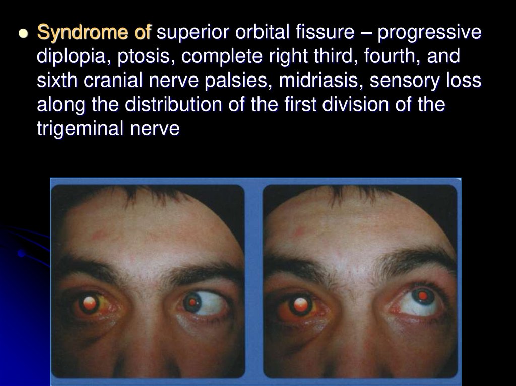
Syndrome of superior orbital fissure – progressivediplopia, ptosis, complete right third, fourth, and
sixth cranial nerve palsies, midriasis, sensory loss
along the distribution of the first division of the
trigeminal nerve
49. Sinuses
50. Clinical and special investigations
Clinical examination, Palpation of anterior orbital tissuesOrbitotonometry
Exophthalmometry
Ultrasonography
Magnetic resonance imaging; fat-suppression techniques
Arteriography
Radionuclide scan
Computed tomography, contrast-enhanced; bone-window
(especially for fractures)
51. ORBITAL DISORDERS
Congenitalorbital
malformations
Acquired
Infections
Trauma
Thyroid-Related
Ophthalmopathy
Tumors
Orbital
periostitis,
Cellulitis,
Cavernous
Sinus
Thrombosis
Vascular
Abnormalities
52. Osteoperiostitis
It may result from injuries or as an extension ofinfection from the surrounding structures
Clinical picture may be in two forms.
Anterior orbital periostitis – it involves the orbital
margin and is characterized by severe pain,
tenderness and swelling of the inflammed area.
Subperiostal abscess when formed, frequently
bursts on the skin surface.
Posterior periostitis is characterized by deep
seated orbital pain, exophthalmos, slight
limitation of ocular movements. Sometimes there
may be anaesthesia of the skin of eyelids and
cornea.
53. Cellulitis - inflammation of the orbit
54. Risk factors:
• Sinus disease: ethmoidal sinusitis (common),maxillary sinusitis.
• Infection of other adjacent structures:
preseptal or facial infection, dacryocystitis,
dental abscess.
• Trauma: septal perforation.
• Surgical: orbital, lacrimal, and vitreoretinal
surgery.
55.
Symptoms include rapid onset of headache,fever, pain, nausea, in some cases –
prostration.
Eyelids are swollen, erythematous, warm
and tender to palpation.
A marked chemosis of conjunctiva.
The eye is proptosed axially (the most
frequently proptosis is lateral and
downwards).
Ocular movements are restricted and
painful.
In advanced cases visual acuity may be
diminished.
56. Potential complications:
Intracranial complications includemeningitis, brain abscess and cavernosus
sinus thrombosis
Subperiorbital abscess (it is relatively rare
in sinus-related orbital cellulitis but may
occur in post-traumatic and postoperative
cases)
Ocular complications (keratopathy, raised
IOP, occlusion of the central retinal artery
or vein, optic neuritis)
57. Principles of treatment of inflammatory diseases of the orbit
It is an emergency requiring hospital admission.Intensive antibiotic therapy (topical (subconjunctival
and retrobulbar injections). Admit for intravenous
antibiotics (e.g., either floxacillin 500–1000 mg
4x/day or cefuroxime 750–1500 mg 3x/day with
metronidazole 500 mg 3x/day).
Analgetics and anti-inflammatory drugs if necessary
Osmotherapy (40% glucosae solution)
Surgical drainage. In most cases it is necessary to
drain both the orbit and the infected paranasal
sinuses.
Physiotherapeutical treatment
58.
Thank you for yourattention!
The lecture is composed by:
prof. doctor of medical sciences
Ivanova N.V.,
Assistant Kondratiuk G.I.