

")


")


")


 Медицина
Похожие презентации:


Chronic Rheumatic Heart Disease
1. r
Chronic Rheumatic Heart Diseaser
ا د ∙ نجوى على
استاذ طب االطفال
2.
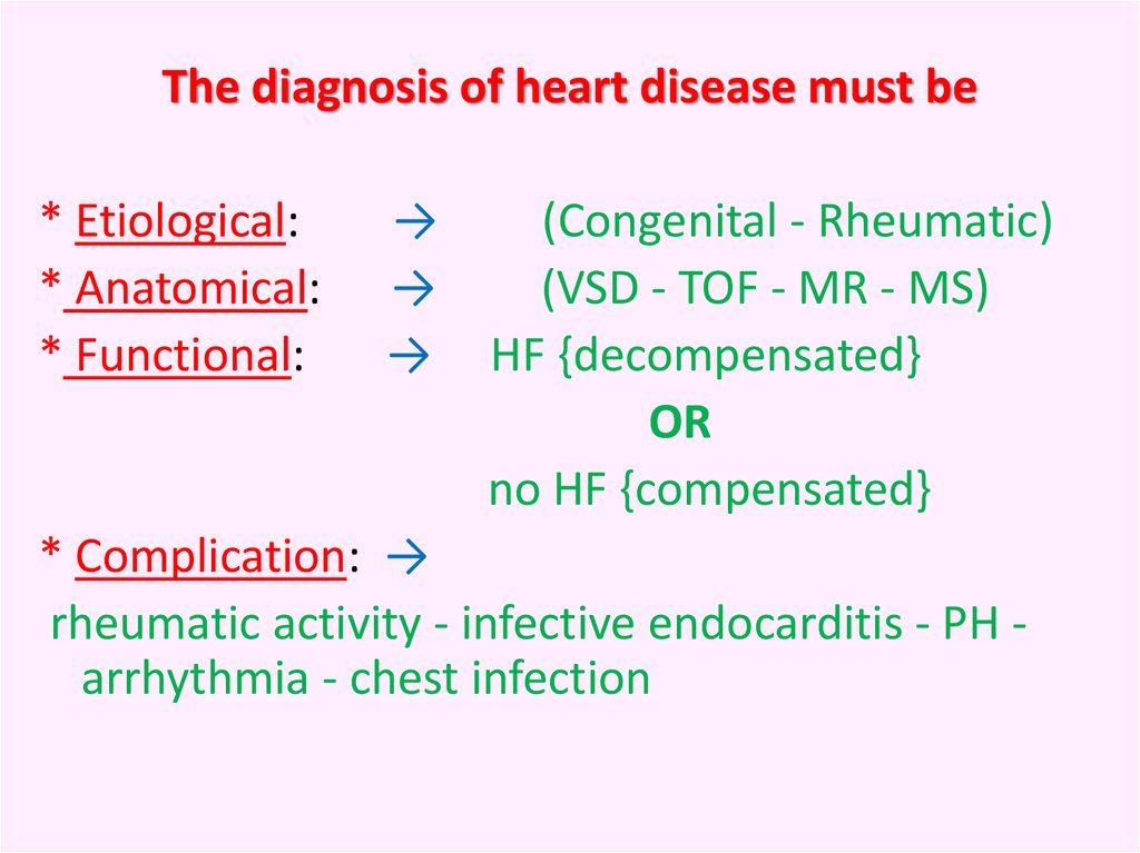
The diagnosis of heart disease must be* Etiological:
* Anatomical:
* Functional:
→
→
→
(Congenital - Rheumatic)
(VSD - TOF - MR - MS)
HF {decompensated}
OR
no HF {compensated}
* Complication: →
rheumatic activity - infective endocarditis - PH arrhythmia - chest infection
3. RHEUMATIC HEART DISEASE
• Occurs in severe cardiacinvolvement during initial or
recurrent attacks of ARF
• Left - sided heart valves are most often
affected, (mitral followed by the aortic valves)
• Mitral regurge is the commonest lesion in
children and adolescent with RHD
4. MITRAL REGURGE (MR, Insufficiency, Regurgitation, Incompetence)
The mitral valve consists of:- an annulus
- 2 leaflets
( anterior & posterior )
- chordae tendinea
- 2 papillary muscles
5. Pathophysiology
Healing ofARF results in
Fibrosis & contracture of leaflets
Shortening & thickening of chordea tendinea.
Leaflets cannot coapt and separated
LA and LV volume overload and enlargement.
Pulmonary venous congestion, PH, RVH
6. Clinical Manifestations: Symptoms
• Mild MR → no symptoms• Severe MR → Symptoms of HF, pulmonary
congestion, pulmonary edema
dyspnea - orthopnea - paroxysmal nocturnal dyspnea
7.
Signs:Apex → (LV apex),
shifted downward, localized, forcible, hyperdynamic
(ill sustained) with systolic thrill.
S1 is usually normal
S2 is usually normal except in PH
Pansystolic murmur maximal intensity at the apex,
radiating to the axilla.
Short middiastolic murmur over the apex may be
heard (functional MS)
Ejection systolic murmur on 2nd Lt ics (PH)
8. Mitral regurge CXR
Mitral regurgeEchocardiography
9. Differential Diagnosis of MR
1- VSD:-maximal intensity over the 3rd &
4th left intercostal spaces
-propagated in fan manner
2- Tricuspid regurge:
- maximal intensity on lower left sternal border
- increases in intensity during inspiration.
3- Mitral regurge of Carditis:
- maximal intensity on the apex
- soft, musical, not associated with thrill, changeable
10. Management
• Prophylaxis→ Against rheumatic recurrences (LONG ACTING PENECILLIN)
→ Against infective endocarditis
-
Medical treatment of
heart failure
arrhythmia
infective endocarditis
Captoprile ( After load reducing agent)
• Surgical treatment (Annuloplasty or valve replacement ) is
indicated in severe mitral regurge with:
- Recurrent heart failure
- cardiomegaly with pulmonary hypertension.
11. MITRAL STENOSIS (MS)
Pathophysiology-Thickening of valve leaflets
- Fusion of commissures
- Shortening & thickening of
chordae tendineae.
- Funnel shaped valve apparatus
→ marked obstruction to blood
flow from LA to LV
- LA enlargement (Not LV),
pulmonary venous congestion,
PH, RV & RA dilation
- Right side HF
12. Clinical manifestations:
The clinical course dependson the severity of MS.
Symptoms:
• Dyspnea on exertion.
• Orthopnea & paroxysmal nocturnal dyspnea.
• Poor growth and development.
• Tachycardia and atrial fibrillation.
• Congestive heart failure may be present.
13.
SignsSigns of RV hypertrophy:
a- The apex is diffuse and
shifted outward (RV apex),
diastolic thrill
b- Left parasternal pulsations
c- Epigastric pulsations
loud S1
Apical, rumbling middiastolic murmur.
14. Mitral stenosis CXR
Mitral stenosis echo15. Differential Diagnosis of MS
• Mitral flow murmur (functional MS)associated with large VSD, PDA, MR, AR (Austin
flint murmur)
- Normal S1
- No presysolic accentuation or opening snap
- Original lesion
-
Carditis (Carey Coombs murmur).
Soft , low pitched
Changeable
Not associated with thrill.
Normal or muffled S1
16. Management:
• Prophylaxis→ Against rheumatic recurrences (LONG ACTING
PENECILLIN)
→ Against infective endocarditis
• Medical treatment:
Heart failure and atrial fibrillation (AF).
• Surgical or baloon trans-catheter valvotomy
.
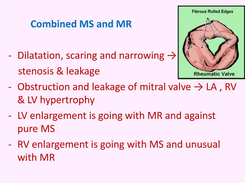
17.
Combined MS and MR- Dilatation, scaring and narrowing →
stenosis & leakage
- Obstruction and leakage of mitral valve → LA , RV
& LV hypertrophy
- LV enlargement is going with MR and against
pure MS
- RV enlargement is going with MS and unusual
with MR
18.
Which of the following pathological change occur inrheumatic mitral stenosis ?
(1) Increased left atrial pressure
(2) Left atrium dilatation
(3) Left ventricular hypertrophy
(4) Left ventricular hypertrophy
(5) Embolization of clots
19. AORTIC REGURGE (AR, Insufficiency, Regurgitation, Incompetence)
• Rheumatic AR is the result offibrosis and contracture of the
aortic valve structure
• Hemodynamically
AR → LV volume overload
• Rheumatic AR is almost always associated with mitral
valve disease.
20. Clinical manifestations:
SymptomsDepend on the severity.
In moderate and severe cases:
• Effort intolerance, palpitation, dyspnea, orthopnea &
paroxysmal nocturnal dyspnea, excessive sweating.
• Manifestations of pulmonary congestion and edema.
21. Signs
The rapid run off of the blood from aortaduring diastole causes the signs of
hyperdynamic circulation:
- The pulse is collapsing (water hammer)
- BP: wide pulse pressure (high systolic & low diastole)
- Corrigan’s sign prominent carotid pulsation in the neck.
- Capillary pulsation is visible (alternative systolic flushing and
diastolic blanching as pressure is applied to finger nails )
- Pistol shots heard over the femoral arteries due opening of
collapsed arteries during systole
- Duroziez’s murmur: a systolic and diastolic murmur detected
by applying mild pressure by the stethoscope over the
femoral artery.
- Musset’s sign: Head movement in time with heart beat.
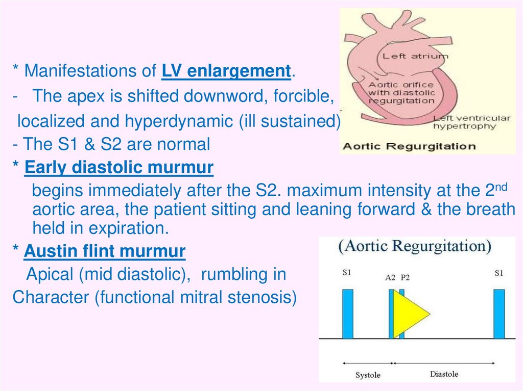
22.
* Manifestations of LV enlargement.- The apex is shifted downword, forcible,
localized and hyperdynamic (ill sustained)
- The S1 & S2 are normal
* Early diastolic murmur
begins immediately after the S2. maximum intensity at the 2nd
aortic area, the patient sitting and leaning forward & the breath
held in expiration.
* Austin flint murmur
Apical (mid diastolic), rumbling in
Character (functional mitral stenosis)
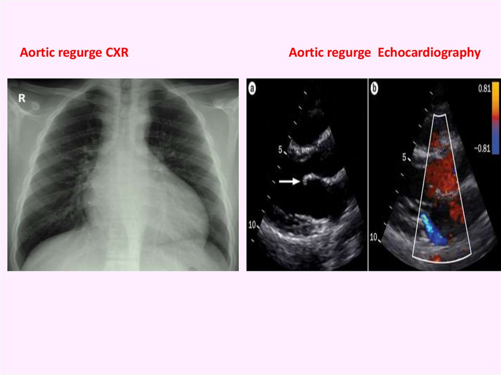
23.
Aortic regurge CXRAortic regurge Echocardiography
24. Management
• Prophylaxis→ Against rheumatic recurrences (LONG ACTING
PENECILLIN)
→ Against infective endocarditis
• Surgery:
Aortic valve replacement. It could be recommended at earlier
stages of the disease.
25. AORTIC STENOSIS
- Commissural adhesions occur slowly and progressive →narrowing and calcification of the orifice leads to significant
aortic stenosis.
- Obstruction of LV emptying results in LV hypertrophy.
26. Clinical manifestations: Symptoms:
In cases with severe stenosis:Chest pain, exercise intolerance, dyspnea, syncope.
27. Signs
- The apex: Localized, forceful & sustained (pressure overload).- Systolic thrill is common on Rt sternal border radiates to the neck.
- Normal S1
- Normal or single S2
Beyond childhood, scarring & calcification decrease mobility of the valve and
thus the intensity of aortic component decreases (single)
- Ejection systolic murmur maximally on 2nd Rt or 3rd Lt ics radiates to the
neck.
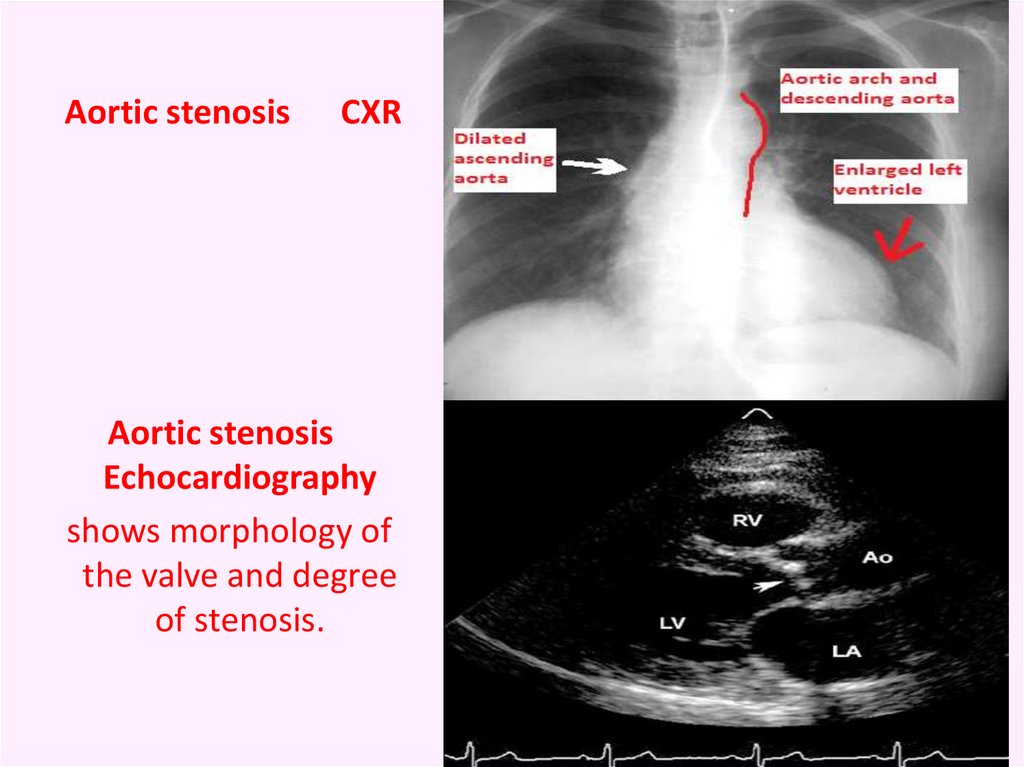
28.
Aortic stenosisCXR
Aortic stenosis
Echocardiography
shows morphology of
the valve and degree
of stenosis.
29. Management
→ Against rheumatic recurrences (LONG ACTINGPENECILLIN)
→ Against infective endocarditis
• Surgical intervention by valve replacement.
• Trans-catheter balloon dilatation is considered
in some cases.
30.
A case with dilated left ventricle and normal size of the otherchamber. The most likely diagnosis is :
(1) mitral stenosis
(2) mitral regurgitation
(3) aortic stenosis
(4) aortic regurgitation
8 year old child with history of rheumatic fever and pansystolic
murmur of mitral regurge. What is your management
(1) prophylaxis against infective endocarditis when indicated
(2) long acting penicillin every 3 weeks
(3) salicylates 70 mg/kg for 6 weeks
(4) Both 1 and 2
(5) All 1, 2 and 3