


 obstruction of a bronchus which creates a valve type of")

: 1-Iatrogenic: as a complication of mechanical ventilation or chest surgery")


 Медицина
Похожие презентации:


")
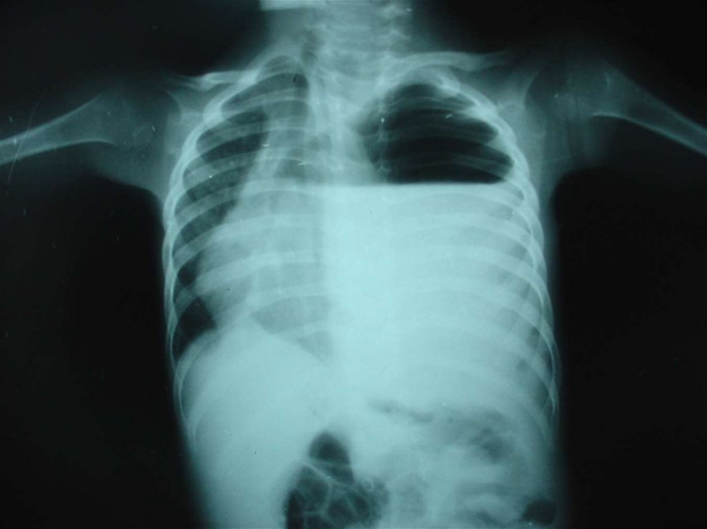
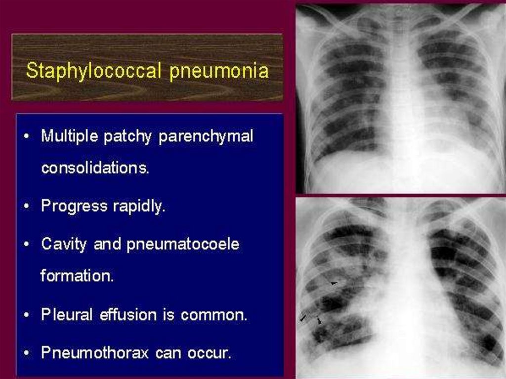
Pediatric chest X-ray
1.
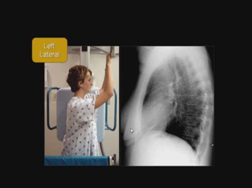
2. Pediatric chest X-ray
3.
4.
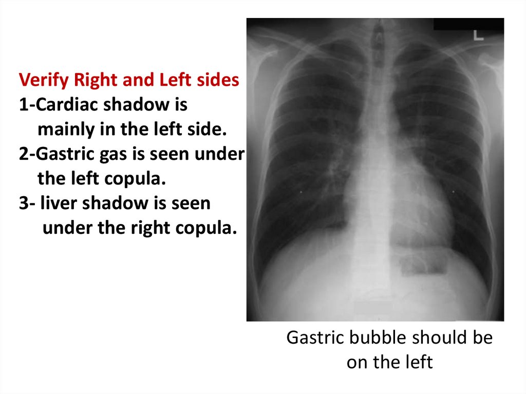
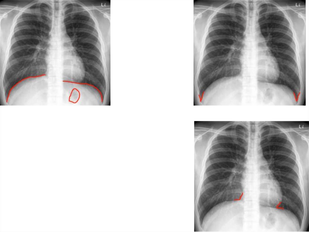
Verify Right and Left sides1-Cardiac shadow is
mainly in the left side.
2-Gastric gas is seen under
the left copula.
3- liver shadow is seen
under the right copula.
Gastric bubble should be
on the left
5.
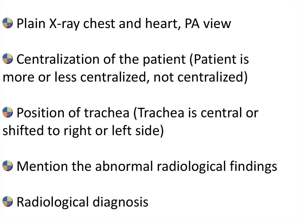
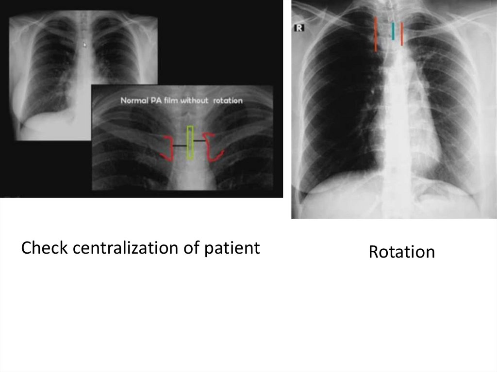
Plain X-ray chest and heart, PA viewCentralization of the patient (Patient is
more or less centralized, not centralized)
Position of trachea (Trachea is central or
shifted to right or left side)
Mention the abnormal radiological findings
Radiological diagnosis
6.
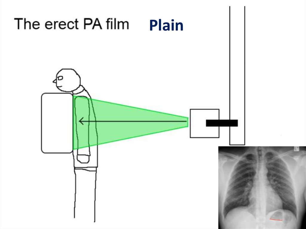
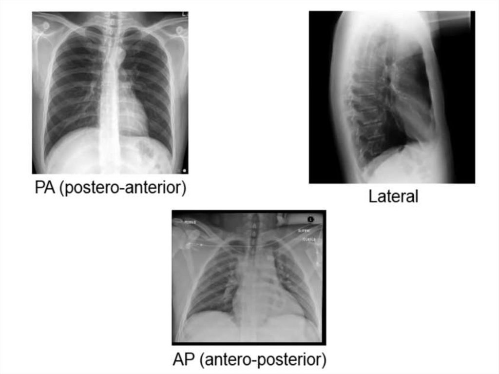
Plain7.
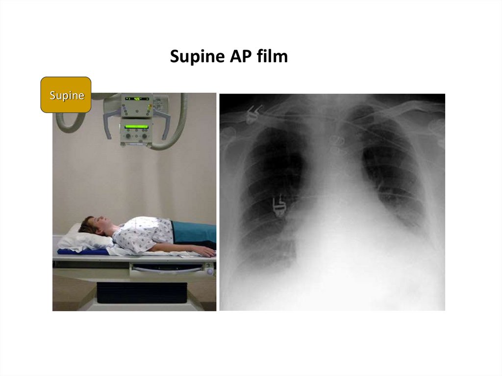
Supine AP filmSupine
8.
9.
10.
Check centralization of patientRotation
11.
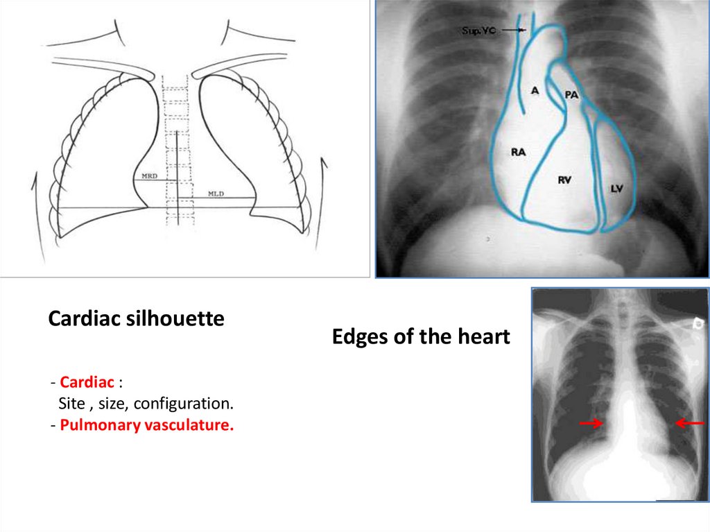
Cardiac silhouette- Cardiac :
Site , size, configuration.
- Pulmonary vasculature.
Edges of the heart
12.
13.
14.
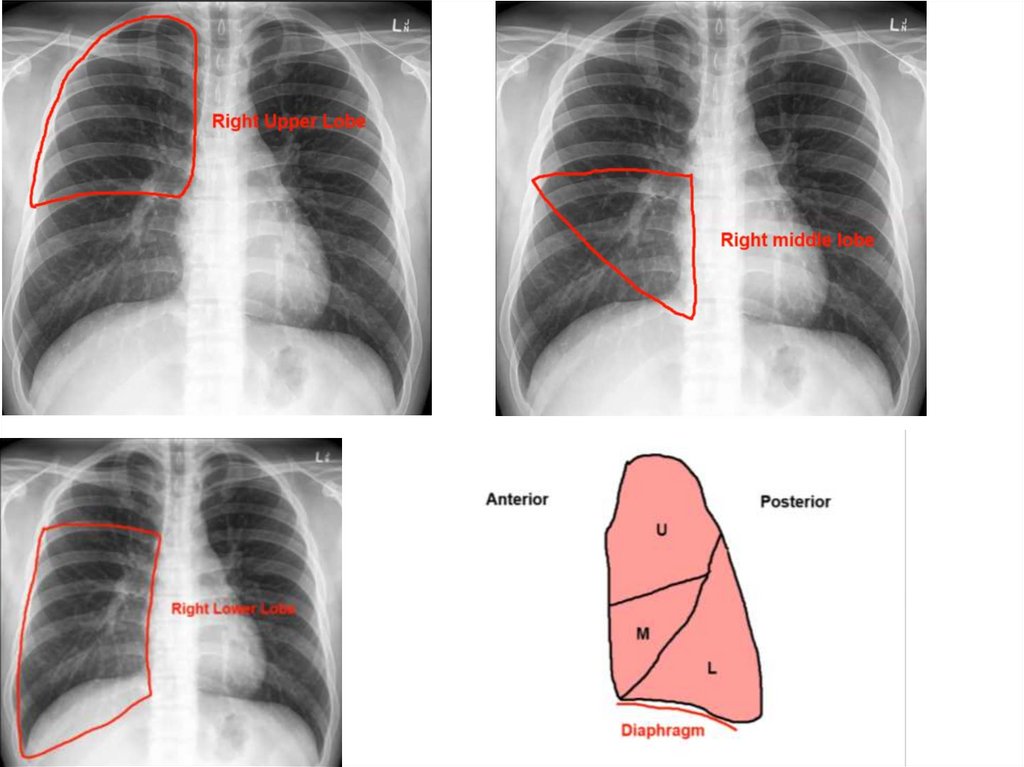
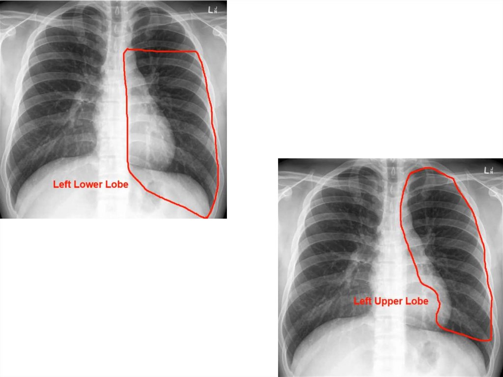
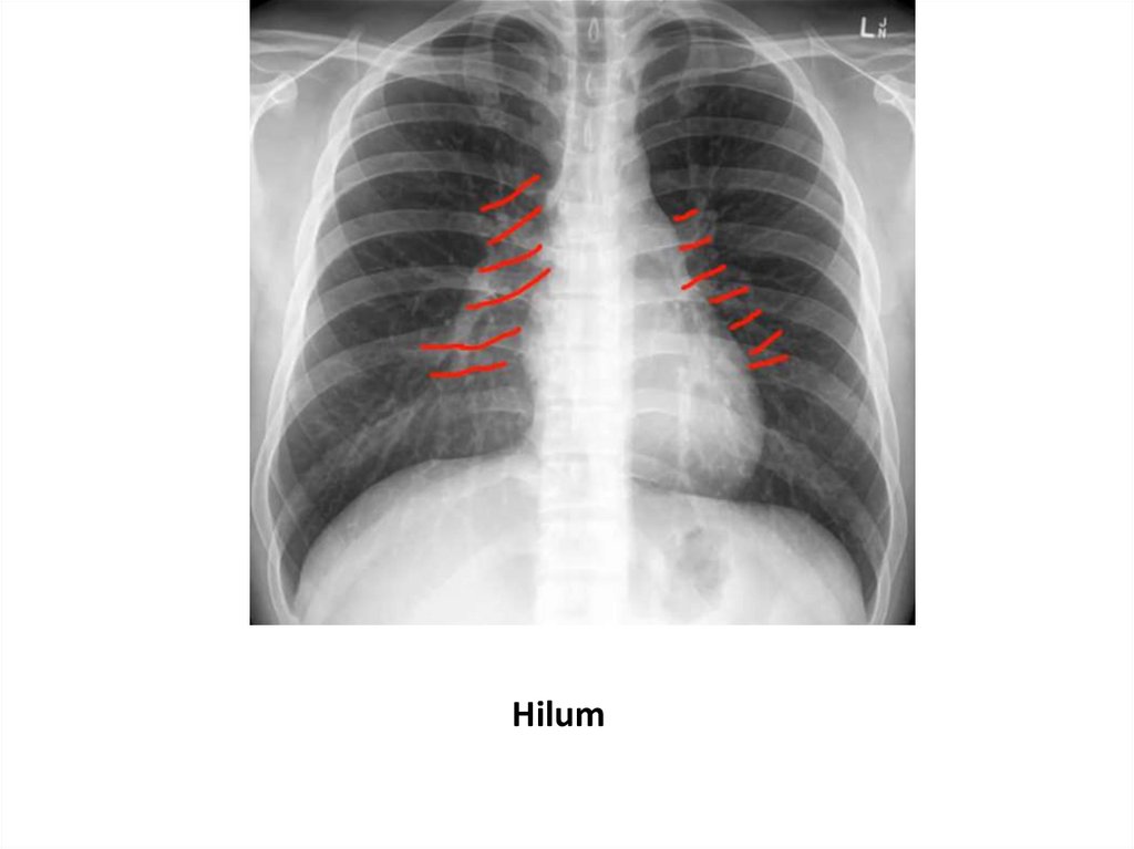
15.
Hilum16.
Look for the abnormalities17.
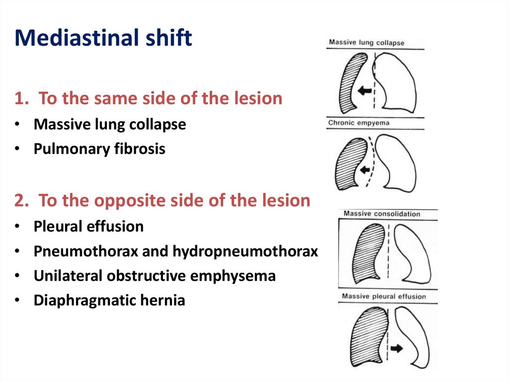
Mediastinal shift1. To the same side of the lesion
• Massive lung collapse
• Pulmonary fibrosis
2. To the opposite side of the lesion
Pleural effusion
Pneumothorax and hydropneumothorax
Unilateral obstructive emphysema
Diaphragmatic hernia
18.
19.
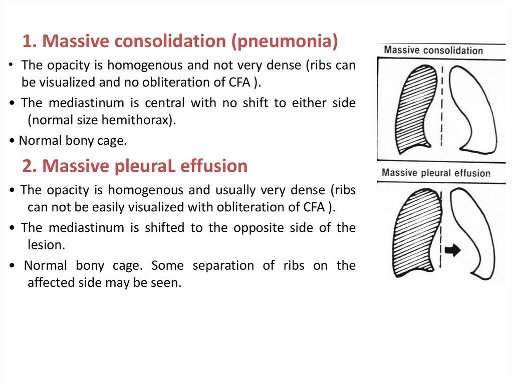
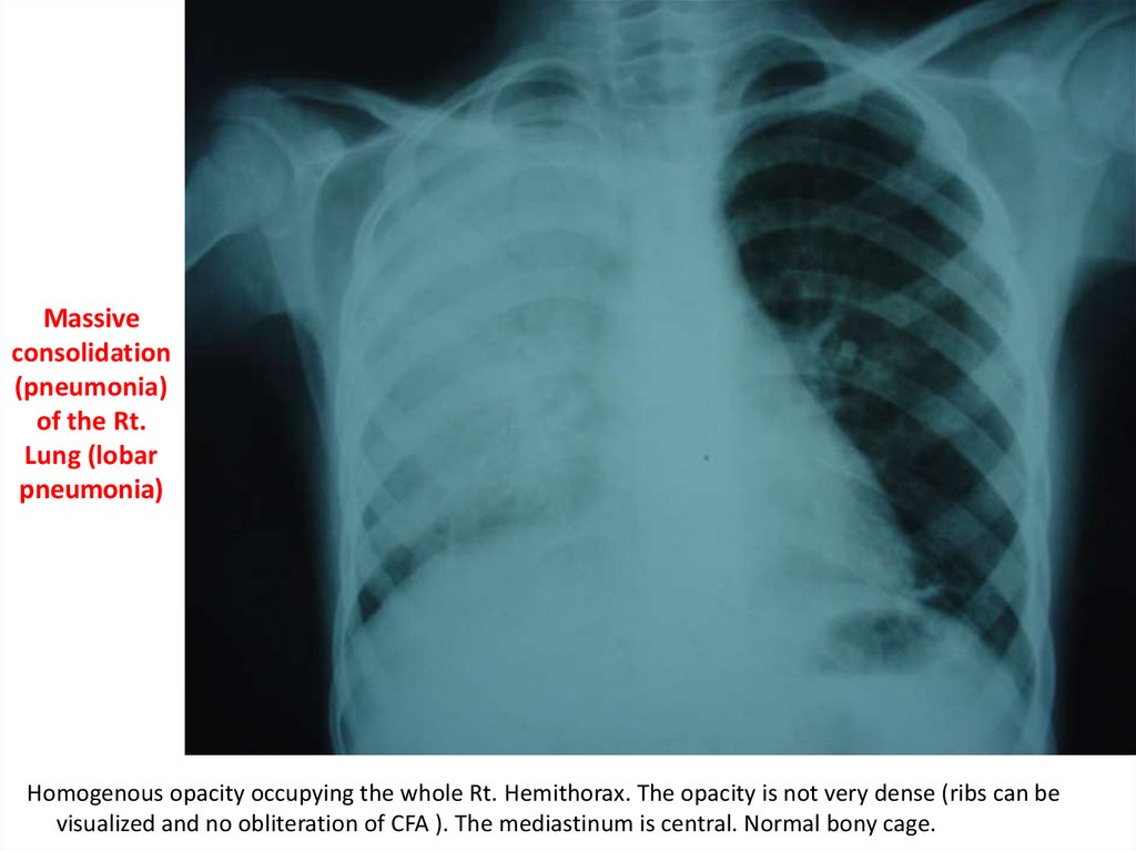
1. Massive consolidation (pneumonia)• The opacity is homogenous and not very dense (ribs can
be visualized and no obliteration of CFA ).
• The mediastinum is central with no shift to either side
(normal size hemithorax).
• Normal bony cage.
2. Massive pleuraL effusion
• The opacity is homogenous and usually very dense (ribs
can not be easily visualized with obliteration of CFA ).
• The mediastinum is shifted to the opposite side of the
lesion.
• Normal bony cage. Some separation of ribs on the
affected side may be seen.
20.
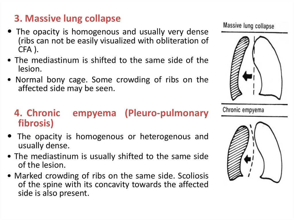
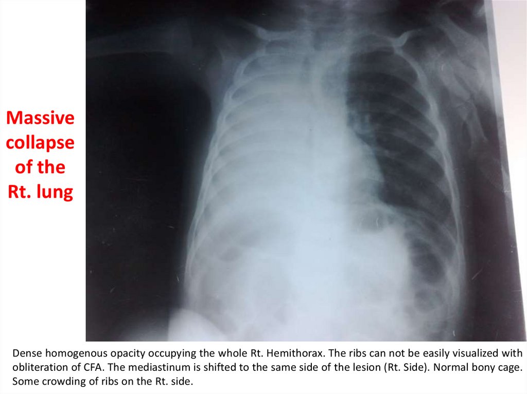
3. Massive lung collapse• The opacity is homogenous and usually very dense
(ribs can not be easily visualized with obliteration of
CFA ).
• The mediastinum is shifted to the same side of the
lesion.
• Normal bony cage. Some crowding of ribs on the
affected side may be seen.
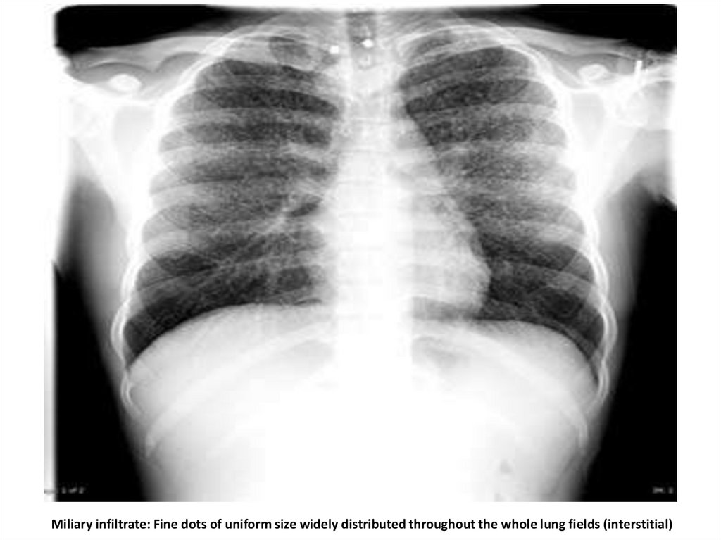
4. Chronic
fibrosis)
empyema (Pleuro-pulmonary
• The opacity is homogenous or heterogenous and
usually dense.
• The mediastinum is usually shifted to the same side
of the lesion.
• Marked crowding of ribs on the same side. Scoliosis
of the spine with its concavity towards the affected
side is also present.
21.
1. Massive consolidation (pneumonia)22.
Massiveconsolidation
(pneumonia)
of the Rt.
Lung (lobar
pneumonia)
Homogenous opacity occupying the whole Rt. Hemithorax. The opacity is not very dense (ribs can be
visualized and no obliteration of CFA ). The mediastinum is central. Normal bony cage.
23.
2. Massive pleuraL effusion24.
Rt. sidedmassive
pleural
effusion
Dense homogeneous opacity occupying the whole Rt. hemithorax and obliterating the right costophrenic
angle, no bronchovascular markings are visible. The mediastinum is markedly shifted to the left side.
Normal bony cage.
25.
26.
Causes of pleural effusion• Empyema (purulent pleurisy)
• Bacterial pneumonias (Staphylococcal, Hemophilus influenza).
• Ruptured lung abscess, mediastinitis, and chest surgery.
Serofibrinous pleurisy
Bacterial pneumonias and tuberculous effusion.
Malignancy: Lymphoma, Neuroblastoma, and metastases.
Rheumatic diseases
• Hydro thorax
• Heart failure, Renal failure, Nephrotic syndrome
• Hemothorax
• Trauma, Tumours
• Chylothorax
• Chest surgery
27.
3. Massive lung collapse28.
Massivecollapse
of the
Rt. lung
Dense homogenous opacity occupying the whole Rt. Hemithorax. The ribs can not be easily visualized with
obliteration of CFA. The mediastinum is shifted to the same side of the lesion (Rt. Side). Normal bony cage.
Some crowding of ribs on the Rt. side.
29.
Massive lung collapseIt results from total obstruction of the main Rt. or
Lt. bronchus.
Causes:
1- FB inhalation.
2- Respiratory paralysis.
3- Postoperative chest surgery.
4- Wrongly placed ETT.
30.
4. Chronic empyema (Pleuro-pulmonary fibrosis).
31.
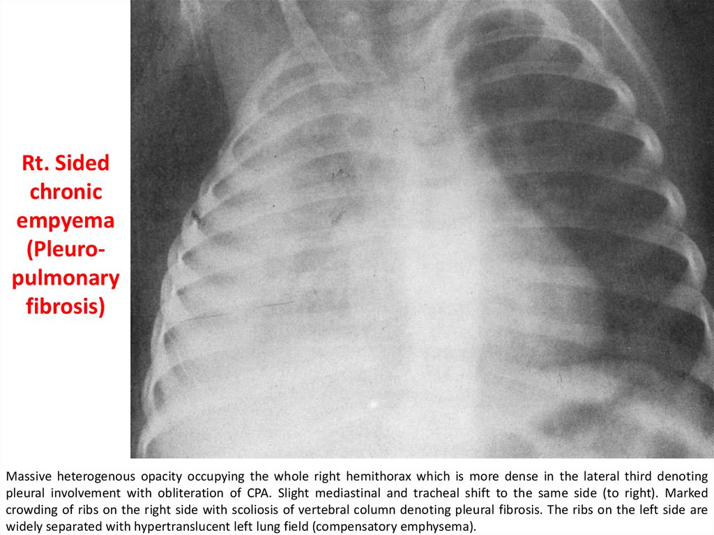
Rt. Sidedchronic
empyema
(Pleuropulmonary
fibrosis)
Massive heterogenous opacity occupying the whole right hemithorax which is more dense in the lateral third denoting
pleural involvement with obliteration of CPA. Slight mediastinal and tracheal shift to the same side (to right). Marked
crowding of ribs on the right side with scoliosis of vertebral column denoting pleural fibrosis. The ribs on the left side are
widely separated with hypertranslucent left lung field (compensatory emphysema).
32.
A-Obstructive emphysemaB-Pneumothorax
33. Massive lung collapse It results from total obstruction of the main Rt. or Lt. bronchus. Causes: 1- FB inhalation. 2-
34.
A-Obstructive emphysema35.
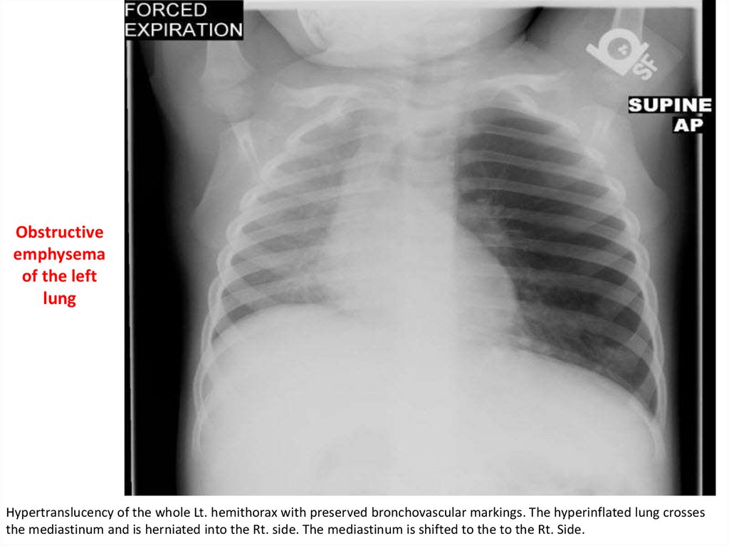
Obstructiveemphysema
of the left
lung
Hypertranslucency of the whole Lt. hemithorax with preserved bronchovascular markings. The hyperinflated lung crosses
the mediastinum and is herniated into the Rt. side. The mediastinum is shifted to the to the Rt. Side.
36.
Obstructive Emphysema-It results from partial (incomplete) obstruction of a
bronchus which creates a valve type of obstruction.
-It can be generalized or localized to one lung.
-Causes of localized obstructive emphysema:
1- In acute conditions: F.B. or viscid secretions.
2- in chronic conditions : T.B. of tracheobronchial LNs.
37.
B-Pneumothorax38.
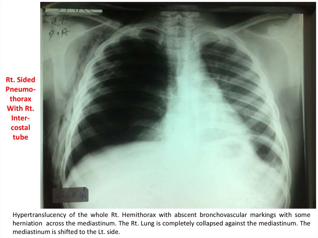
Rt. SidedPneumothorax
With Rt.
Intercostal
tube
Hypertranslucency of the whole Rt. Hemithorax with abscent bronchovascular markings with some
herniation across the mediastinum. The Rt. Lung is completely collapsed against the mediastinum. The
mediastinum is shifted to the Lt. side.
39.
40. Obstructive Emphysema -It results from partial (incomplete) obstruction of a bronchus which creates a valve type of
41.
Causes of pneumothorax (free air in pleural space):1-Iatrogenic: as a complication of mechanical ventilation or
chest surgery (commonest).
2-Spontaneous : with acute conditions as acute
bronchiolitis, bronchial asthma, pertussis and interstitial
pneumonias.
42.
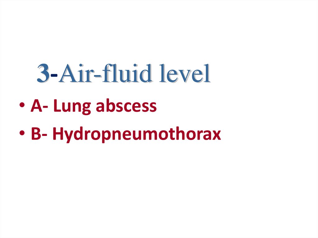
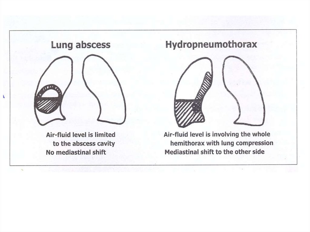
3-Air-fluid level• A- Lung abscess
• B- Hydropneumothorax
43.
44.
• A- Lung abscess45. Causes of pneumothorax (free air in pleural space): 1-Iatrogenic: as a complication of mechanical ventilation or chest surgery
Lung abscess(Solitary lung abscess of
the right lower lobe(
Dense homogenous opacity in the lower zone of the right lung field (clear costophrenic angle)
with horizontal upper level (fluid level) and hypertranslucent area devoid of lung markings
above it (air). Note the following :
1. The hypertranslucent area is not reaching to the apex of the right lung but surrounded by a
dense opacity (wall of the abscess).
2. The fluid level is not involving the whole hemithorax.
3. The lung is not collapsed against the mediastinum.
4. The mediastinum is not shifted to the other side.
46.
47.
Lung Abscess• It results from suppurative destruction of lung
parenchyma and formation of a cavity containing
purulent material.
• It occurs with aspiration of infected material or with
bacterial pneumonias.
48.
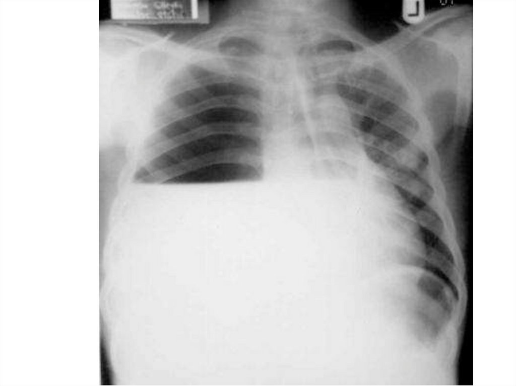
• B- Hydropneumothorax49.
50.
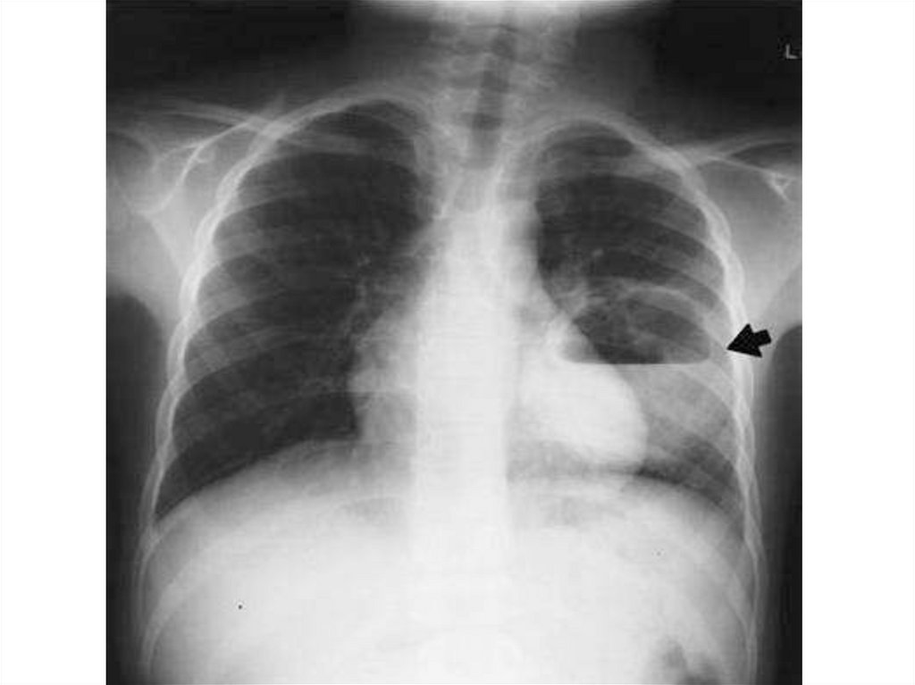
* Plain x-ray of a chest and heart, posteroanterior view.• The patient is not centralized.
• The mediastinum is markedly shifted to the Rt side.
* There is dense homogeneous opacity obliterating the left
costophrenic angle and occupying the lower ⅔ of the left
hemithorax, with horizontal fluid level, and the upper ⅓ of
the left hemithrax is occupied by a jet black colour
(hypertranslucent) without bronchovascular markings with
collapsed lt. lung.
** The radiological diagnosis: Lt sided hydropneumothorax.
51.
52.
Hydropneumothorax• It occurs mostly with cases of pleural effusion due to
one of 2 causes:
-Iatrogenic introduction of air into the pleural space
during diagnostic aspiration (thoracocentesis).
-Bronchopleural fistula allowing air entry from a
bronchus into the pleural space.
53.
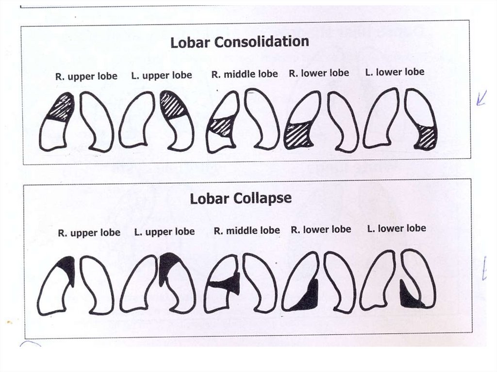
4-Partial unilateral opacity• Lobar consolidation (pneumonia)
• Lobar collapse (atelectasis)
• Solitary patch or nodule
54.
55.
4-Partial unilateral opacity• A--Lobar consolidation (pneumonia)
56.
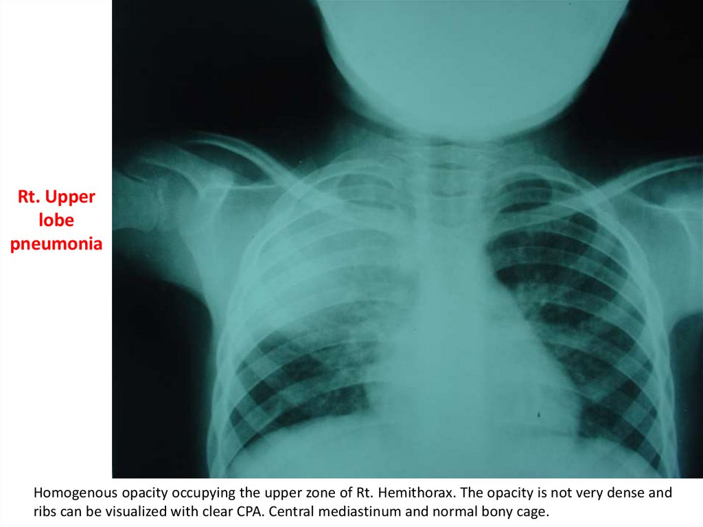
Rt. Upperlobe
pneumonia
Homogenous opacity occupying the upper zone of Rt. Hemithorax. The opacity is not very dense and
ribs can be visualized with clear CPA. Central mediastinum and normal bony cage.
57.
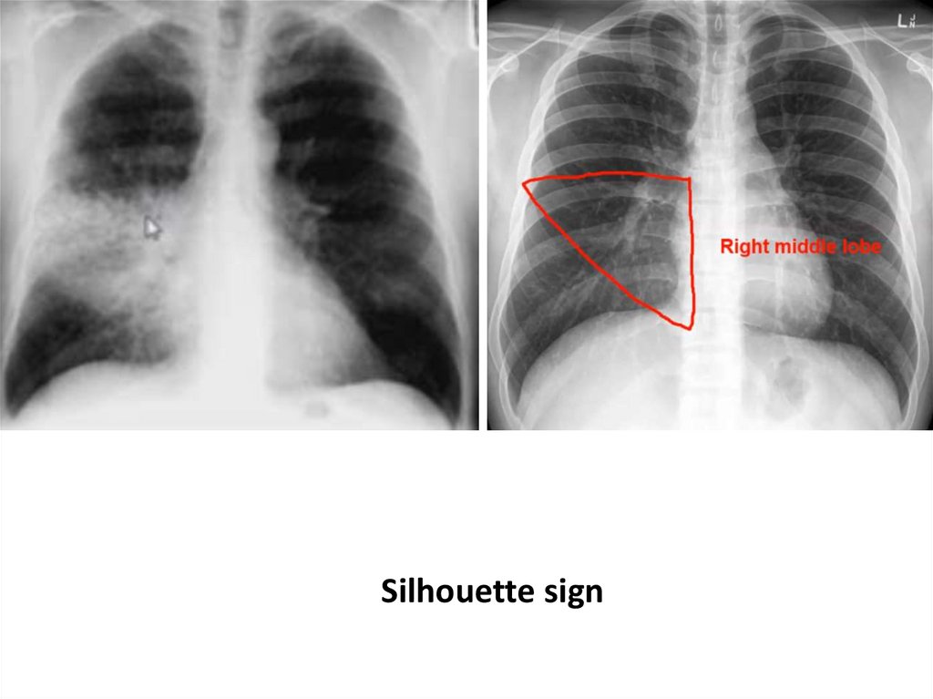
Silhouette sign58.
59.
Right middleand lower
lobe
consolidation
60.
Dense homogeneous opacity occupying the Lower zone of Rt. hemithorax and obliterating the rightcostophrenic angle, with concave upper border raising to the axilla. The mediastinum is shifted to the left side.
Normal bony cage (Rt. side moderate pleural effusion).
61.
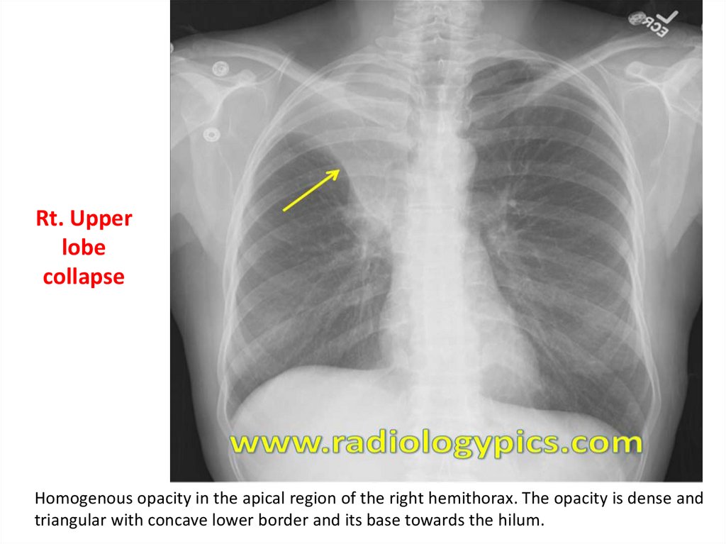
4-Partial unilateral opacity• B- Lobar collapse (atelectasis)
62.
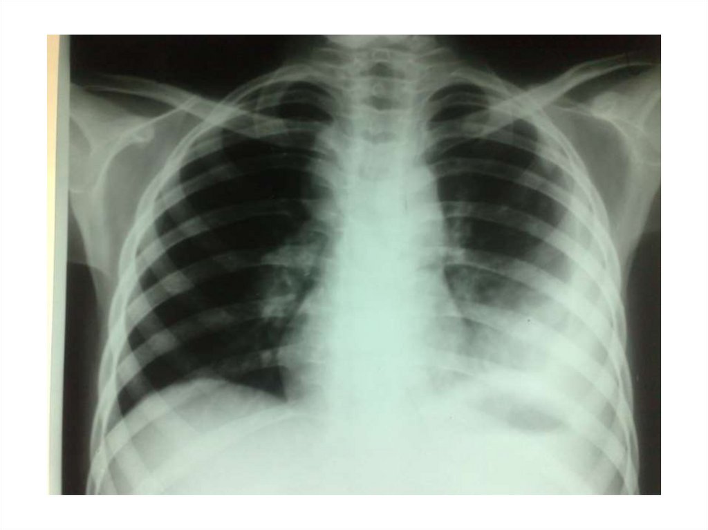
Rt. Upperlobe
collapse
Homogenous opacity in the apical region of the right hemithorax. The opacity is dense and
triangular with concave lower border and its base towards the hilum.
63.
64.
65.
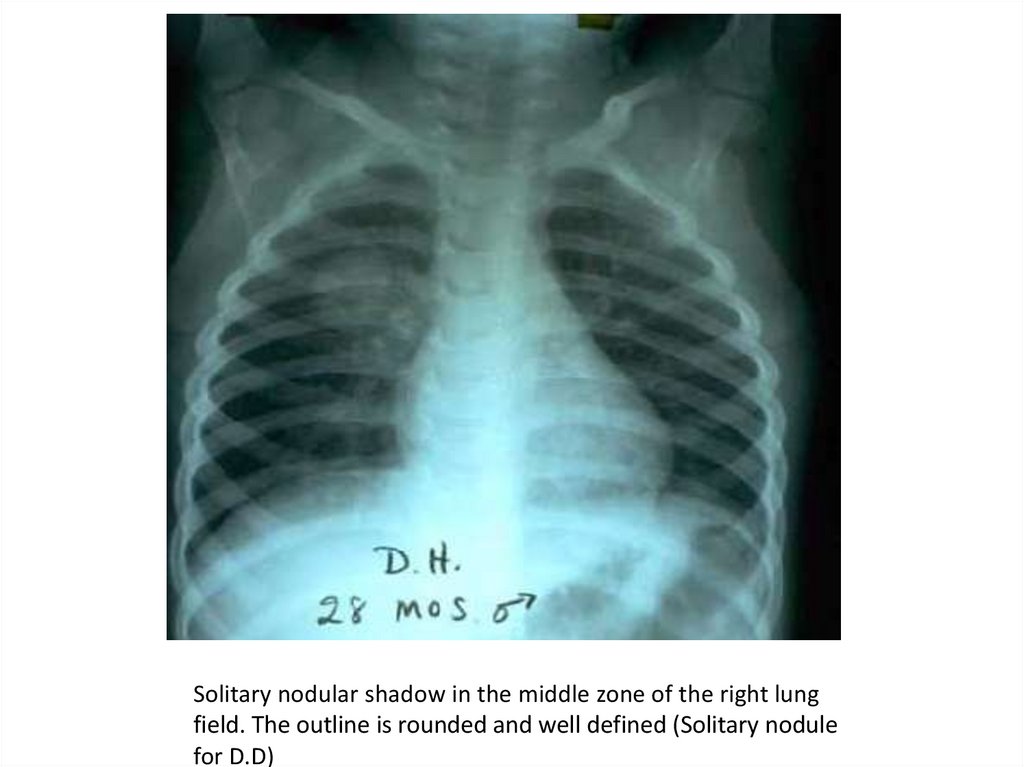
4-Partial unilateral opacity• C- Solitary patch or nodule
66.
Solitary nodular shadow in the middle zone of the right lungfield. The outline is rounded and well defined (Solitary nodule
for D.D)
67.
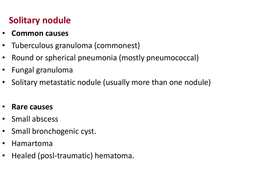
Solitary noduleCommon causes
Tuberculous granuloma {commonest}
Round or spherical pneumonia (mostly pneumococcal)
Fungal granuloma
Solitary metastatic nodule (usually more than one nodule)
Rare causes
Small abscess
Small bronchogenic cyst.
Hamartoma
Healed (posl-traumatic) hematoma.
68.
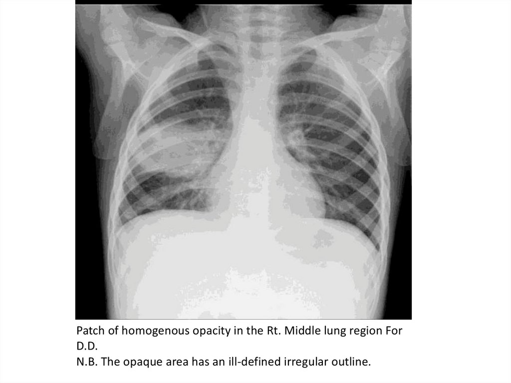
Patch of homogenous opacity in the Rt. Middle lung region ForD.D.
N.B. The opaque area has an ill-defined irregular outline.
69.
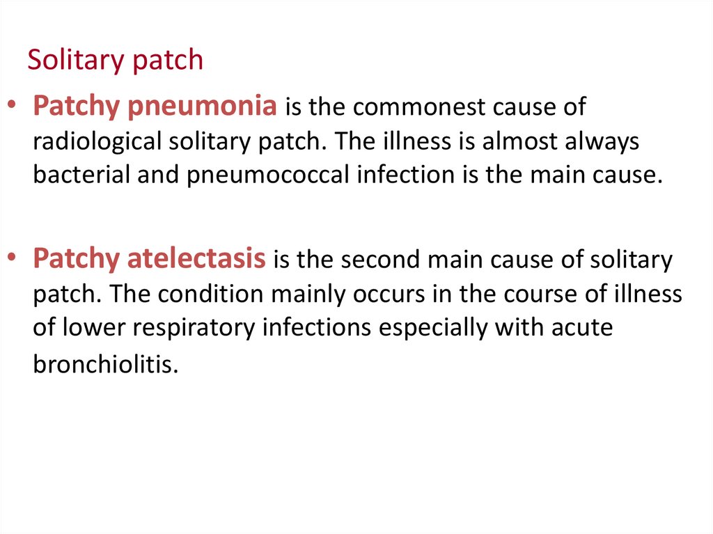
Solitary patch• Patchy pneumonia is the commonest cause of
radiological solitary patch. The illness is almost always
bacterial and pneumococcal infection is the main cause.
• Patchy atelectasis is the second main cause of solitary
patch. The condition mainly occurs in the course of illness
of lower respiratory infections especially with acute
bronchiolitis.
70.
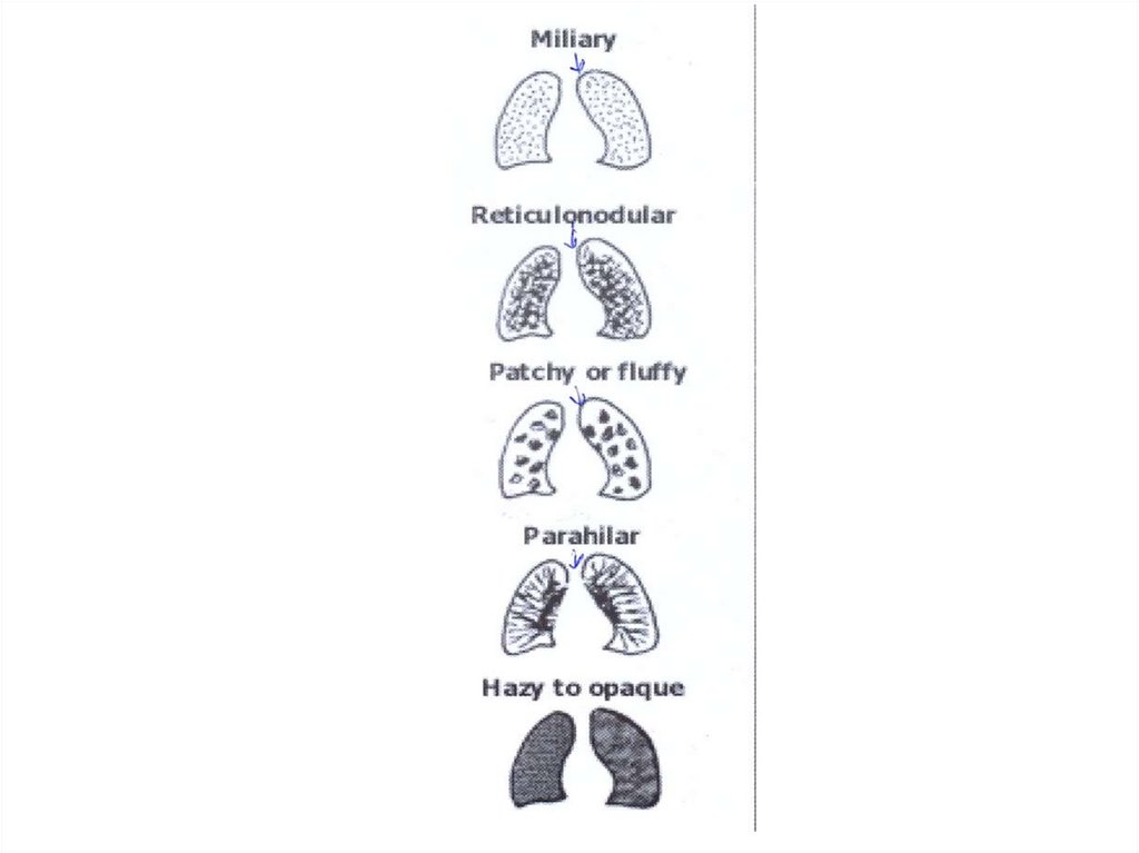
5-Pulmonary infiltrateMiliary infiltrate
Recticulonodular infiltrate
Patchy or fluffy infiltrate
Parahilar peribronchial infiltrate (most common)
Hazy to opaque infiltrate (most serious)
71.
72.
A- Miliary infiltrate73.
Miliary infiltrate: Fine dots of uniform size widely distributed throughout the whole lung fields (interstitial)74.
Causes of miliary infiltrate1-Infectious conditions
• Miliary tuberculosis (commonest)
• Viral interstitial pneumonias
• Pulmonary fungal infections.
2- Noninfectious conditions
• Idiopathic pulmonary hemosiderosis
• Histiocytosis
• Metastatic diseases to the lung as Leukemia and
lymphoma.
75.
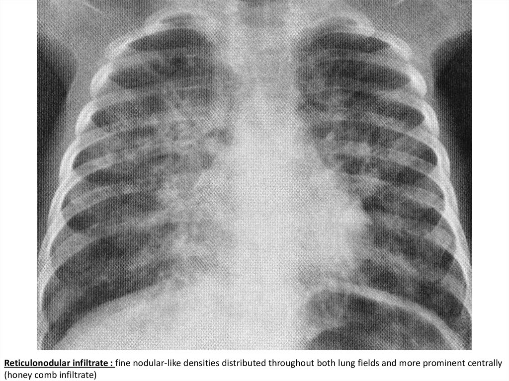
B- Recticulonodular infiltrate76.
Reticulonodular infiltrate : fine nodular-like densities distributed throughout both lung fields and more prominent centrally(honey comb infiltrate)
77.
Causes of reticulonodular infiltrate1-Infectious conditions
• Viral interstitial pneumonia (commonest)
• Mycoplasma pneumonia
• Pneumocystis carinii pneumonia
• Pulmonary fungal infections.
2- Noninfectious conditions
• Histiocytosis
• Idiopathic pulmonary hemosiderosis
• Pulmonary lymphangiectasia
78.
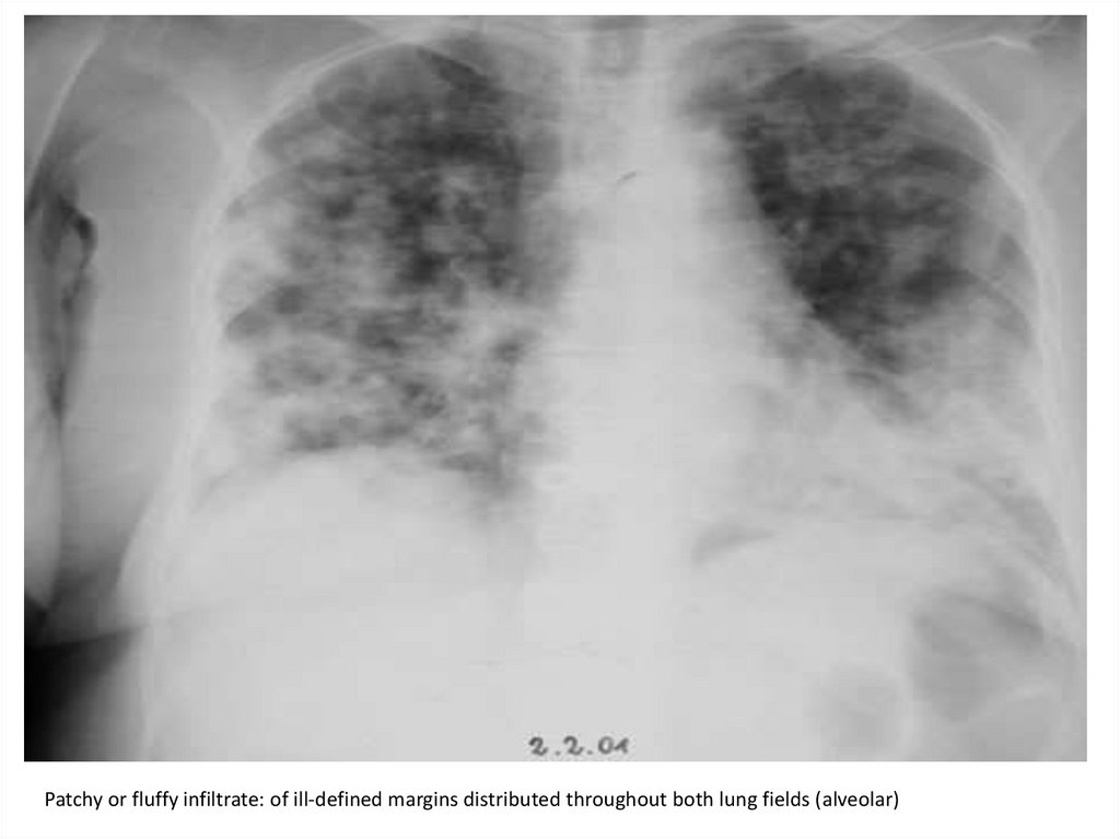
C- Patchy or fluffy infiltrate79.
Patchy or fluffy infiltrate: of ill-defined margins distributed throughout both lung fields (alveolar)80.
Causes of patchy / fluffy infiltrate1-Infectious conditions
• Bacterial bronchopneumonia (commonest),
staphylococcal and hemophilus influenza
• Aspiration pneumonias
• Pulmonary fungal infections
2- Noninfectious conditions
• Pulmonary hemorrhage
• Near drowning
81.
82.
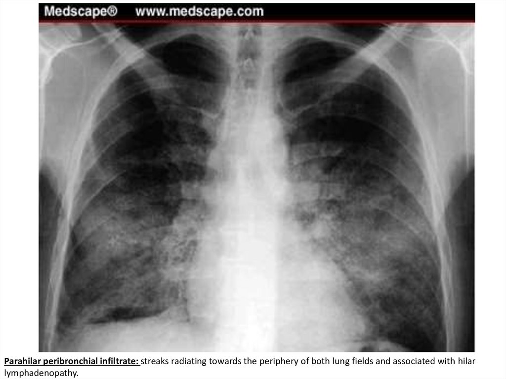
D-Parahilar peribronchial infiltrate(most common)
83.
Parahilar peribronchial infiltrate: streaks radiating towards the periphery of both lung fields and associated with hilarlymphadenopathy.
84.
Causes of parahilar peribronchial infiltrate1-Infectious conditions
• Viral lower respiratory infections as bronchitis
(commonest)
• Bronchial asthma especially when associated with viral
respiratory infections
2- Noninfectious conditions
• Interstitial pulmonary fibrosis
• Cystic fibrosis
85.
E- Hazy to opaque infiltrate (most serious)86.
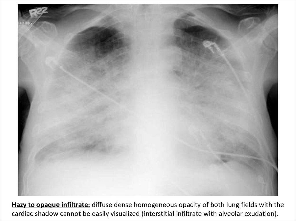
Hazy to opaque infiltrate: diffuse dense homogeneous opacity of both lung fields with thecardiac shadow cannot be easily visualized (interstitial infiltrate with alveolar exudation).
87.
Causes of hazy to opaque infiltrate• Pulmonary edema (commonest):
-Cardiac causes: myocarditis, CHD with Lt. to Rt. shunt
-Non-cardiac causes: ARF, iatrogenic fluid overload,
fulminant pneumonia or ARDS and neurogenic
Pulmonary edema
• Pneumocystis carinii & viral interstitial pneumonia
• Pulmonary hemorrhage/hemosiderosis
88.
6- Dense Hilar ShadowHilar lymphadenopathy
• Bilateral:
• Viral lower respiratory infections
• Chronic aspiration
• Malignancies as Lymphoma or leukemia
• Unilateral:
• Tuberculosis of trachiobronchial LNs
• Mycoplasma pneumonia
Pulmonary Hypertension
• Dense hilar shadow and large convex pulmonary
segment
89.
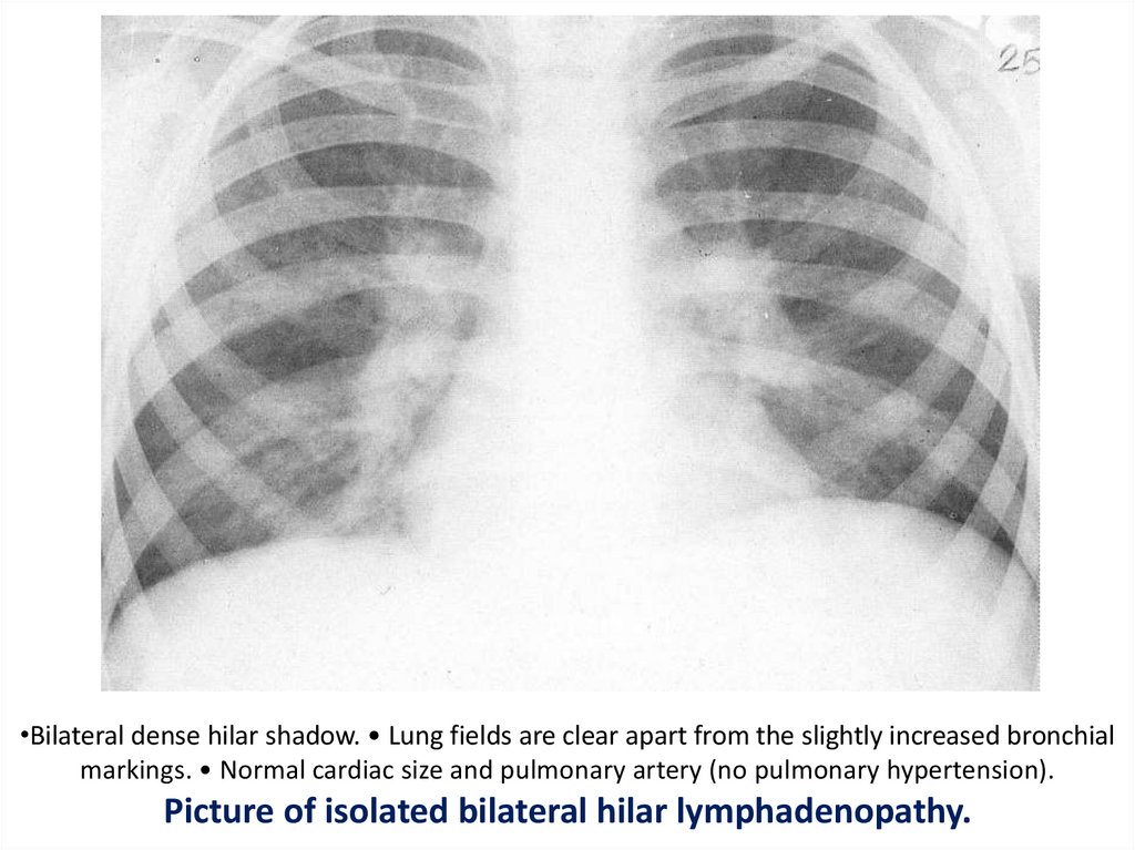
•Bilateral dense hilar shadow. • Lung fields are clear apart from the slightly increased bronchialmarkings. • Normal cardiac size and pulmonary artery (no pulmonary hypertension).
Picture of isolated bilateral hilar lymphadenopathy.
90.
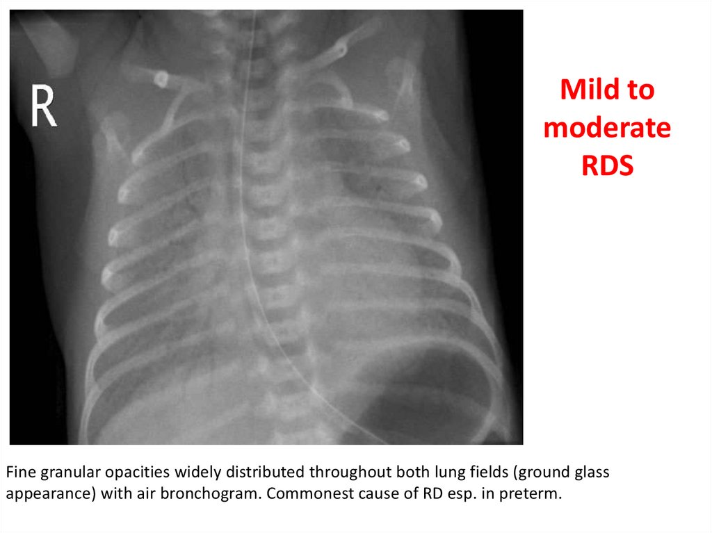
Mild tomoderate
RDS
Fine granular opacities widely distributed throughout both lung fields (ground glass
appearance) with air bronchogram. Commonest cause of RD esp. in preterm.
91.
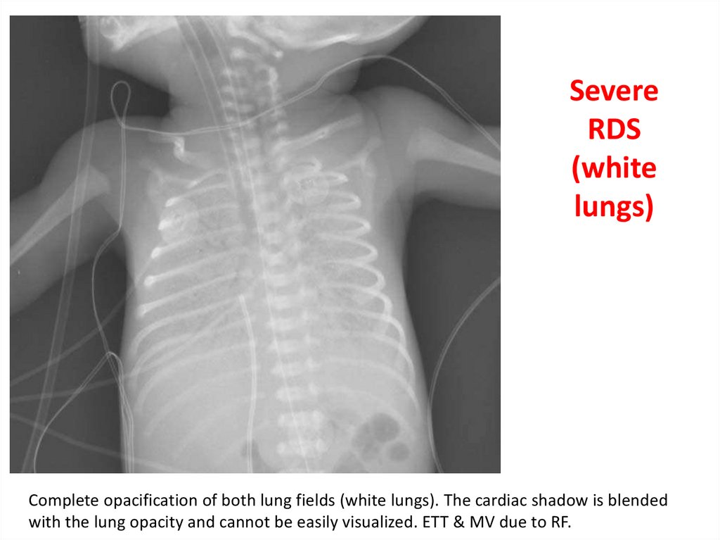
SevereRDS
(white
lungs)
Complete opacification of both lung fields (white lungs). The cardiac shadow is blended
with the lung opacity and cannot be easily visualized. ETT & MV due to RF.
92.
MAS with pneumothorax93.
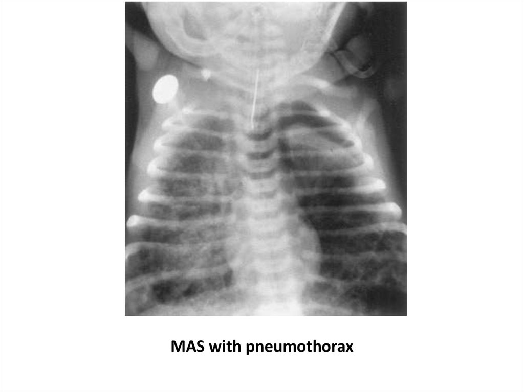
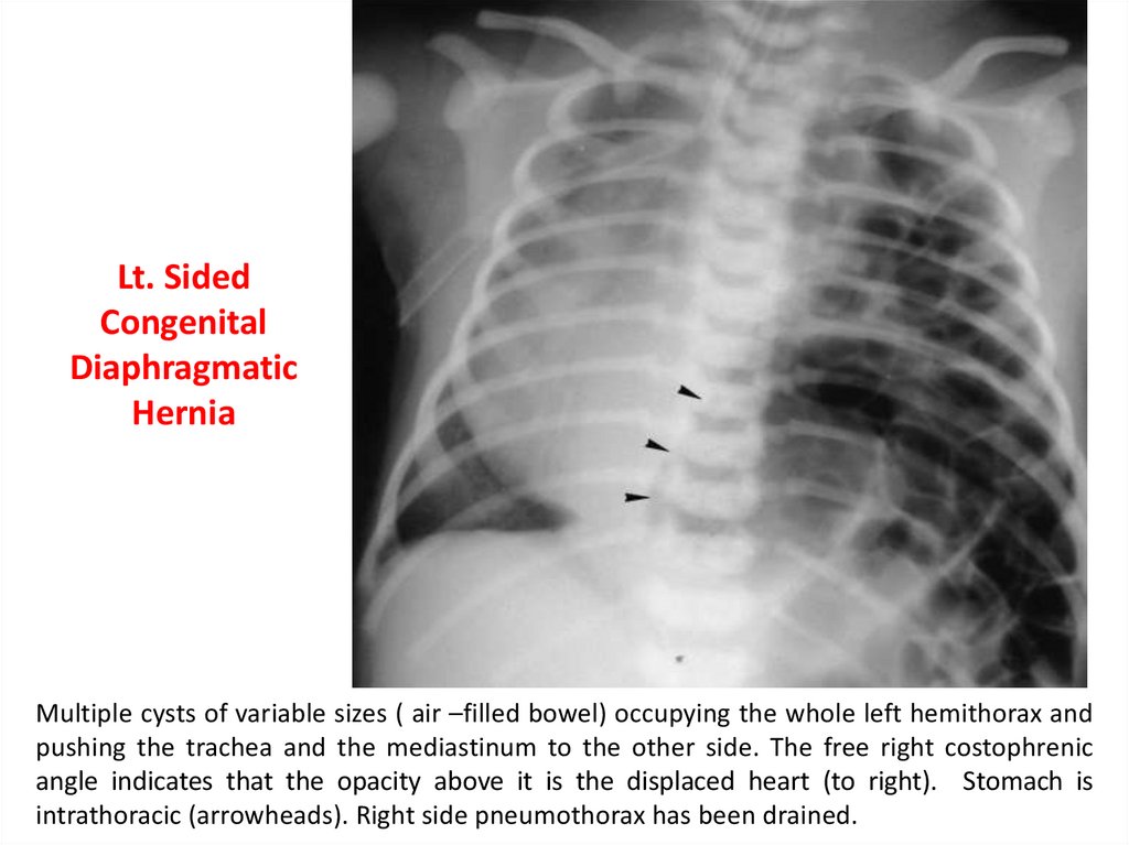
Lt. SidedCongenital
Diaphragmatic
Hernia
Multiple cysts of variable sizes ( air –filled bowel) occupying the whole left hemithorax and
pushing the trachea and the mediastinum to the other side. The free right costophrenic
angle indicates that the opacity above it is the displaced heart (to right). Stomach is
intrathoracic (arrowheads). Right side pneumothorax has been drained.
94.
95.
96.
97.
Thankyou
سبحانك اللهم و بحمدك نشهد ان ال اله اال انت نستغفرك و نتوب اليك