
 Медицина
Похожие презентации:


Alzheimer's disease. Болезнь Альцгеймера
1.
SIWALZHEIMER’S DISEASE. ASL.
MULTIPLE SCLEROSIS
DEPARTMENT OF PATHOLOGICAL ANATOMY AND JUDICIAL MEDICINE NAME
OF PRUGLO Y.V.
PREPARED BY: KAMZINA LYAZZAT , 327 g,. GM
CHECKED BY:
2.
PLAN❏Introduction
❖ Alzheimer’s disease
● Etiology and patogenesis
● Macroscopy
● Microscopy
● Outcomes
❖ ASL
❖ Multiple sclerosis
➔Conclusion
★ Bibliography
3.
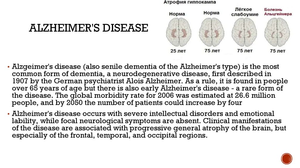
ALZHEIMER'S DISEASE▪ Alzgeimer's disease (also senile dementia of the Alzheimer's type) is the most
common form of dementia, a neurodegenerative disease, first described in
1907 by the German psychiatrist Alois Alzheimer. As a rule, it is found in people
over 65 years of age but there is also early Alzheimer's disease - a rare form of
the disease. The global morbidity rate for 2006 was estimated at 26.6 million
people, and by 2050 the number of patients could increase by four
▪ Alzheimer's disease occurs with severe intellectual disorders and emotional
lability, while focal neurological symptoms are absent. Clinical manifestations
of the disease are associated with progressive general atrophy of the brain, but
especially of the frontal, temporal, and occipital regions.
4.
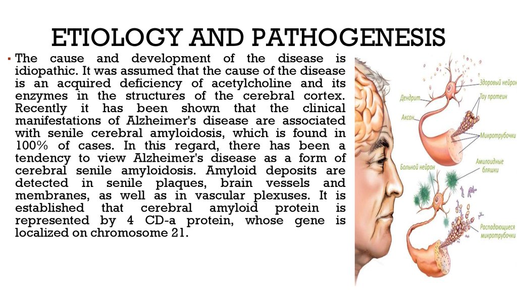
▪ TheETIOLOGY AND PATHOGENESIS
cause and development of the disease is
idiopathic. It was assumed that the cause of the disease
is an acquired deficiency of acetylcholine and its
enzymes in the structures of the cerebral cortex.
Recently it has been shown that the clinical
manifestations of Alzheimer's disease are associated
with senile cerebral amyloidosis, which is found in
100% of cases. In this regard, there has been a
tendency to view Alzheimer's disease as a form of
cerebral senile amyloidosis. Amyloid deposits are
detected in senile plaques, brain vessels and
membranes, as well as in vascular plexuses. It is
established that cerebral amyloid protein is
represented by 4 CD-a protein, whose gene is
localized on chromosome 21.
5.
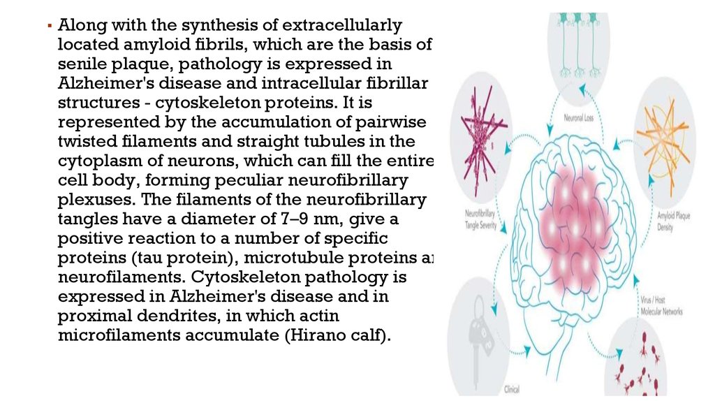
▪ Along with the synthesis of extracellularlylocated amyloid fibrils, which are the basis of
senile plaque, pathology is expressed in
Alzheimer's disease and intracellular fibrillar
structures - cytoskeleton proteins. It is
represented by the accumulation of pairwise
twisted filaments and straight tubules in the
cytoplasm of neurons, which can fill the entire
cell body, forming peculiar neurofibrillary
plexuses. The filaments of the neurofibrillary
tangles have a diameter of 7–9 nm, give a
positive reaction to a number of specific
proteins (tau protein), microtubule proteins and
neurofilaments. Cytoskeleton pathology is
expressed in Alzheimer's disease and in
proximal dendrites, in which actin
microfilaments accumulate (Hirano calf).
6.
STAGES OF ALZHEIMER'SDISEASE
Early stage Alzheimer's
Not remembering episodes of forgetfulness
Forgets names of family or friends
Changes may only be noticed by close
friends or relatives
Some confusion in situations outside the
familiar
Late stage Alzheimer's
Poor ability to think
Problems speaking
Repeats same conversations
More abusive, anxious, or paranoid
Middle stage Alzheimer's
Greater difficulty remembering recently learned
information
Deepening confusion in many circumstances
Problems with sleep
Trouble determining their location
7.
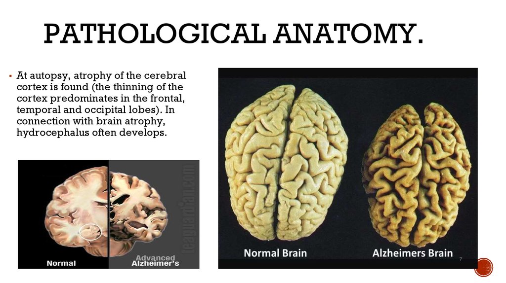
PATHOLOGICAL ANATOMY.▪ At autopsy, atrophy of the cerebral
cortex is found (the thinning of the
cortex predominates in the frontal,
temporal and occipital lobes). In
connection with brain atrophy,
hydrocephalus often develops.
8.
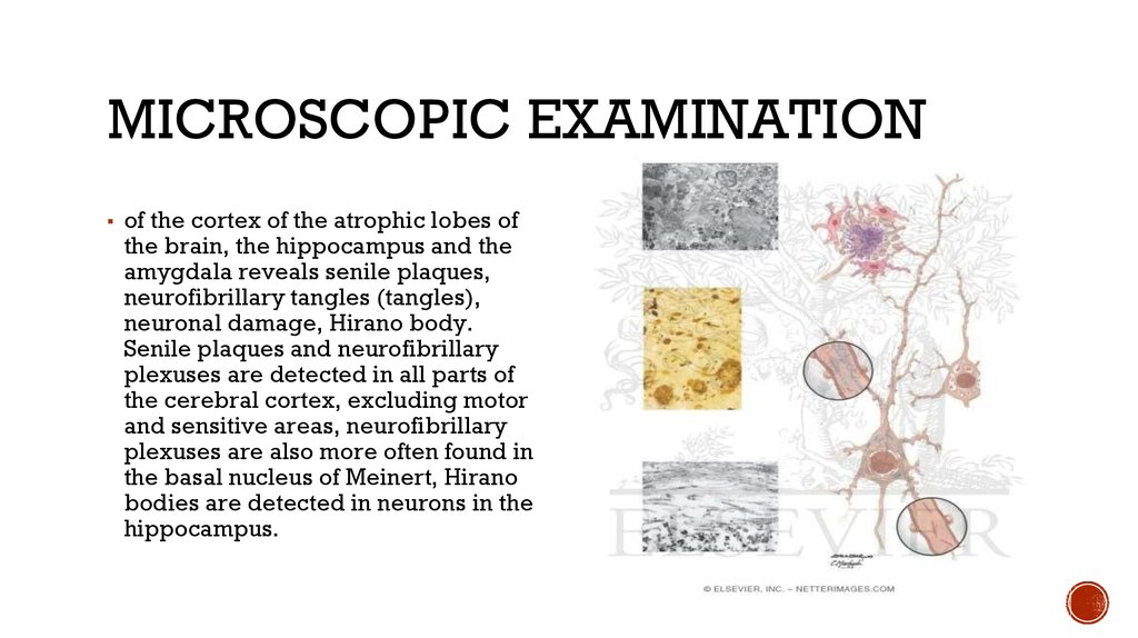
MICROSCOPIC EXAMINATION▪ of the cortex of the atrophic lobes of
the brain, the hippocampus and the
amygdala reveals senile plaques,
neurofibrillary tangles (tangles),
neuronal damage, Hirano body.
Senile plaques and neurofibrillary
plexuses are detected in all parts of
the cerebral cortex, excluding motor
and sensitive areas, neurofibrillary
plexuses are also more often found in
the basal nucleus of Meinert, Hirano
bodies are detected in neurons in the
hippocampus.
9.
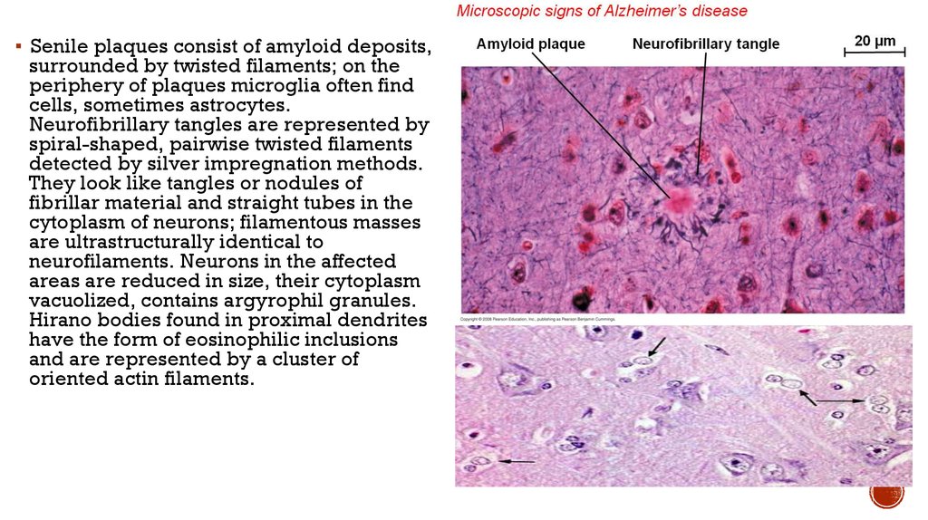
▪ Senile plaques consist of amyloid deposits,surrounded by twisted filaments; on the
periphery of plaques microglia often find
cells, sometimes astrocytes.
Neurofibrillary tangles are represented by
spiral-shaped, pairwise twisted filaments
detected by silver impregnation methods.
They look like tangles or nodules of
fibrillar material and straight tubes in the
cytoplasm of neurons; filamentous masses
are ultrastructurally identical to
neurofilaments. Neurons in the affected
areas are reduced in size, their cytoplasm
vacuolized, contains argyrophil granules.
Hirano bodies found in proximal dendrites
have the form of eosinophilic inclusions
and are represented by a cluster of
oriented actin filaments.
10.
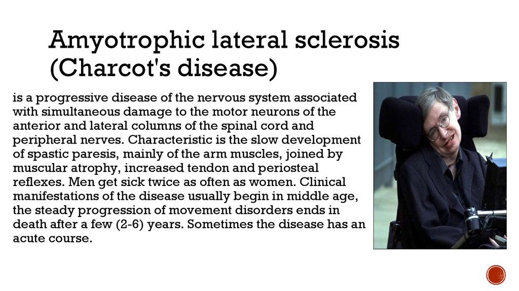
Amyotrophic lateral sclerosis(Charcot's disease)
is a progressive disease of the nervous system associated
with simultaneous damage to the motor neurons of the
anterior and lateral columns of the spinal cord and
peripheral nerves. Characteristic is the slow development
of spastic paresis, mainly of the arm muscles, joined by
muscular atrophy, increased tendon and periosteal
reflexes. Men get sick twice as often as women. Clinical
manifestations of the disease usually begin in middle age,
the steady progression of movement disorders ends in
death after a few (2-6) years. Sometimes the disease has an
acute course.
11.
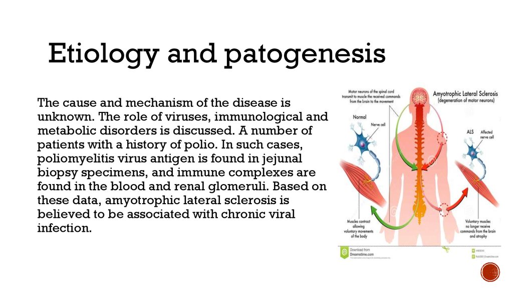
Etiology and patogenesisThe cause and mechanism of the disease is
unknown. The role of viruses, immunological and
metabolic disorders is discussed. A number of
patients with a history of polio. In such cases,
poliomyelitis virus antigen is found in jejunal
biopsy specimens, and immune complexes are
found in the blood and renal glomeruli. Based on
these data, amyotrophic lateral sclerosis is
believed to be associated with chronic viral
infection.
12.
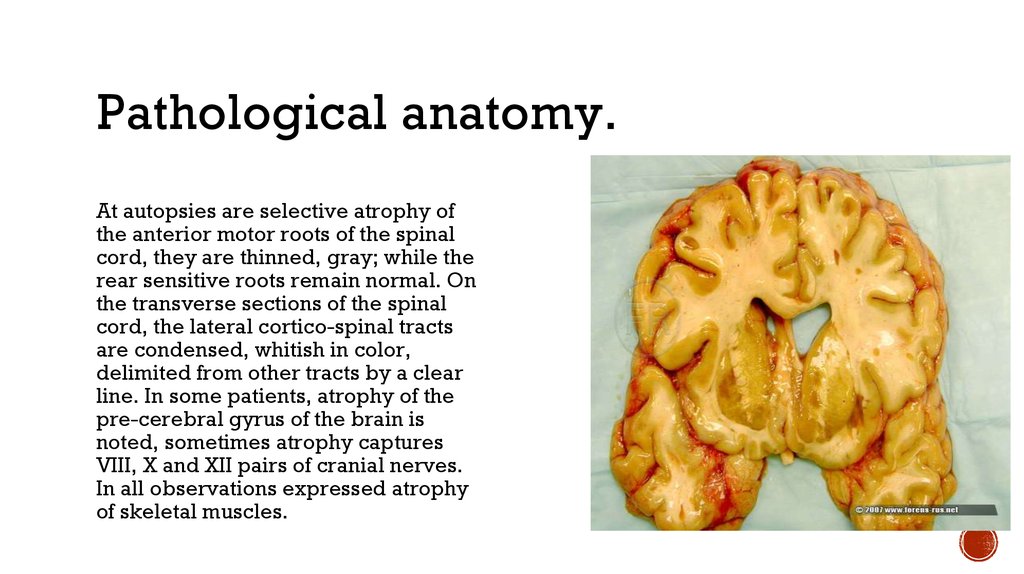
Pathological anatomy.At autopsies are selective atrophy of
the anterior motor roots of the spinal
cord, they are thinned, gray; while the
rear sensitive roots remain normal. On
the transverse sections of the spinal
cord, the lateral cortico-spinal tracts
are condensed, whitish in color,
delimited from other tracts by a clear
line. In some patients, atrophy of the
pre-cerebral gyrus of the brain is
noted, sometimes atrophy captures
VIII, X and XII pairs of cranial nerves.
In all observations expressed atrophy
of skeletal muscles.
13.
Microscopic examinationin the anterior horns of the spinal cord find pronounced changes in nerve
cells; they are wrinkled or in the form of shadows; extensive fields of
neuron loss are found. Sometimes foci of neuron prolapse are found in the
brain stem and precentral gyrus. In the nerve fibers of the affected areas
of the spinal cord are determined demi-linisation, uneven swelling with
subsequent disintegration and death of axial cylinders. The demyelination
of nerve fibers usually extends to peripheral nerves. Often, the pyramidal
paths are involved in the process throughout their length - the spinal cord
and the medulla, up to the cortex of the cerebral hemispheres. As a rule,
reactive proliferation of glial cells is noted. Some observations describe
minor lymphoid infiltrates in the spinal cord, its lining and peripheral
nerves along the vessels.
14.
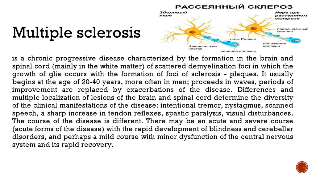
Multiple sclerosisis a chronic progressive disease characterized by the formation in the brain and
spinal cord (mainly in the white matter) of scattered demyelination foci in which the
growth of glia occurs with the formation of foci of sclerosis - plaques. It usually
begins at the age of 20-40 years, more often in men; proceeds in waves, periods of
improvement are replaced by exacerbations of the disease. Differences and
multiple localization of lesions of the brain and spinal cord determine the diversity
of the clinical manifestations of the disease: intentional tremor, nystagmus, scanned
speech, a sharp increase in tendon reflexes, spastic paralysis, visual disturbances.
The course of the disease is different. There may be an acute and severe course
(acute forms of the disease) with the rapid development of blindness and cerebellar
disorders, and perhaps a mild course with minor dysfunction of the central nervous
system and its rapid recovery.
15.
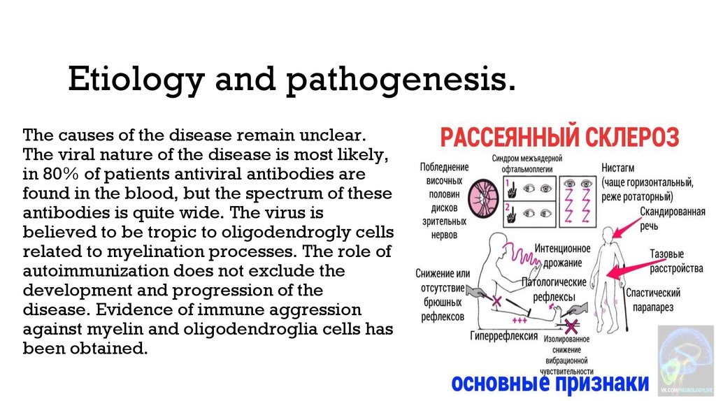
Etiology and pathogenesis.The causes of the disease remain unclear.
The viral nature of the disease is most likely,
in 80% of patients antiviral antibodies are
found in the blood, but the spectrum of these
antibodies is quite wide. The virus is
believed to be tropic to oligodendrogly cells
related to myelination processes. The role of
autoimmunization does not exclude the
development and progression of the
disease. Evidence of immune aggression
against myelin and oligodendroglia cells has
been obtained.
16.
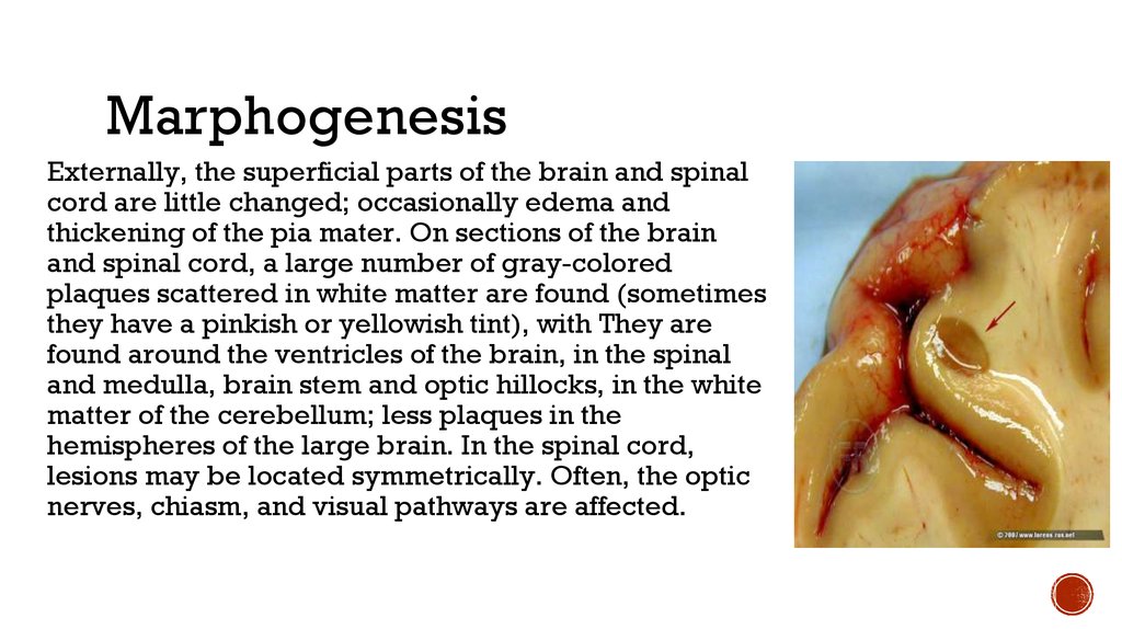
MarphogenesisExternally, the superficial parts of the brain and spinal
cord are little changed; occasionally edema and
thickening of the pia mater. On sections of the brain
and spinal cord, a large number of gray-colored
plaques scattered in white matter are found (sometimes
they have a pinkish or yellowish tint), with They are
found around the ventricles of the brain, in the spinal
and medulla, brain stem and optic hillocks, in the white
matter of the cerebellum; less plaques in the
hemispheres of the large brain. In the spinal cord,
lesions may be located symmetrically. Often, the optic
nerves, chiasm, and visual pathways are affected.
17.
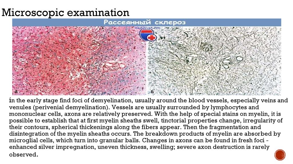
Microscopic examinationin the early stage find foci of demyelination, usually around the blood vessels, especially veins and
venules (perivenial demyelination). Vessels are usually surrounded by lymphocytes and
mononuclear cells, axons are relatively preserved. With the help of special stains on myelin, it is
possible to establish that at first myelin sheaths swell, tinctorial properties change, irregularity of
their contours, spherical thickenings along the fibers appear. Then the fragmentation and
disintegration of the myelin sheaths occurs. The breakdown products of myelin are absorbed by
microglial cells, which turn into granular balls. Changes in axons can be found in fresh foci enhanced silver impregnation, uneven thickness, swelling; severe axon destruction is rarely
observed.
18.
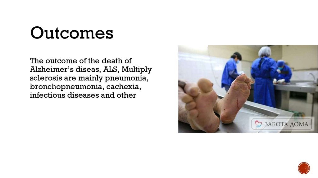
OutcomesThe outcome of the death of
Alzheimer’s diseas, ALS, Multiply
sclerosis are mainly pneumonia,
bronchopneumonia, cachexia,
infectious diseases and other
19.
ConclusionDementia is a loss of cognitive functioning — thinking, remembering,
and reasoning — and behavioral abilities to the extent that it interferes
with everyday life and human activities. These functions include
memory, language skills, visual perception, problem solving, selfmanagement, and the ability to focus and pay attention. Some people
with dementia cannot control their emotions, and their personalities may
change. The severity of dementia varies from the easiest stage, when it
only begins to affect the functioning of a person, to the most severe
stage, when a person must be completely dependent on others for the
main types of life activity. And we need to better understand why this is
happening to fight it.
20.
Bibliography1. Патологическая анатомия : учебник / А. И. Струков, В. В. Серов. - 5-е
изд., 356 б
2. Патологиялық анатомия : [Текст] : оқулық / Ж. Б. Ахметов. 143 б
3. https://www.eurolab.ua/encyclopedia/morbid-anatomy/32639/
4. https://www.alzheimers.org.uk/about-dementia/typesdementia/alzheimers-disease
5. http://www.medical-enc.ru/m/16/rasseyannyj-sklerozpatologicheskaya-anatomiya.shtml
6. https://www.nia.nih.gov/health/what-dementia
7. https://forens.ru/forum/204
8. https://www.guttmann.com/ru/treatment/bokovoy-amiotroficheskiyskleroz-bas