


 - - -fibrous (present")


")
")


 and myelin incisions are distinguished")


 Медицина
Похожие презентации:


Nerve tissue
1.
Nerve tissueAssociate Professor Kharchenko S.V.
Department of Histology and Embryology
Medical Academy named after S.I. Georgievsky
22/06/20
1
2. Nerve tissue is a system of nerve cells and neuroglia that provide specific functions of perception of stimulation, excitation,
22/06/202
3. Structural components of nerve tissue
Nerve cells - neurons - the maincomponent of nerve tissue.
Neuroglia - provides the existence
and functioning of nerve cells,
implements supporting, trophic,
barier, secretory and protective
function.
22/06/20
3
4.
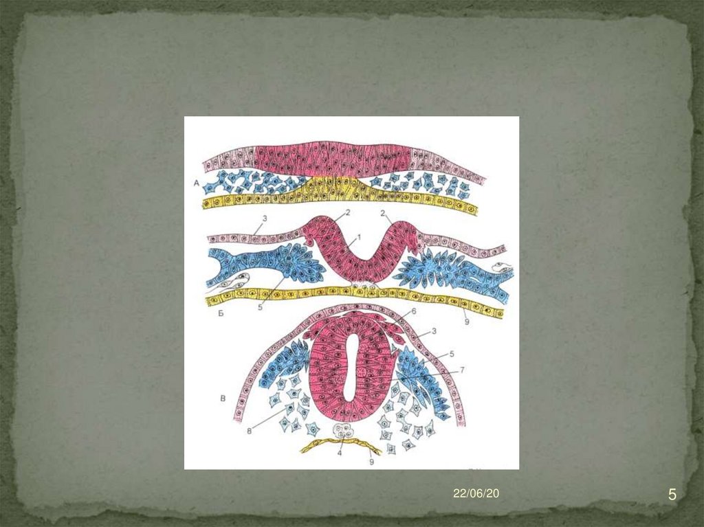
Histogenesis of nerve tissueNT develops from ectoderm
For 18 day the neural plate (thickening of the
dorsal ectoderm) is differentiated, then the nerve
folds (the thickened edges of the neural plate,
which rise and close) form the neural tube
Part of the cells of the N. plate forms the N. crest
(ganglionic plate).
CNS neurons and macroglia of the central
nervous system are formed from the N. tube
From the neural crest neurons of sensitive and
autonomic ganglia, brain membrane cells,
neurolemmocytes, ganglia satellite, medulla of
adrenal medulla, melanocytes of the skin are
formed.
22/06/20
4
5.
22/06/205
6. In the cranial part of the embryo, thickening of the ectoderm is formed - placodes from which the ganglia of V, VII, IX, X
22/06/206
7.
The ventricular zone consists of ependymocyteprogenitor cells
The intermediate zone consists of neuroblasts
and glioblasts. Neuroblasts differentiate into
neurons, glioblasts → into astrocytes and
oligodendrocytes. From the cells of this zone, gray
matter s / m and part of the gray matter g / m are
formed.
22/06/20
7
8.
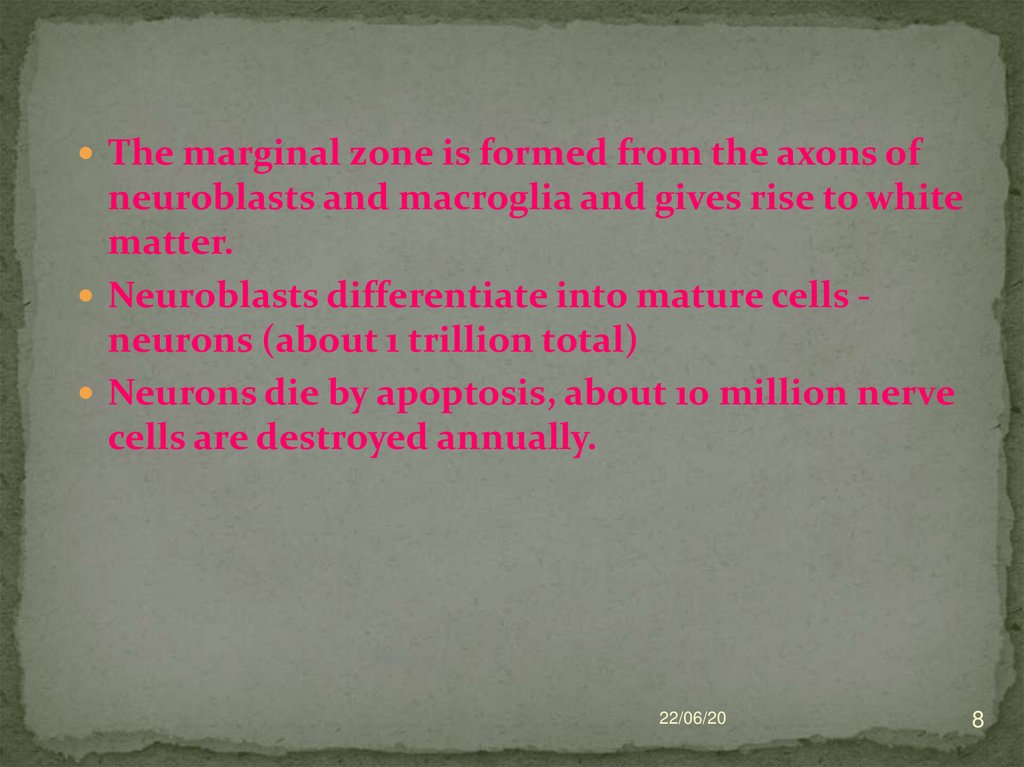
The marginal zone is formed from the axons ofneuroblasts and macroglia and gives rise to white
matter.
Neuroblasts differentiate into mature cells neurons (about 1 trillion total)
Neurons die by apoptosis, about 10 million nerve
cells are destroyed annually.
22/06/20
8
9. Characteristic of neurons
These are specialized cells,responsible for the reception, conduction,
processing of the impulse and its transmission
to other neurons, muscle or secretory cells.
With the help of their processes, neurons form
contacts with other neurons (reflex arcs).
22/06/20
9
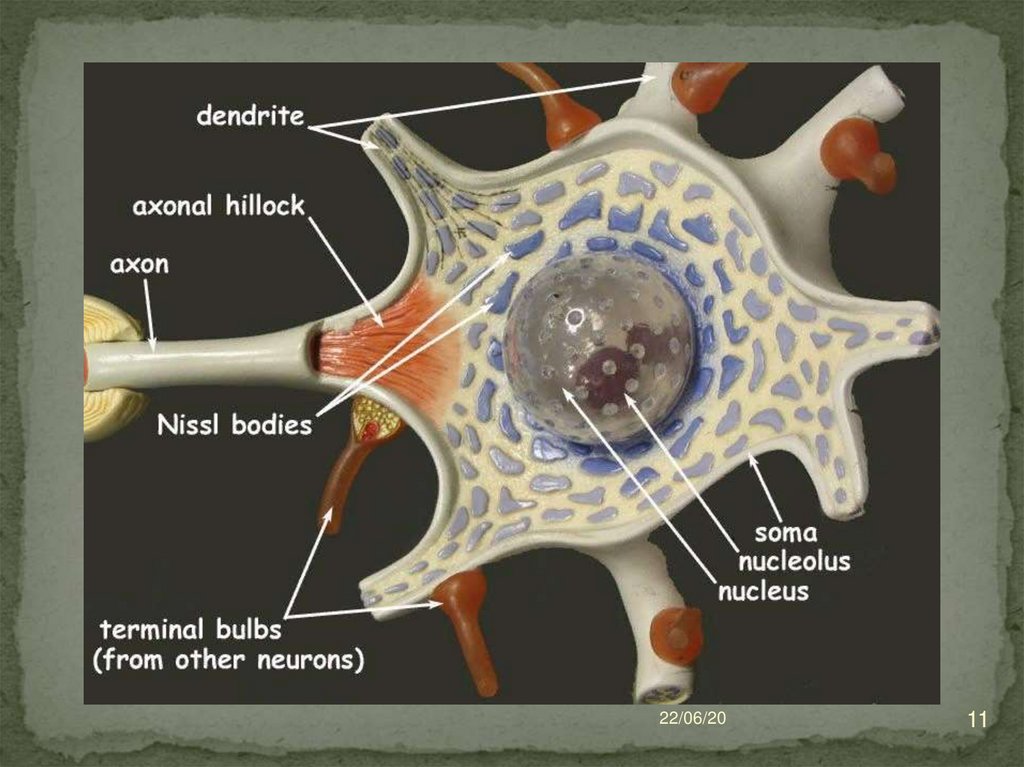
10. Structure of neuron
Neurons are composed of thebody (pericarion) and processes
(1 axon and dendrites)
Axon (neuritis) is the central
process along which the
impulse is transmitted from the
body of the neuron.
Dendrites - transmit nerve
impulses to the body of a
neuron
The sizes of neurons range from
4-6 microns (certebellar cortexgranuk cells) to 130-150 microns
(Betz pyramidal cells in the
cortex of brain).
22/06/20
10
11.
22/06/2011
12. Нейроны
22/06/2012
13.
22/06/2013
14.
22/06/2014
15.
Plasmolemma has the ability to generate andconduct impulse
The nucleus is usually one
Among other organelles are well developed: CG,
mitochondria, lysosomes.
With age lipofuscin - an aging pigment
accumulates in neurons. These are residual
bodies.
22/06/20
15
16.
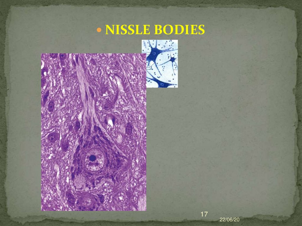
Chromatophilic substance (tigroid orNissl's body) - detected in the
cytoplasm in the form of basophilic
clumps or grains of various sizes.
Formed by rEPR cisternas.
Basophilia of Nissle body is associated
with a high content of RNA.
THIS IS THE MOST IMPORTANT
NEURON STRUCTURE!
22/06/20
16
17.
NISSLE BODIES17
22/06/20
18.
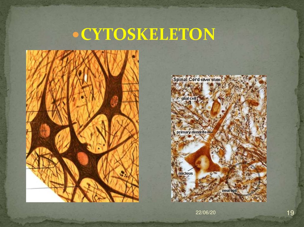
The cytoskeleton is represented by neurofibrils(12 nm) and neurotubules (24-27 nm). In the body
of the neuron they are located in the form of a
network, and in the processes - in parallel.
THIS IS THE SECOND IMPORTANT NEURON
STRUCTURE
Neurotubules are involved in maintaining cell
shape and axonal transport.
Axonal transport - the movement of substances
from the body to the processes - and vice versa
(retrograde - to the body of a neuron, anterograde
- from the body of a neuron - to the processes;
fast - 400-2000 mm per day, slow -1-2 mm per
day).
22/06/20
18
19.
CYTOSKELETON22/06/20
19
20. Morphological classification of neurons
Unipolar (with one process - axon) - a personhas only embryogenesis
Bipolar (with two processes - one axon and
one dendrite) – it is retinal photoreceptors
Multipolar (with many processes)
Pseudo-unipolar (the common process
departs from the body of such neurons, then
subdivided into axon and dendrite) –present
in the dorsal root ganglion
22/06/20
20
21.
2122/06/20
22. Functional classification of neurons
Sensitive (afferent, receptor) - located in the spinalnode. They generate n. impulse and spend it in the
dorsal horn of the spinal cord).
Motor (motor, efferent) -they carry out n. impulse
from the ventral horns of the spinal cord to the
working organ.
Interneurons (associative) - located in the horn
horns. Spend n impulse inside spinal cord.
22
22/06/20
23. SECRETORY NEURONS
In the cytoplasm and axons are large granulesof neurosecrete, which are excreted into the
blood or cerebrospinal fluid.
Similar neurons are localized in the
neurosecretory nuclei of the hypothalamic
region.
22/06/20
23
24. GLIAL CELLS
CNSPNS
22/06/20
24
25. NEUROGLIA
CNS glial cells are divided into:1) macroglia (originates from the glioblast of
the neural tube)
ependymocytes,
astrocytes
(fibrous and protoplasmic)
oligodendrocytes
2) microglia (from PHSC-monocytes of
blood)
22/06/20
25
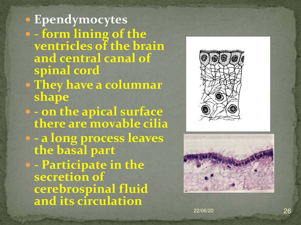
26.
Ependymocytes- form lining of the
ventricles of the brain
and central canal of
spinal cord
They have a columnar
shape
- on the apical surface
there are movable cilia
- a long process leaves
the basal part
- Participate in the
secretion of
cerebrospinal fluid
and its circulation
22/06/20
26
27. Astrocytes: - protoplasmic (present in the gray central nervous system, have short branching processes) - - -fibrous (present
22/06/2027
28. отростки А тя-нутся к капилля-рам, телам и ден-дритам нейронов, к мягкой мозговой оболочке. Эти клетки входят в состав
22/06/2028
29. Oligodendrocytes
Have few processusPresent in gray matter near perikarions of neurons
In white, they are part of the myelin and non-myelin
nerve fibers.
22/06/20
29
30. Microglia (glial macrophages)
Come from blood monocyte!Function - protecting brain tissue from
infection
Microglia cells are motile, capable of
phagocytosis.
Types:
Resting - in adults, low activity
Amoeboid - in newborns with high
phagocytic activity
Reactive - after damage
22/06/20
30
31. Glia of the peripheral nervous system (originates from the neural crest)
Neurolemmocytes (Schwann cells) form thesheaths of the processes of nerve cells in the
nerve fibers of the peripheral nervous
system
Ganglial glyocytes (surround the bodies of
neurons in the nerve nodes)
22/06/20
31
32. NERVE FIBERS
- are processes of nerve cells which are coveredwith sheath.
- Process is almost always AXON (axial cylinder)
In the central nervous system fiber sheaths are
formed using oligodendrocytes,
In the peripheral - with the help of
neurolemmocytes.
Distinguish:
myelinated nerve fibers
unmyelinated nerve fibers
------------------------------------------- 22/06/20
32
33. Non-myelinated nerve fibers
Are part of vegetative NS.The axial cylinders of several
neurons take part in the structure
of the fiber. They are located on
the periphery of the fiber.
Mesaxones are short. There are no
gaps between adjacent
neurolemmocytes.
The resulting fibers are called
cable-type fibers.
33
22/06/20
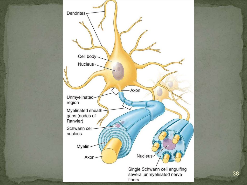
34. MYELINATED NERVE FIBER
They are found both in the central nervous systemand in peripheral NS.
They consist of one axial cylinder located in the
center of the fiber.
Covered with a complex membrane consisting of
Schwann cells.
Two layers are distinguished in the shell:
- internal - myelin
- external - consists of the cytoplasm and
the nucleus of a neurolemmocyte.
22/06/20
34
35.
22/06/2035
36. In myelin fiber of nodesRanvier (after 1-2 mm) and myelin incisions are distinguished
during myelinisation theaxon is immersed in to the
cytoplasm of the
neurolemmocyte.
In this case, mesaxone is
formed (duplication of the
Schwann cell cytolemma).
Mesaxon is layered on the
36
22/06/20
37. MYELINISATION
The speed of impulse transmission alongmyelin fibers (5-120 m / s), along
bezmyelinovyh - (1-2 m / s).
22/06/20
37
38.
22/06/2038
39. NERVE ENDINGD are terminal parts of nerve fibers
They are divided into 3 groups accordingto functions:
- motor (effector)
- sensitive (receptor)
- synapses
22/06/20
39
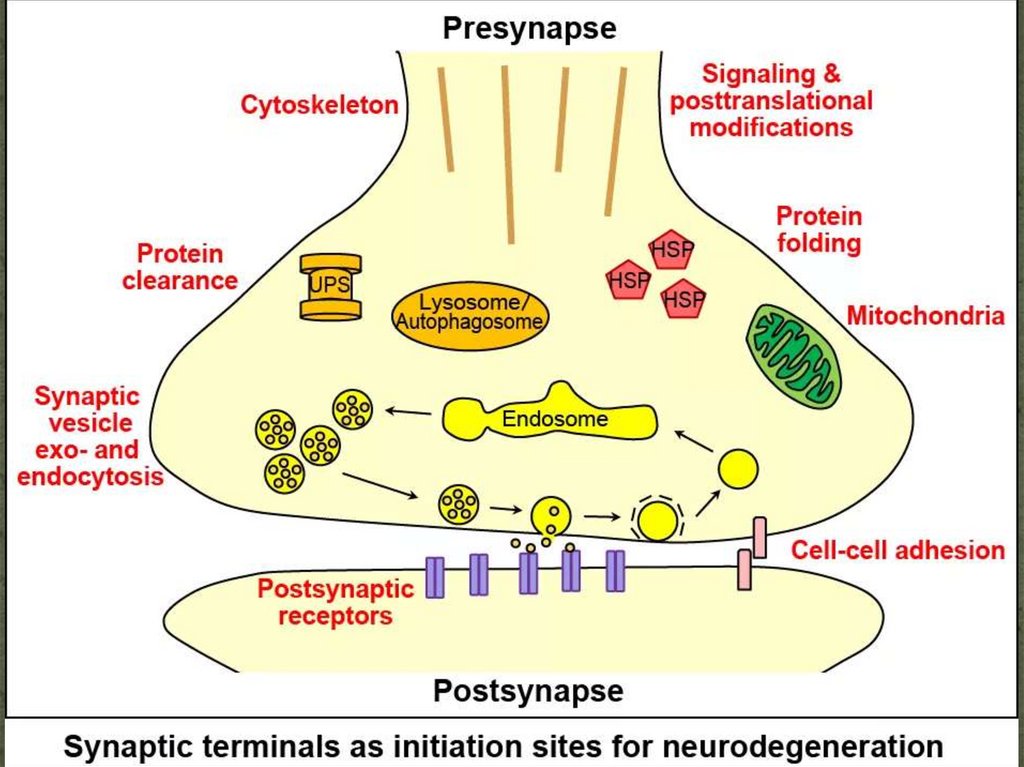
40. SYNAPSE
Structurally:- axodendritic
- axosomatic
- axoaxial
muscle or motor plaques
By transmission method:
- chemical (due to mediators or
neurotransmiters)
- electrical (contribute to the
synchronization of activity).
22/06/20
40
41. Chemical
Transmit impulse using mediatorsThe axon terminal is the presynaptic
part . It contains synaptic vesicles,
mitochondria, neurofilaments,
calcium ions.
The postsynaptic part is represented
by the membrane of the second
neuron with which it is in contact.
Contains receptors, a recognizable
mediator.
Synaptic cleft = 20-30 nm
22/06/20
41
42.
22/06/2042
43.
Low molecular weight mediators:- Acetylcholin, norepinephrine,
serotonin, histamine, glutamate, glycine,
GABA, dopamine,
Neuropeptides:
- endorphins, enkephalins, dinorins,
substance R.
Brain synapse mediators:
dopamine, glycine, GABA
22/06/20
43
44.
The processes in the synapse are developed asfollows:
Depolarization wave reaches presynaptic
membrane
Ca channels open
Ca causes neurotransmitter exocytosis
Diffusion of the neurotransmitter through the
synaptic cleft
Ion channels open in the postsynaptic
membrane
The postsynaptic potential is created..
22/06/20
44
45. Effector nerve endings
They are terminal apparatuses of axons of motorcells of somatic or vegetative
With their participation the impulse is
transmitted to the tissues of the working organs.
The neuromuscular ending consists of the
terminal branching of the axial cylinder of the
nerve fiber and the muscle fiber site.
22/06/20
45
46. Neuromuscular nerve ending
Myelin ed nerve fiber losesthe myelin layer and is
submerged in muscle fiber.
Plasmolemma and sarcolema
are separated by a synaptic
cleft of about 50 nm.
In the postsynaptic part folds
are formed
Skeletal fiber loses striation
in the contact area
22/06/20
46
47.
In the smooth muscle tissue nerve endingsare clearly distinct thickenings occurring
among smooth myocytes.
Secretory nerve endings are thickening of
the terminals along the nerve fiber.
22/06/20
47
48. RECEPTORS
1) By localization:extero- and interoreceptors
2) By the specificity of perception:
chemoreceptors, mechanoreceptors,
baroreceptors, thermoreceptors, etc.
3) According to the features of the structure:
a) - free nerve endings (consist of branching of
the axial cylinder)
b) - non-free nerve endings (conteins axon and
sheath)
- encapsulated (covered with a capsule)
- unencapsulated (not having capsules).
22/06/20
48
49. FREE N.E.
PRESENT in the epitheliumMyelin fibers approach the
epithelial layer, lose
myelin, axial cylinders
enter the epithelium and
break up between cells into
terminal branches.
22/06/20
49
50. A variety of receptors is found in connective tissue.
Lamellar bodies of Fater-Pacini (0.5-2 mm) are found in the skin and int.
organs.
In the center is ext. bulbs
The myelin fiber loses myelin,
penetrates the bulb and branches.
Outside, the body is surrounded by a
layered capsule consisting of
fibroblasts and spiral fibers.
Taurus FP perceives pressure and
vibration.
22/06/20
50
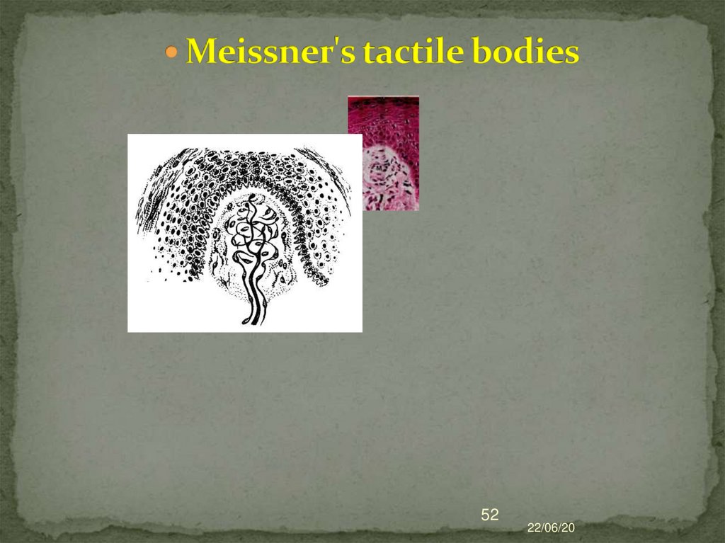
51. Meissner's tactile bodies
They are located at the apex of the connectivetissue papillae of the skin.
They consist of modified neurolemocytes - tactile
cells.
Outside surrounded by a thin capsule
The myelin fiber enters from below loses the
myelin layer and branches. Any displacement of
the epidermis is transmitted to the tactile body.
22/06/20
51
52.
5222/06/20
53. Благодарю за внимание !
22/06/2053