 Биология
 Английский язык
Похожие презентации:


Anatomical structure of the esophagus
1.
Балхашский Медицинский КолледжEnglish
Тема:
Выполнила:
Медведева София –
студентка 2 курса группы
ЛД209Б
Преподаватель :
Омаркулова Сапура
2.
Балхашский Медицинский КолледжEnglish
Тема:
3.
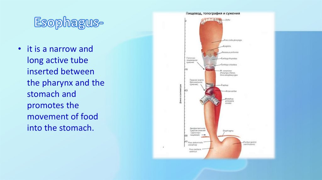
• it is a narrow andlong active tube
inserted between
the pharynx and the
stomach and
promotes the
movement of food
into the stomach.
4.
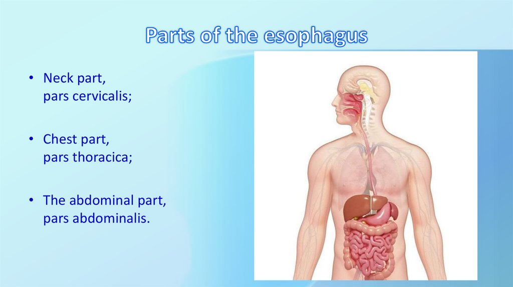
• Neck part,pars cervicalis;
• Chest part,
pars thoracica;
• The abdominal part,
pars abdominalis.
5.
• Sagittal curves correspond to the • Frontal bends depend on thecurves of the spine
relative position of the esophagus
with the organs of the neck and
chest.
6.
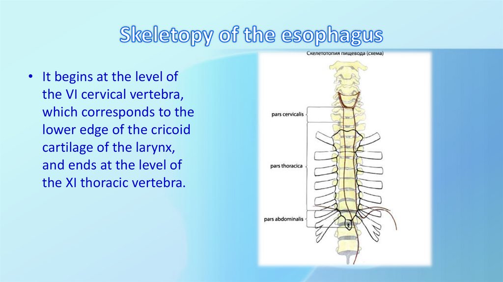
• It begins at the level ofthe VI cervical vertebra,
which corresponds to the
lower edge of the cricoid
cartilage of the larynx,
and ends at the level of
the XI thoracic vertebra.
7.
• The cervical part, pars cervicalis, is located between the VI cervicalvertebrae behind the cricoid cartilage and the third thoracic vertebra, its
length is 5-6 cm. The front of the esophagus is adjacent to the trachea.
In the intervals between the esophagus and trachea, recurrent laryngeal
nerves pass, which should be taken into account during surgical
interventions in this area. The side surfaces of the esophagus touch the
thyroid gland. In the lower part of the left side of the esophagus is
adjacent to the thoracic duct. Behind the esophagus is the
extraesophageal space, which is a continuation of the pharyngeal space.
It connects to the posterior mediastinum.
8.
• The thoracic part, pars thoracica, is contained in the posteriormediastinum in the space from the II thoracic vertebra to the esophageal
opening of the diaphragm. Its length is 15-18 cm. Anterior to the thoracic
part of the adjacent left recurrent laryngeal nerve, branches of the left
vagus nerve, esophageal nerve plexus, left common carotid artery,
bifurcation of the artery, left pulmonary bronchus. To the left of the
esophagus are: the left subclavian artery, the left vagus nerve, the
thoracic duct, the aortic arch, and the thoracic aorta. The right contains
branches of the vagus nerve and an unpaired vein. The long neck muscles,
cervical vertebrae, thoracic duct, semiseparous vein, and thoracic aorta
are adjacent to the thoracic esophagus from behind.
9.
• The abdominal part, pars abdominalis, occupies the space from theesophagusthe opening of the diaphragm to the entrance to the
stomach, its length is from 2 to 4 cm. In this area, the esophagus is
covered with peritoneum in front and on the sides. The left lobe of the
liver is adjacent to it in front, and the upper pole of the spleen is on the
left. First, the esophagus passes on the neck to the left of the midline to
the VI thoracic vertebra. Starting from the level of the V thoracic
vertebra, the esophagus is directed along the midline, then bends to the
right to the VIII thoracic vertebra, and then again to the left.