


 Медицина
Похожие презентации:


Appendectomy By Mohan Krishna Redlapalle
1.
AppendectomyBy Mohan Krishna Redlapalle
2.
Outline• Let us revise vermiform Appendix
• Definition of Appendectomy
• Indications
• Types
• Open Appendectomy
• Laparoscopic (Key hole) Appendectomy
• Complications
• References
3.
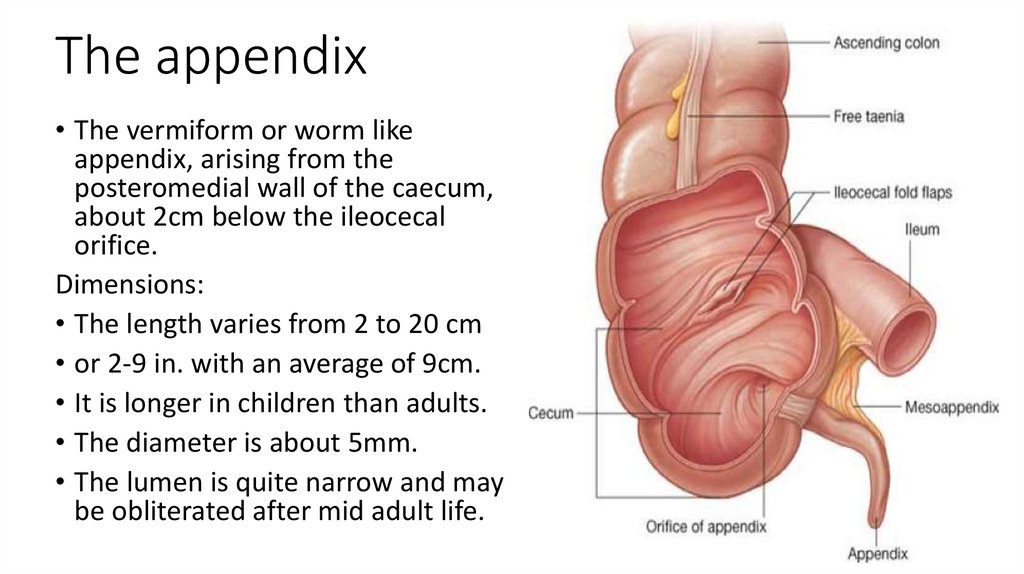
The appendix• The vermiform or worm like
appendix, arising from the
posteromedial wall of the caecum,
about 2cm below the ileocecal
orifice.
Dimensions:
• The length varies from 2 to 20 cm
• or 2-9 in. with an average of 9cm.
• It is longer in children than adults.
• The diameter is about 5mm.
• The lumen is quite narrow and may
be obliterated after mid adult life.
4.
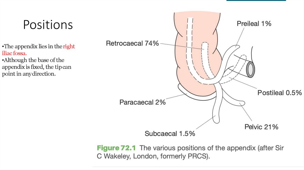
Positions•The appendix lies in the right
iliac fossa.
•Although the base of the
appendix is fixed, the tip can
point in any direction.
5.
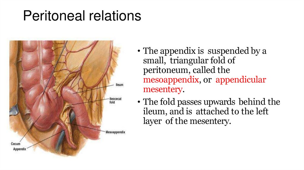
Peritoneal relations• The appendix is suspended by a
small, triangular fold of
peritoneum, called the
mesoappendix, or appendicular
mesentery.
• The fold passes upwards behind the
ileum, and is attached to the left
layer of the mesentery.
6.
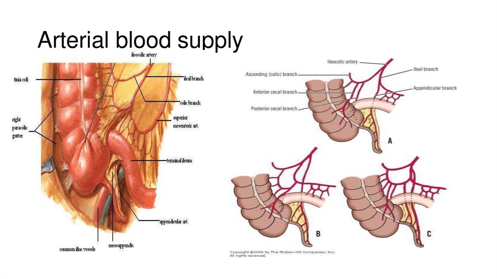
Arterial blood supply7.
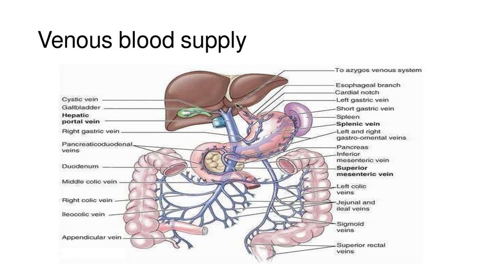
Venous blood supply8.
Nerve supplySympathetic nerves are derived from segments T9 to T10 through
the celiac plexus.
Parasympathetic nerves are derived from the Vegas N.
9.
Now,What is
Appendectomy?
10.
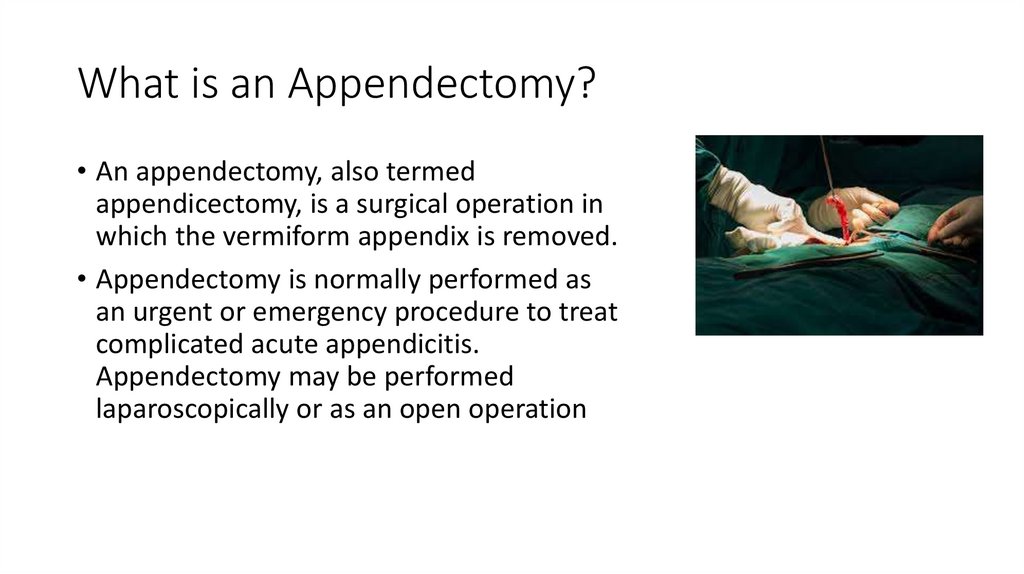
What is an Appendectomy?• An appendectomy, also termed
appendicectomy, is a surgical operation in
which the vermiform appendix is removed.
• Appendectomy is normally performed as
an urgent or emergency procedure to treat
complicated acute appendicitis.
Appendectomy may be performed
laparoscopically or as an open operation
11.
Types of Appendectomy•Open
•Laparoscopic
• General anesthesia.
• Laparoscopic: nasogastric tube & empty bladder.
• Palpation for mass in R.I.F.
12.
INDICATIONS• Acute appendicitis
• Recurrent appendicitis, Stump Appendicitis
• As Interval appendectomy after drainage of abscess or in
appendiceal mass
• Carcinoid tumor : at the tip <2cm
• Mucocele of the appendix
• Appendicular graft; ileal conduit
• On table colonic lavage
13.
Contraindications• Extensive adhesions
• Radiation or immunosuppressive therapy,
• severe portal hypertension
• Gross coagulopathies.
• Laparoscopic appendectomy is contraindicated in the first trimester
of pregnancy
• Concerns for Crohn’s disease or Meckel’s diverticulum should be of
priority.
14.
If an acutely inflamed appendix had been found and removed, the restof the abdomen does not need to be explored.
Local lavage
• However, if the appendix is not inflamed, the surgeon needs to
exclude other pathologic processes;
Terminal ileitis
Meckel’s diverticulum
Tubal or ovarian cause in female
Crohn’s disease
15.
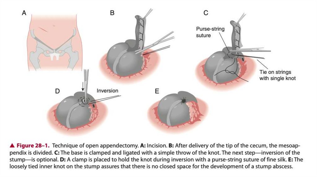
Open Appendectomy (Conventional)- Anoverview
Under general anesthesia, skin is incised. Two layers of superficial fascia are cut.
External oblique aponeurosis is opened in the line of incision.
Internal oblique and transverse muscles are split in the line of fibres.
Peritoneum is opened in the line of incision.
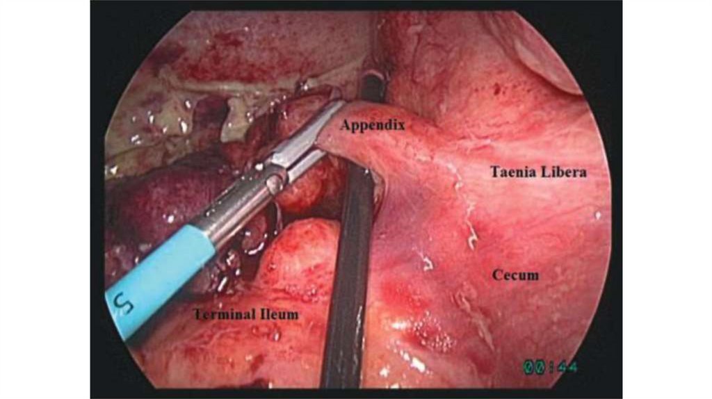
Caecum is identified by taeniae, and ileocaecal junction.
Omentum when adherent is separated.
Appendix is held with Babcock’s forceps.
Mesoappendix with appendicular artery is ligated. Using thread or silk, a purse—
string suture is placed around the base of the appendix.
• Base of the appendix is crushed with artery forceps and transfixed using vicryl
(absorbable). Appendix is cut distal to the suture ligature and removed.
• Stump is cleaned with antiseptics. Purse string suture is tightened so as to bury
the stump.
16.
Special circumstances:-Edema of the cecal wall.
-Base of the app. severely inflamed.
-Gangrenous app. base.
-Retrograde appendectomy.
-Drainage of the peritoneal cavity ??
17.
PRE-OP PREPARATION• INVESTIGATION
Urinalysis- exclude infection
Full blood count- leukocytosis
Ultrasound scan – non compressible diameter of > 6mm
Rehydrate patient with IV fluids; N/S
Pass urethral catheter
N-G tube
• IV antibiotics prophylaxis- broad Prophylactic antibiotics are
indicated preoperatively with a single-drug regimen, usually a
cephalosporin.
18.
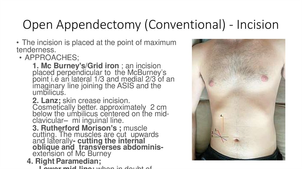
Open Appendectomy (Conventional) - Incision• The incision is placed at the point of maximum
tenderness.
• APPROACHES;
1. Mc Burney’s/Grid iron ; an incision
placed perpendicular to the McBurney’s
point i.e an lateral 1/3 and medial 2/3 of an
imaginary line joining the ASIS and the
umbilicus.
2. Lanz; skin crease incision.
Cosmetically better. approximately 2 cm
below the umbilicus centered on the midclavicular– mi inguinal line.
3. Rutherford Morison’s ; muscle
cutting. The muscles are cut upwards
and laterally- cutting the internal
oblique and transverses abdominisextension of Mc Burney
4. Right Paramedian;
19.
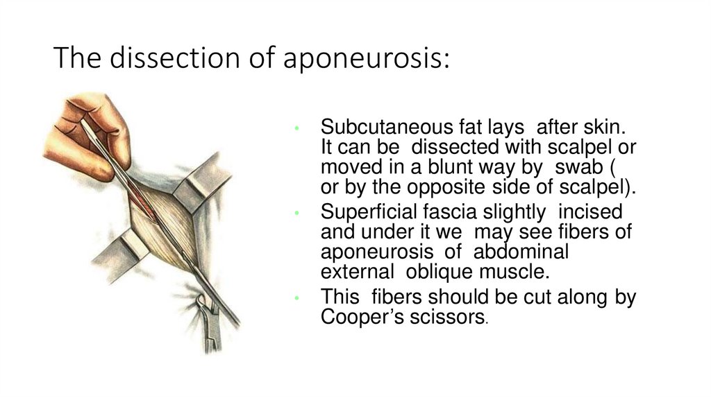
The dissection of aponeurosis:Subcutaneous fat lays after skin.
It can be dissected with scalpel or
moved in a blunt way by swab (
or by the opposite side of scalpel).
Superficial fascia slightly incised
and under it we may see fibers of
aponeurosis of abdominal
external oblique muscle.
This fibers should be cut along by
Cooper’s scissors.
20.
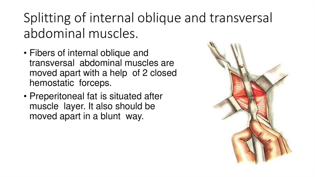
Splitting of internal oblique and transversalabdominal muscles.
• Fibers of internal oblique and
transversal abdominal muscles are
moved apart with a help of 2 closed
hemostatic forceps.
• Preperitoneal fat is situated after
muscle layer. It also should be
moved apart in a blunt way.
21.
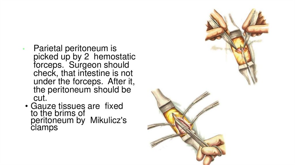
Parietal peritoneum is
picked up by 2 hemostatic
forceps. Surgeon should
check, that intestine is not
under the forceps. After it,
the peritoneum should be
cut.
• Gauze tissues are fixed
to the brims of
peritoneum by Mikulicz's
clamps
22.
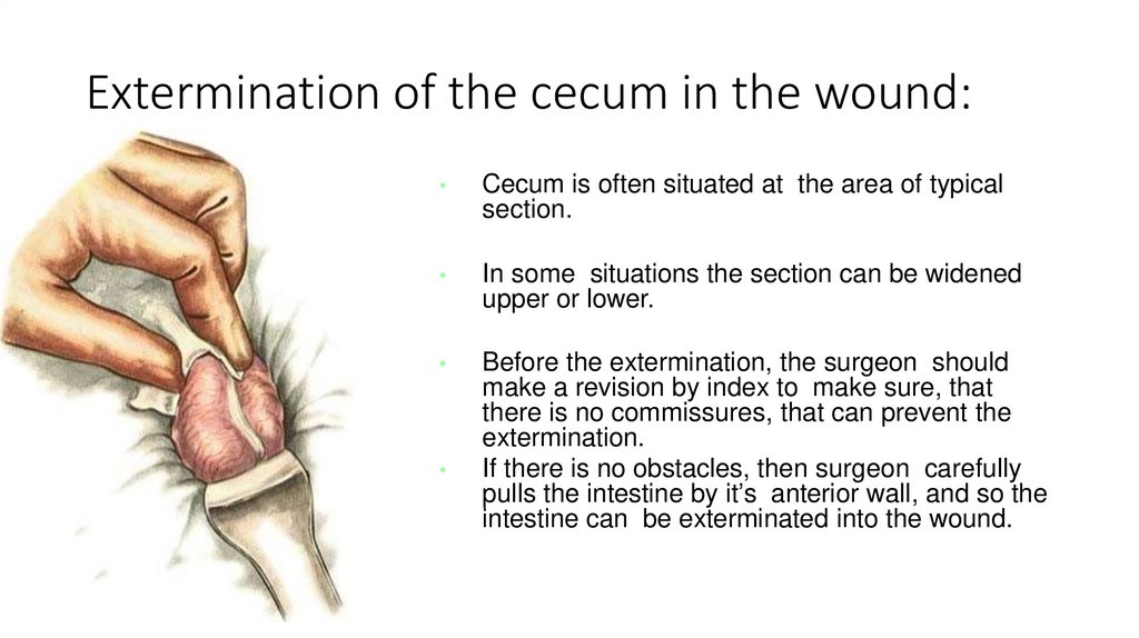
Extermination of the cecum in the wound:Cecum is often situated at the area of typical
section.
In some situations the section can be widened
upper or lower.
Before the extermination, the surgeon should
make a revision by index to make sure, that
there is no commissures, that can prevent the
extermination.
If there is no obstacles, then surgeon carefully
pulls the intestine by it’s anterior wall, and so the
intestine can be exterminated into the wound.
23.
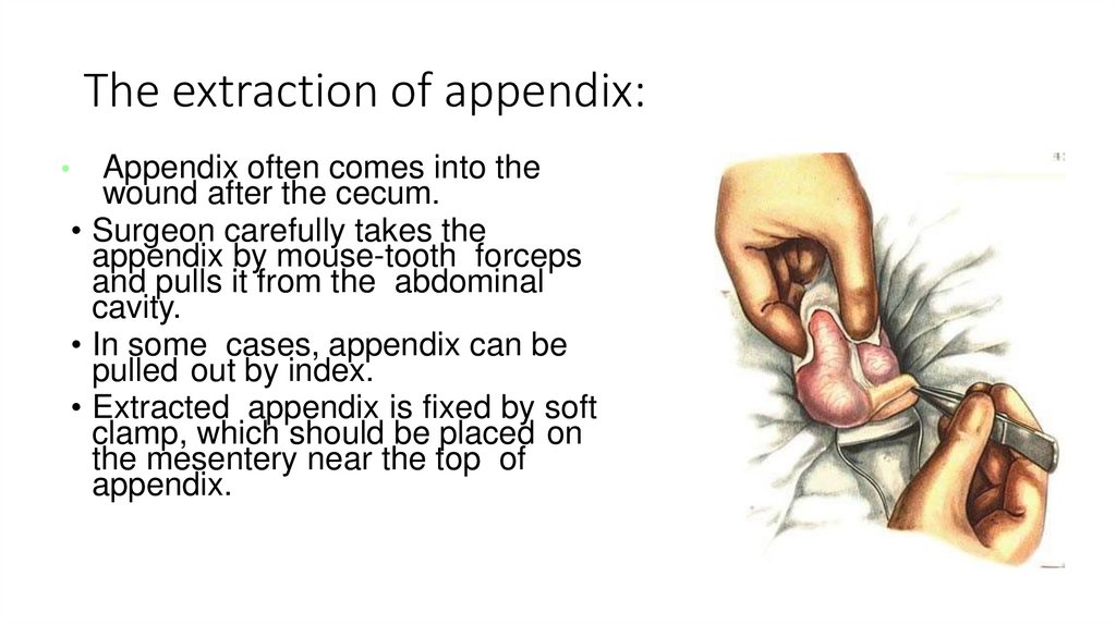
The extraction of appendix:Appendix often comes into the
wound after the cecum.
• Surgeon carefully takes the
appendix by mouse-tooth forceps
and pulls it from the abdominal
cavity.
• In some cases, appendix can be
pulled out by index.
• Extracted appendix is fixed by soft
clamp, which should be placed on
the mesentery near the top of
appendix.
24.
Methods of appendectomy• Antegrade (in the case of mobile cecum)
• Retrograde (in the case of immobile cecum)
25.
Anterograde Open Appendectomy26.
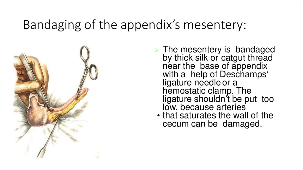
Bandaging of the appendix’s mesentery:The mesentery is bandaged
by thick silk or catgut thread
near the base of appendix
with a help of Deschamps’
ligature needle or a
hemostatic clamp. The
ligature shouldn’t be put too
low, because arteries
• that saturates the wall of the
cecum can be damaged.
27.
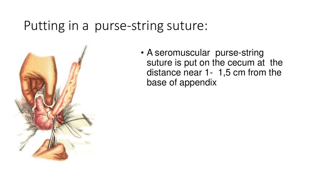
Putting in a purse-string suture:• A seromuscular purse-string
suture is put on the cecum at the
distance near 1- 1,5 cm from the
base of appendix
28.
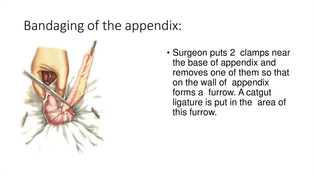
Bandaging of the appendix:• Surgeon puts 2 clamps near
the base of appendix and
removes one of them so that
on the wall of appendix
forms a furrow. A catgut
ligature is put in the area of
this furrow.
29.
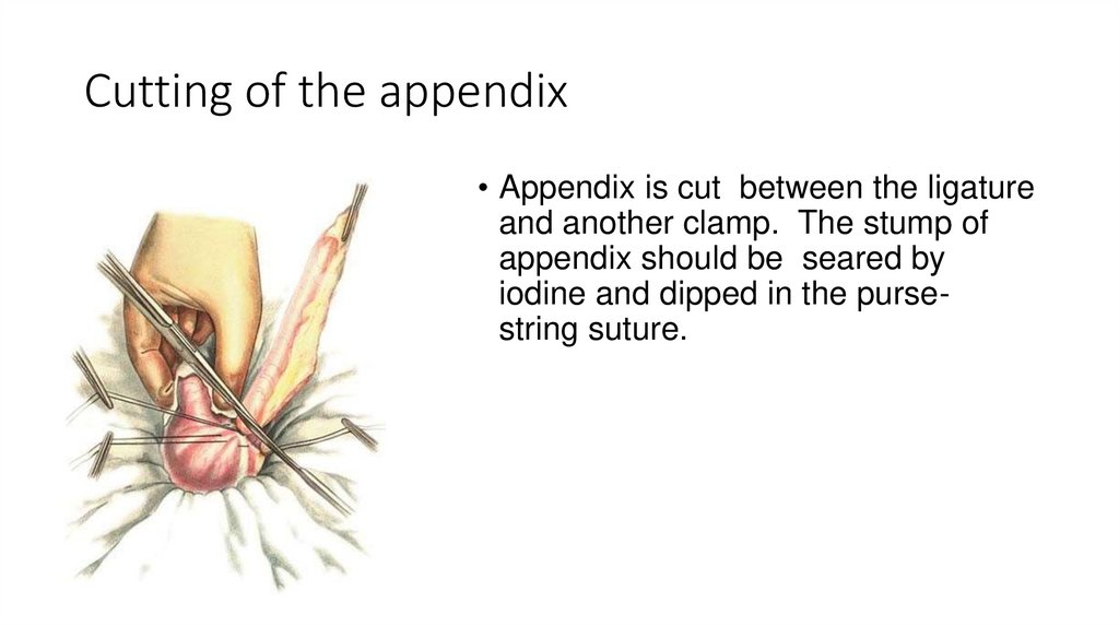
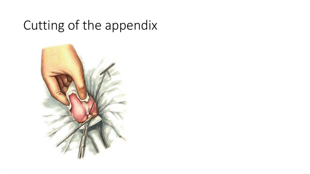
Cutting of the appendix• Appendix is cut between the ligature
and another clamp. The stump of
appendix should be seared by
iodine and dipped in the pursestring suture.
30.
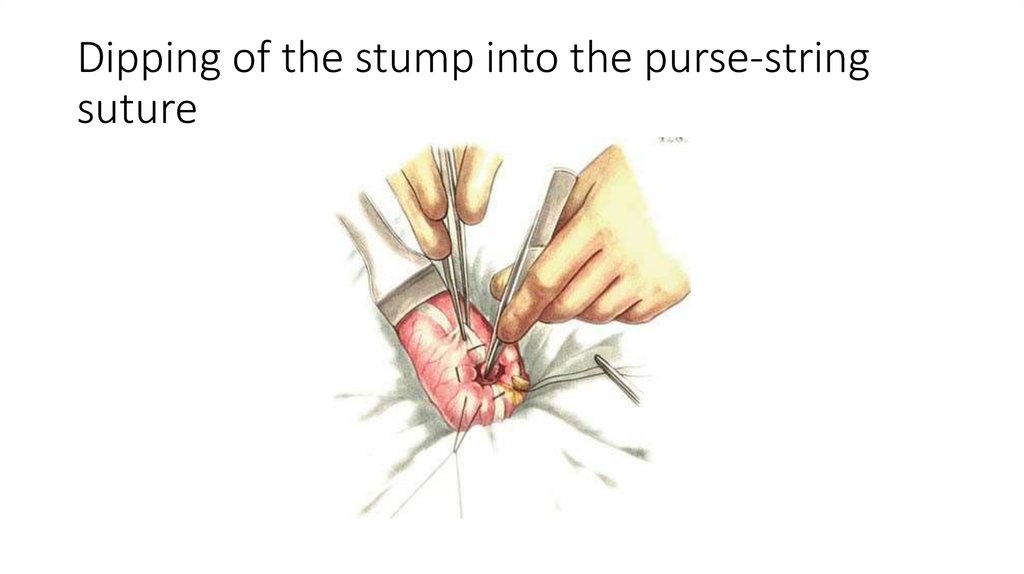
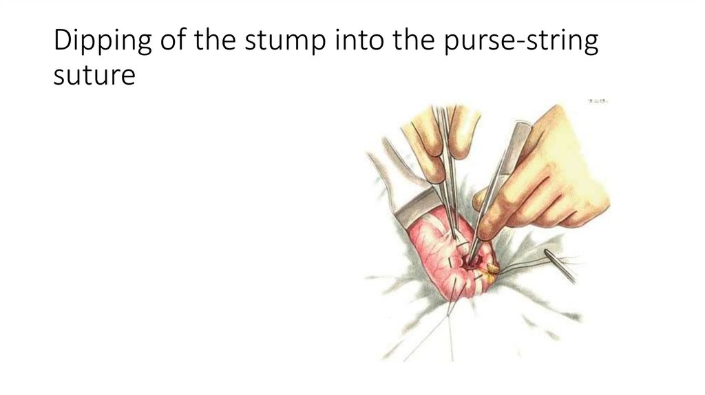
Dipping of the stump into the purse-stringsuture
31.
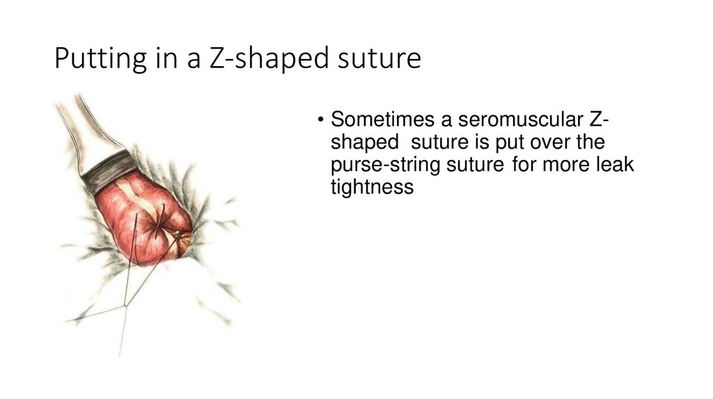
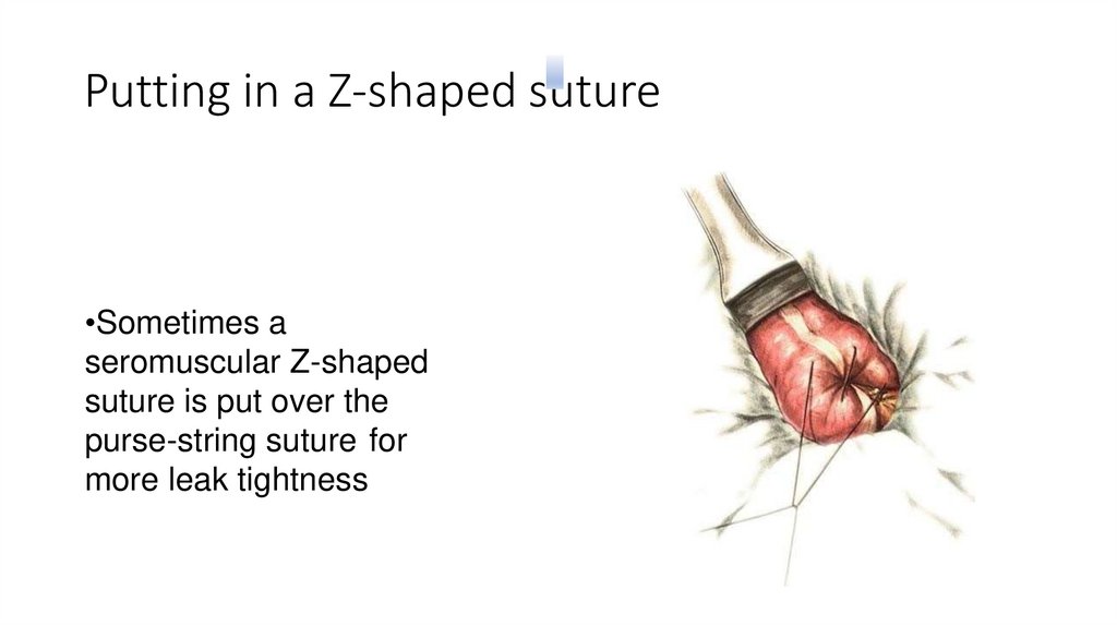
Putting in a Z-shaped suture• Sometimes a seromuscular Zshaped suture is put over the
purse-string suture for more leak
tightness
32.
33.
Retrograde Open Appendectomy34.
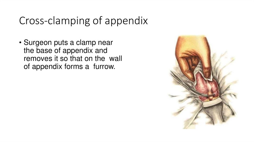
Cross-clamping of appendix• Surgeon puts a clamp near
the base of appendix and
removes it so that on the wall
of appendix forms a furrow.
35.
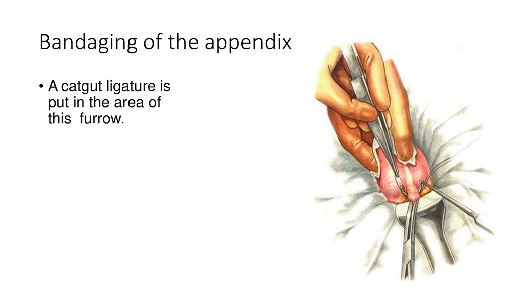
Bandaging of the appendix• A catgut ligature is
put in the area of
this furrow.
36.
Cutting of the appendix37.
Dipping of the stump into the purse-stringsuture
38.
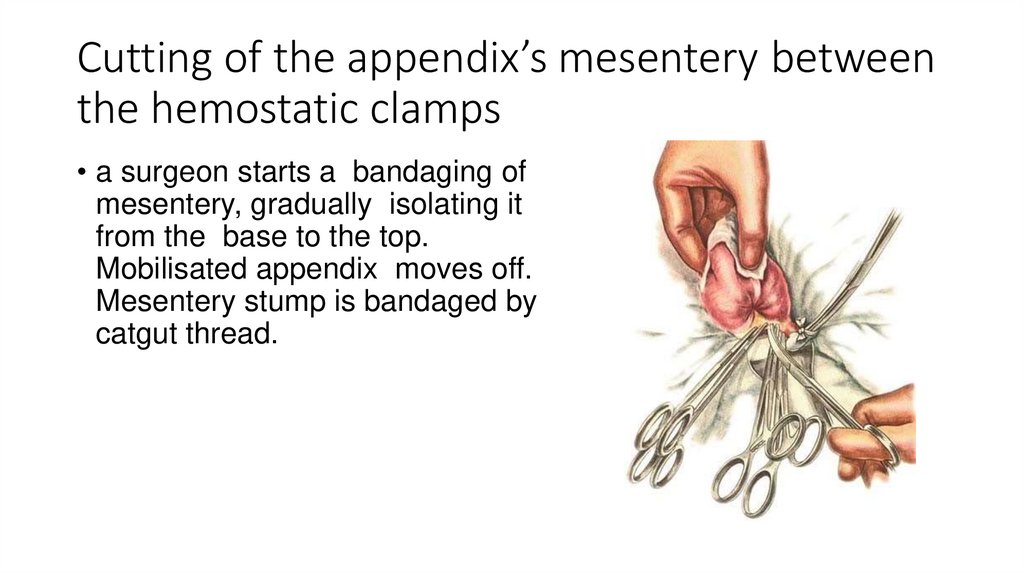
Cutting of the appendix’s mesentery betweenthe hemostatic clamps
• a surgeon starts a bandaging of
mesentery, gradually isolating it
from the base to the top.
Mobilisated appendix moves off.
Mesentery stump is bandaged by
catgut thread.
39.
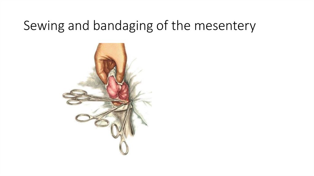
Sewing and bandaging of the mesentery40.
Putting in a Z-shaped suture•Sometimes a
seromuscular Z-shaped
suture is put over the
purse-string suture for
more leak tightness
41.
Appendectomy. Retroperitonealposition of appendix
• If there is no commissures in the abdominal cavity and the
appendix can not be found, then a surgeon should think about
the retroperitoneal position of appendix. In this case appendix is
situated behind the ascending colon and it’s top can reach the
lower pole of kidney
42.
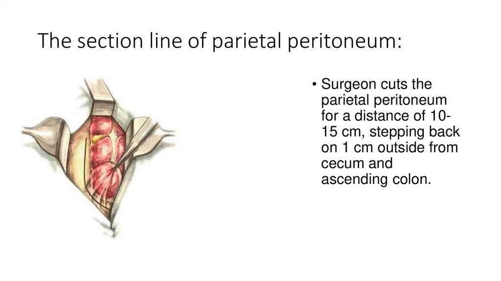
The section line of parietal peritoneum:• Surgeon cuts the
parietal peritoneum
for a distance of 1015 cm, stepping back
on 1 cm outside from
cecum and
ascending colon.
43.
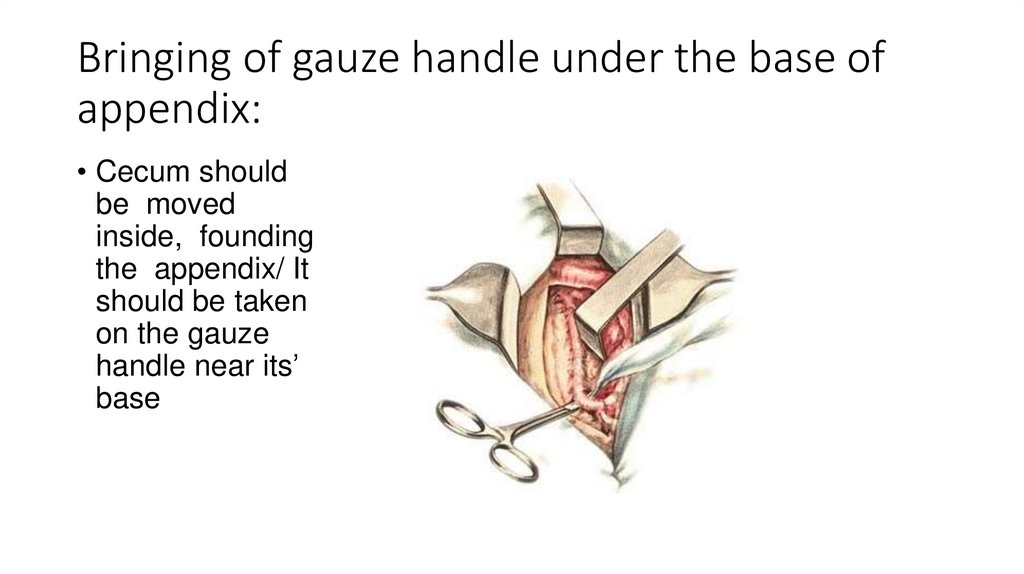
Bringing of gauze handle under the base ofappendix:
• Cecum should
be moved
inside, founding
the appendix/ It
should be taken
on the gauze
handle near its’
base
44.
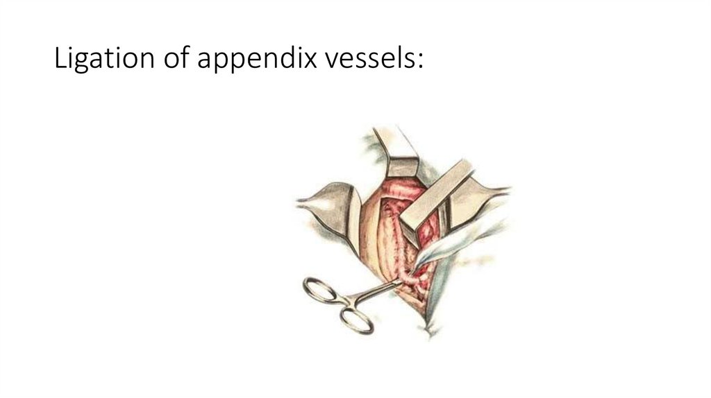
Ligation of appendix vessels:45.
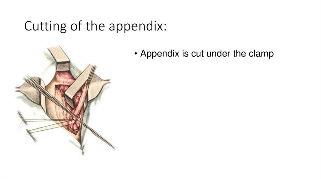
Cutting of the appendix:• Appendix is cut under the clamp
46.
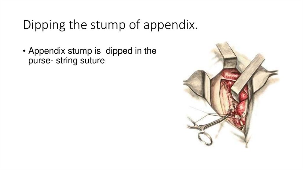
Dipping the stump of appendix.• Appendix stump is dipped in the
purse- string suture
47.
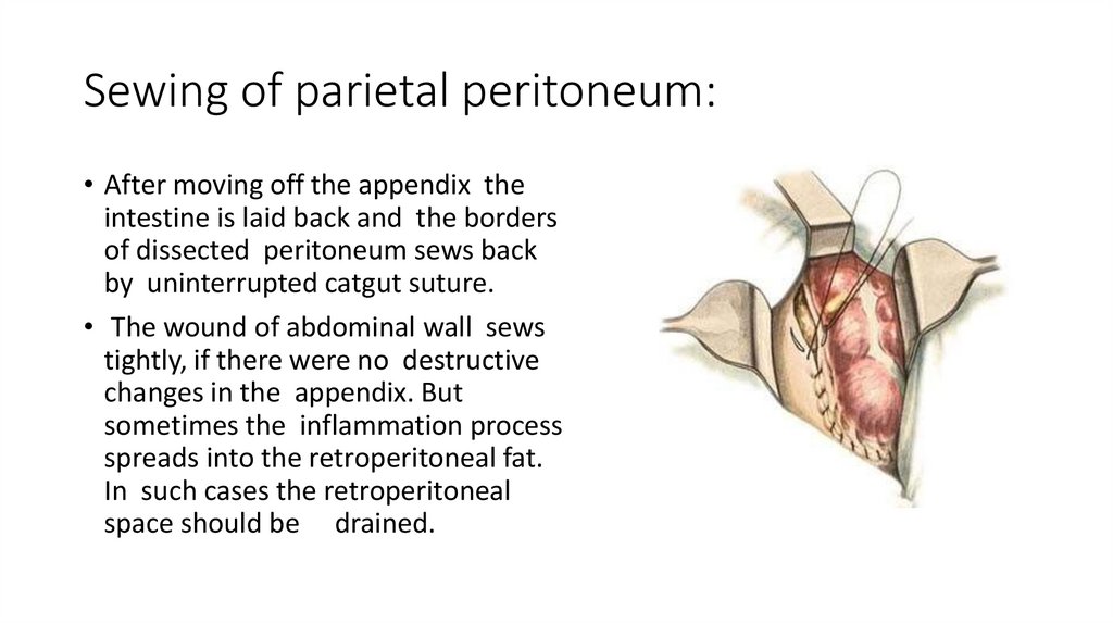
Sewing of parietal peritoneum:• After moving off the appendix the
intestine is laid back and the borders
of dissected peritoneum sews back
by uninterrupted catgut suture.
• The wound of abdominal wall sews
tightly, if there were no destructive
changes in the appendix. But
sometimes the inflammation process
spreads into the retroperitoneal fat.
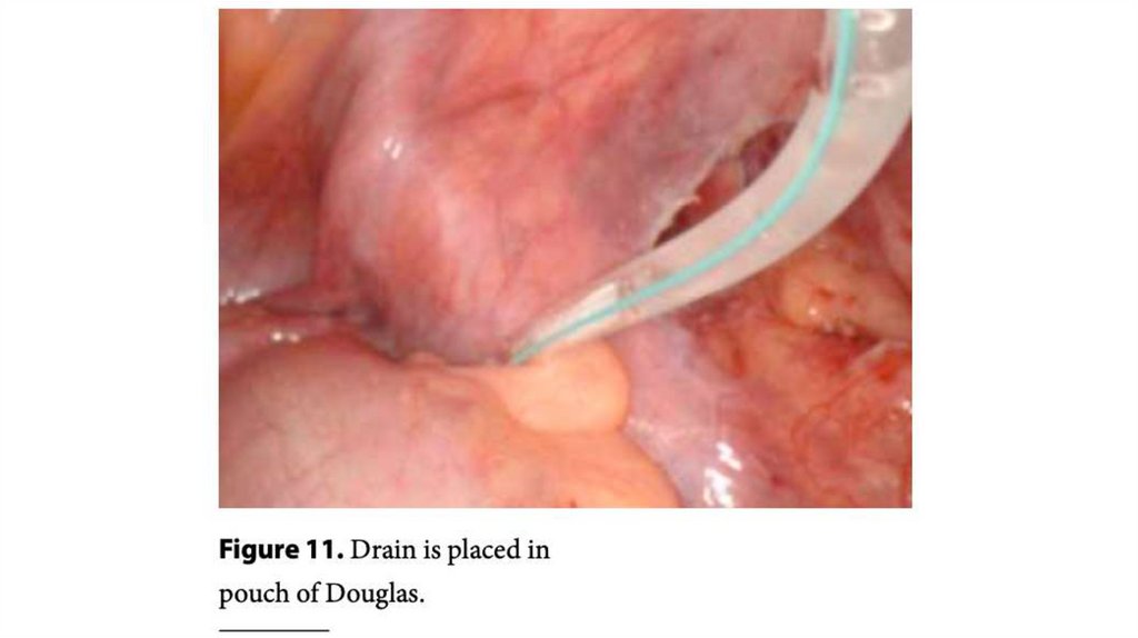
In such cases the retroperitoneal
space should be drained.
48.
CLOSURE• The peritoneum is grasped with curved Kelly clamps and
approximated with 3-0 continuous absorbable sutures.
• The transversus and internal oblique muscle layers are
irrigated and loosely approximated with 2-0 absorbable
sutures
• The external oblique fascia is repaired with continuous 0-0
absorbable sutures
• The subcutaneous tissue is irrigated, and the skin is
approximated with staples.
• If there had been excessive contamination of the wound, it
should be left open and the subcutaneous tissue packed with
saline-soaked gauze. A delayed primary closure can be
performed by day 3 to 4.
49.
The final stage:After moving out the appendix cecum moves back in the
abdominal cavity. Surgeon should check that there is no
bleeding from the mesentery and then the wound of the
abdominal wall sews tightly in layers. Peritoneum sews by
uninterrupted catgut suture, muscles, aponeurosis and
subcutaneous fat - by nodal catgut suture, skin – by nodal
silk suture.
In some cases abdominal cavity should be drained by thin
rubber or polyvinyl chloride tube.
Putting in a rubber tube is indicated in such cases, when there
was purulent exudate in the abdominal cavity of
phlegmonous changes of cecum.
50.
51.
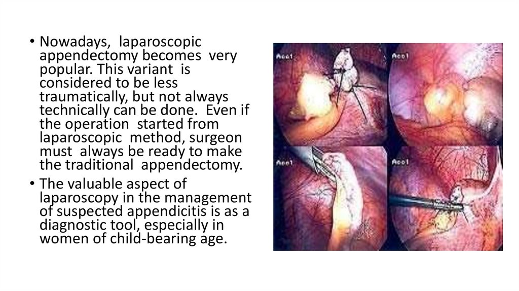
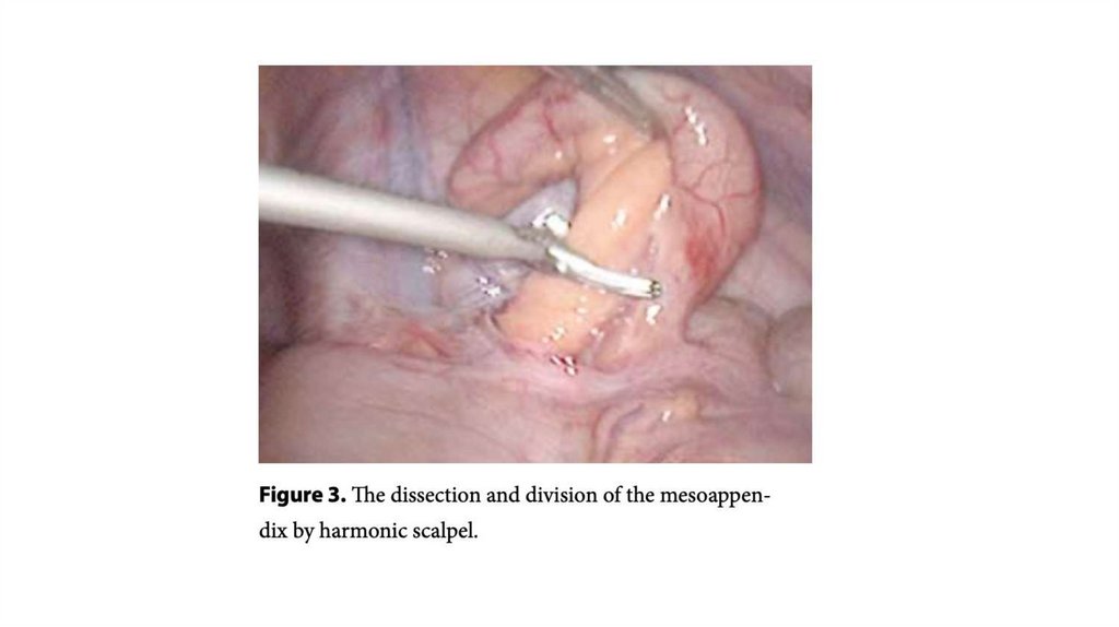
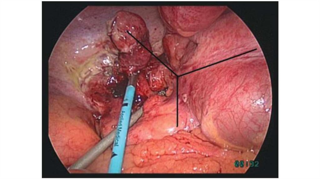
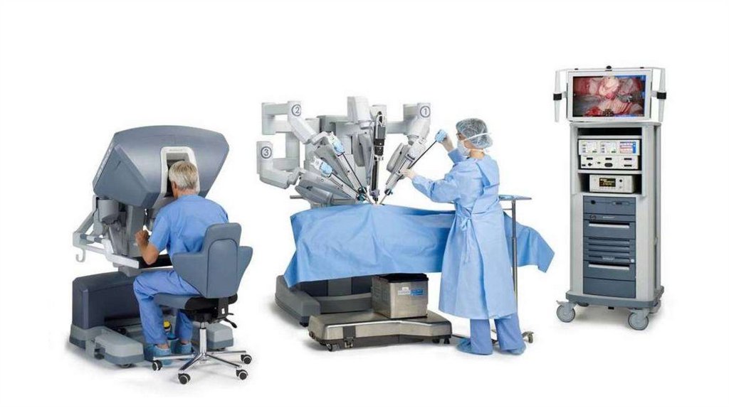
• Nowadays, laparoscopicappendectomy becomes very
popular. This variant is
considered to be less
traumatically, but not always
technically can be done. Even if
the operation started from
laparoscopic method, surgeon
must always be ready to make
the traditional appendectomy.
• The valuable aspect of
laparoscopy in the management
of suspected appendicitis is as a
diagnostic tool, especially in
women of child-bearing age.
52.
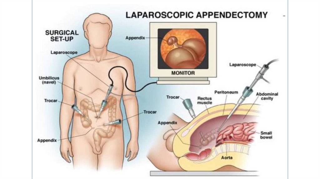
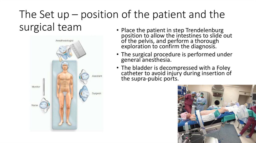
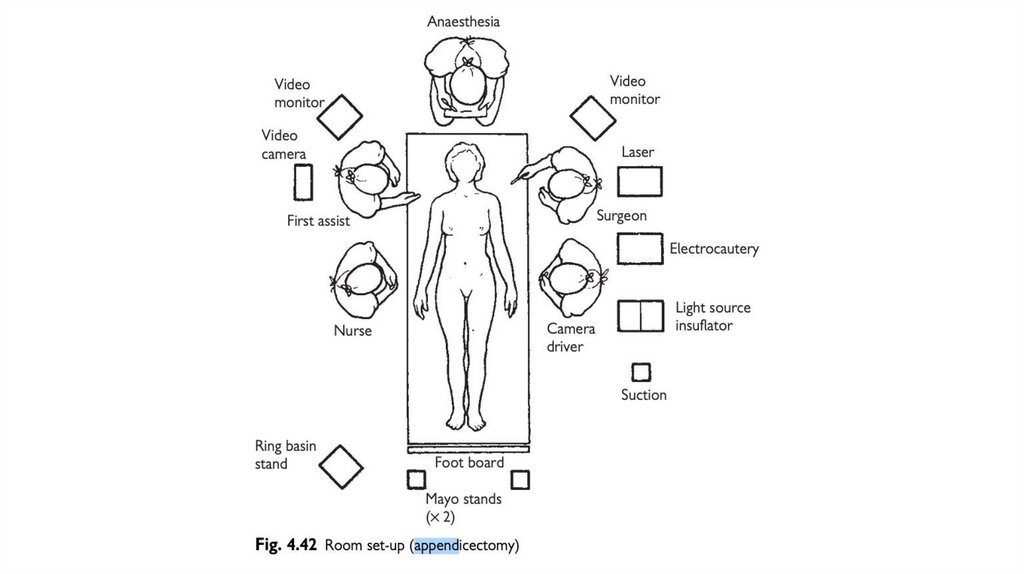
The Set up – position of the patient and thesurgical team
• Place the patient in step Trendelenburg
position to allow the intestines to slide out
of the pelvis, and perform a thorough
exploration to confirm the diagnosis.
• The surgical procedure is performed under
general anesthesia.
• The bladder is decompressed with a Foley
catheter to avoid injury during insertion of
the supra-pubic ports.
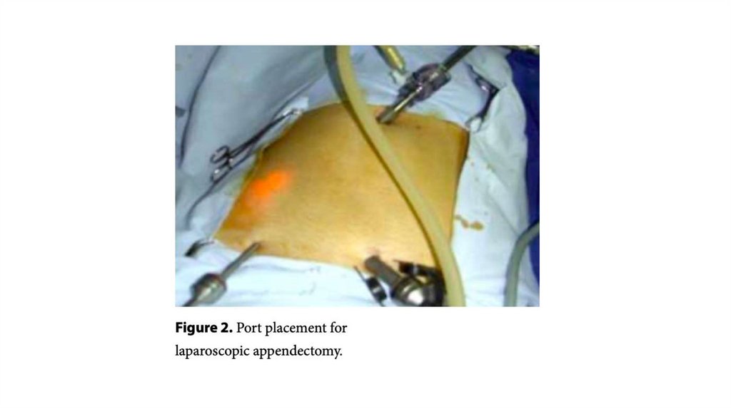
53.
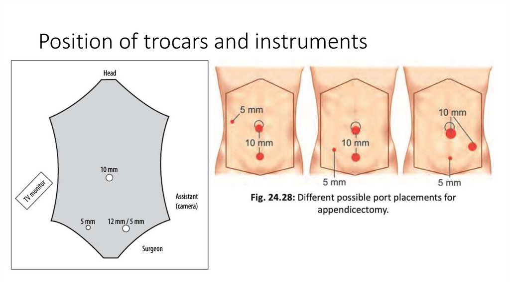
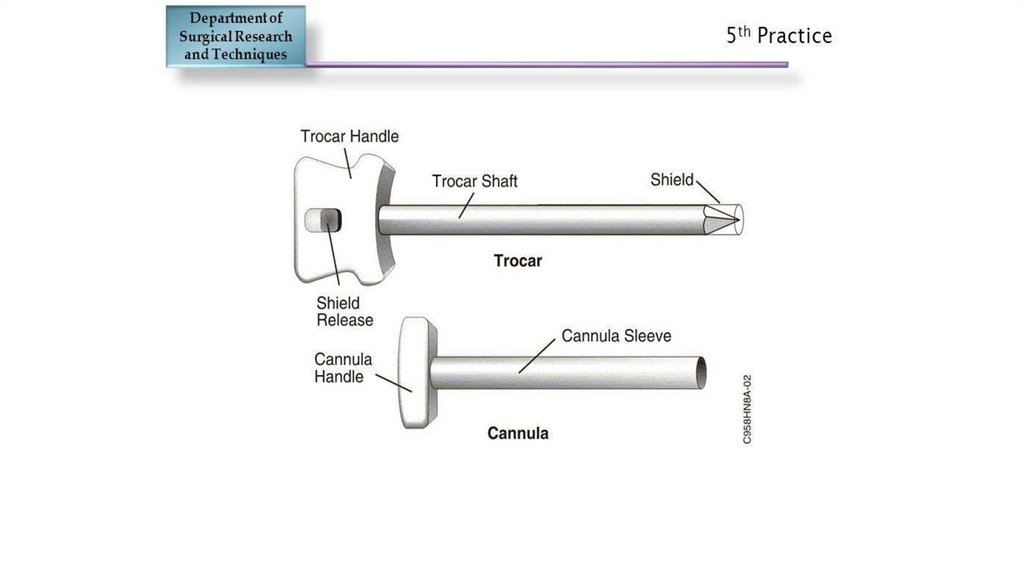
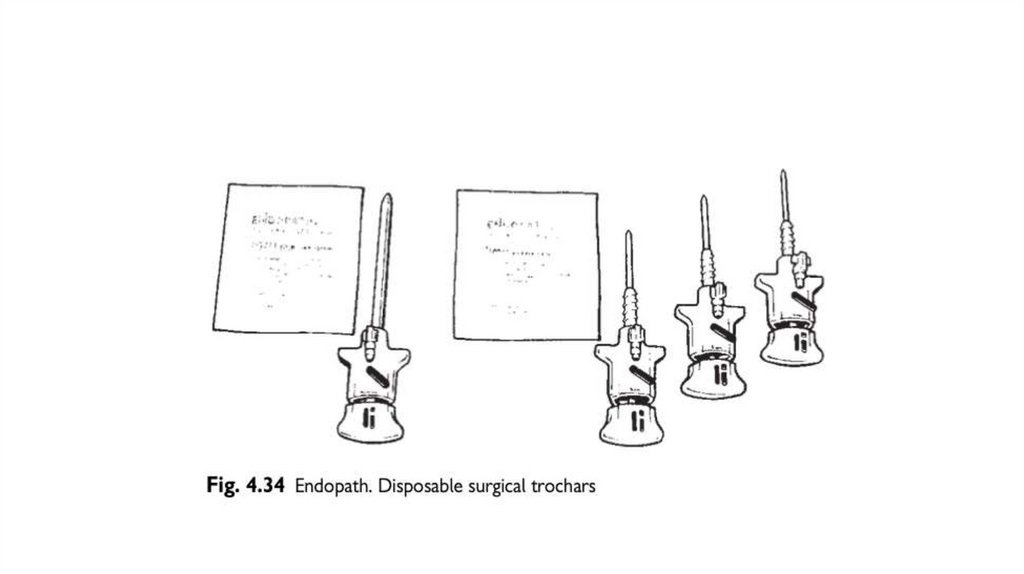
Position of trocars and instruments54.
55.
56.
57.
58.
59.
60.
61.
62.
63.
64.
65.
66.
67.
68.
69.
70.
71.
72.
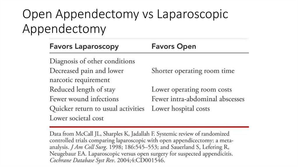
Open Appendectomy vs LaparoscopicAppendectomy
73.
POST-OP MANAGEMENT• In uncomplicated case, antibiotics should be continued up to 24
hours post-operatively ,oral fluid are started 12hrs after
recovery followed by light diet 24hrs later.
• In complicated antibiotics should be continued for anywhere
between 3 and 7 days, iv fluids, iv antibiotics and NPO with NG
tube drainage until bowel activity recommence and
temperature subsides
• An interval appendectomy is generally performed 6-8 weeks after
conservative management with antibiotics for special cases, such as
perforated appendicitis
• Stiches removed in 7-10days
74.
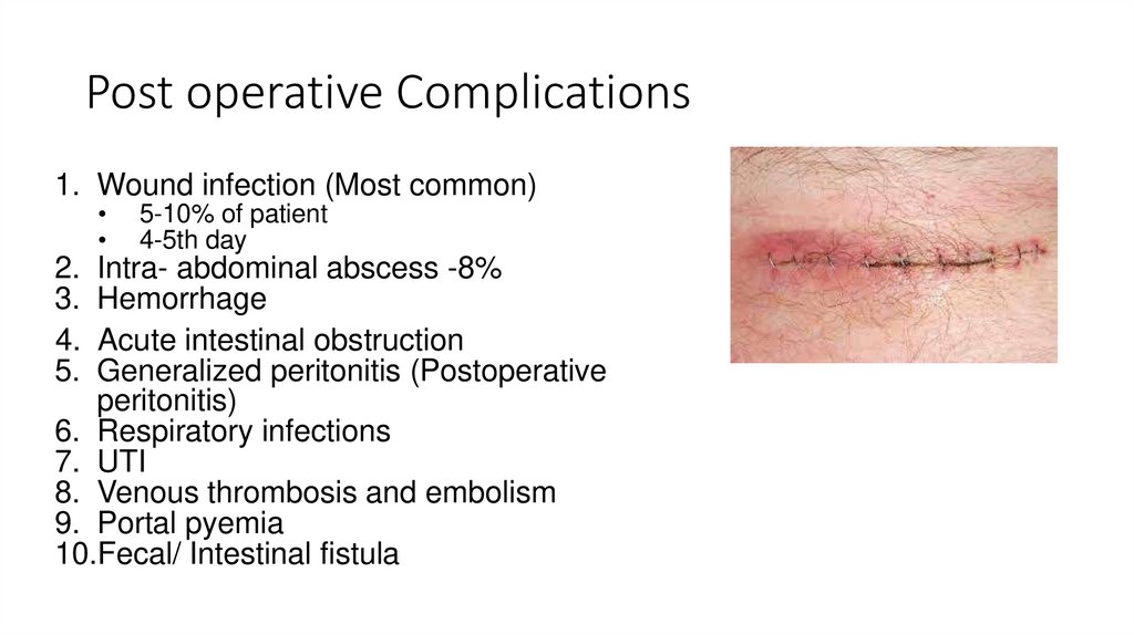
Post operative Complications1. Wound infection (Most common)
2.
3.
4.
5.
5-10% of patient
4-5th day
Intra- abdominal abscess -8%
Hemorrhage
Acute intestinal obstruction
Generalized peritonitis (Postoperative
peritonitis)
6. Respiratory infections
7. UTI
8. Venous thrombosis and embolism
9. Portal pyemia
10.Fecal/ Intestinal fistula
75.
Alternative Methods of Appendectomy• Laparoscopic Single-Incision Appendectomy
• Natural orifice transluminal endoscopic surgery (NOTES)
76.
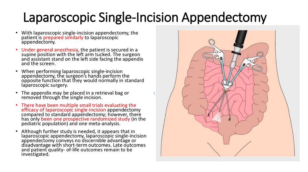
Laparoscopic Single-Incision Appendectomy• With laparoscopic single-incision appendectomy, the
patient is prepared similarly to laparoscopic
appendectomy.
• Under general anesthesia, the patient is secured in a
supine position with the left arm tucked. The surgeon
and assistant stand on the left side facing the appendix
and the screen.
• When performing laparoscopic single-incision
appendectomy, the surgeon’s hands perform the
opposite function that they would normally in standard
laparoscopic surgery.
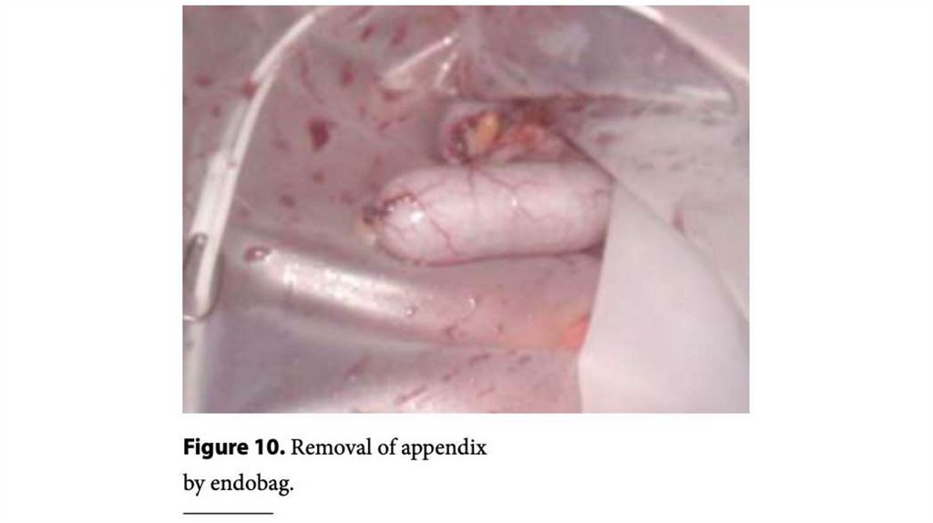
• The appendix may be placed in a retrieval bag or
removed through the single incision.
• There have been multiple small trials evaluating the
efficacy of laparoscopic single-incision appendectomy
compared to standard appendectomy; however, there
has only been one prospective randomized study (in the
pediatric population) and one meta-analysis.
• Although further study is needed, it appears that in
laparoscopic appendectomy, laparoscopic single-incision
appendectomy conveys no discernible advantage or
disadvantage with short-term outcomes. Late outcomes
and patient quality- of-life outcomes remain to be
investigated.
77.
Natural Orifice Transluminal EndoscopicSurgery
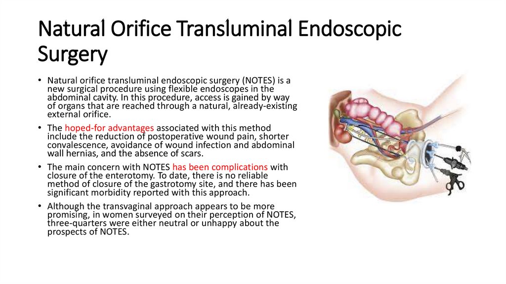
• Natural orifice transluminal endoscopic surgery (NOTES) is a
new surgical procedure using flexible endoscopes in the
abdominal cavity. In this procedure, access is gained by way
of organs that are reached through a natural, already-existing
external orifice.
• The hoped-for advantages associated with this method
include the reduction of postoperative wound pain, shorter
convalescence, avoidance of wound infection and abdominal
wall hernias, and the absence of scars.
• The main concern with NOTES has been complications with
closure of the enterotomy. To date, there is no reliable
method of closure of the gastrotomy site, and there has been
significant morbidity reported with this approach.
• Although the transvaginal approach appears to be more
promising, in women surveyed on their perception of NOTES,
three-quarters were either neutral or unhappy about the
prospects of NOTES.
78.
REFERENCES• Schwartz's Principles of Surgery ;Textbook by F. Charles Brunicardi and Seymour I.
Schwartz
• SRB's Manual of Surgery 5th edition.
• Washington's manual of surgery 7th edition.
• Curet MJ et al. (2009). Laparoscopic General Surgery. In Jaffe RA, Samuels SI
(Eds.), Anesthesiologist’s Manual of Surgical Procedures (4th Ed., pp. 569-608).
Philadelphia: Lippincott Williams and Wilkins.
• Jeong J et al. Laparoscopic appendectomy is a safe and beneficial procedure in
pregnant women. Surg Laparosc Endosc Percutan Tech 2011;21:1, 24-27.
• Sauerland S, Jaschinski T, Neugebauer EA. Laparoscopic versus open surgery for
suspected appendicitis. Cochrane Database Syst Rev. 2010 Oct 6;(10):CD001546.
• Dershwitz M, ed. The MGH Board Review of Anesthesiology, 5th ed. New York:
Appelton & Lange, 1999.
• Atlas of Surgical Operations ;Book by Jr Robert Zollinger