


 Медицина
Похожие презентации:


Specific Hernia Types
1.
University of Al-Ameed / College of MedicineDepartment of Surgery
Specific Hernia Types
Ass. Prof Dr. Abdulrazzak Kalaf Hassan
Consultant General Surgeon / Head of surgical department
E-mail: abdulrazzak2006@yahoo.com
2020-2021
1
2.
Inguinal Hernia2
3.
Is the most common hernia in men & women but much morecommon in men.
There are two basic types which are fundamentally different in
anatomy, causation & complications.
However, they are anatomically very close to one another,
surgical repair techniques are very similar & ultimate
reinforcement of the weakened anatomy is identical so they are
often referred to together as inguinal hernia.
3
4.
Basic anatomy of inguinal canal:Superficial inguinal ring:
Triangular opening in external oblique aponeurosis 1.25 cm above
the public tubercle (Normally the ring will not admit the tip of the
little finger).
Deep inguinal ring:
U-shaped opening in transversalis fascia 1.25cm above the mid
inguinal ligament.
4
5.
Inguinal canal:In infant deep & superficial inguinal ring are almost
superimposed, but in adult it is 3.75cm long & directed
downwards & medially from deep to superficial ring.
5
6.
67.
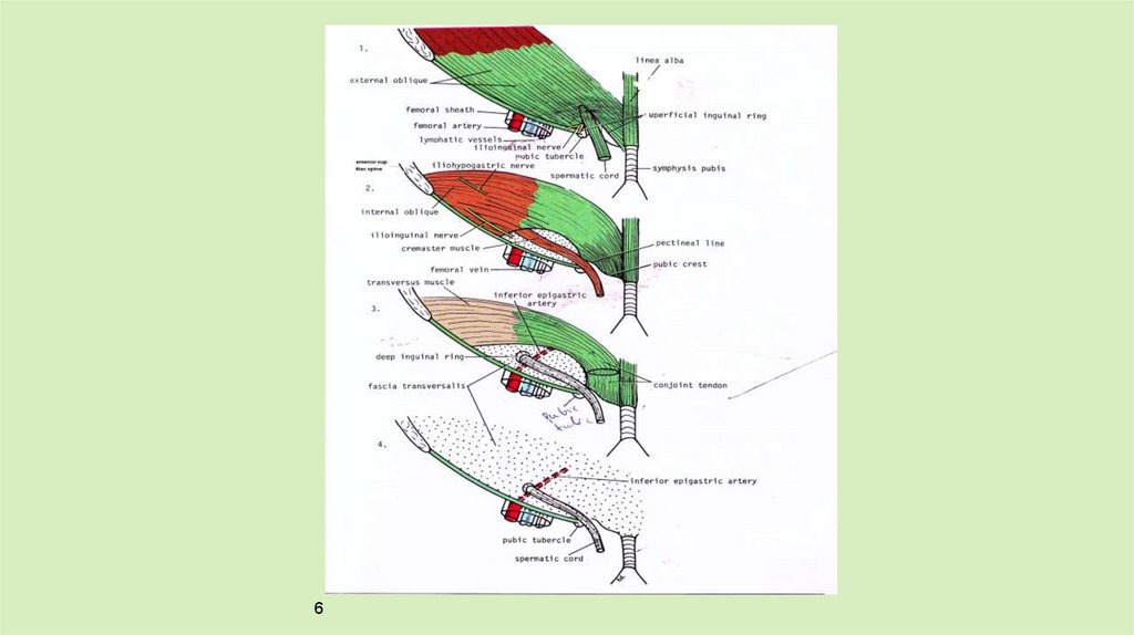
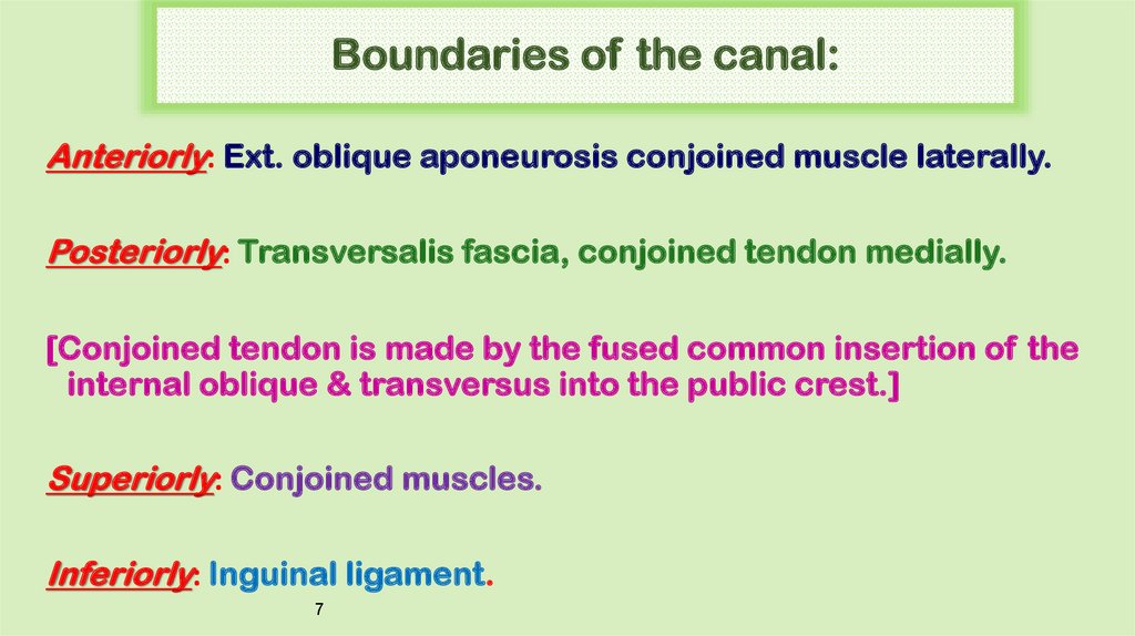
Boundaries of the canal:Anteriorly: Ext. oblique aponeurosis conjoined muscle laterally.
Posteriorly: Transversalis fascia, conjoined tendon medially.
[Conjoined tendon is made by the fused common insertion of the
internal oblique & transversus into the public crest.]
Superiorly: Conjoined muscles.
Inferiorly: Inguinal ligament.
7
8.
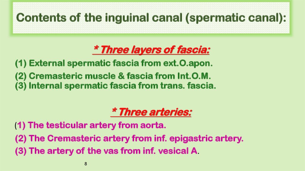
Contents of the inguinal canal (spermatic canal):* Three layers of fascia:
(1) External spermatic fascia from ext.O.apon.
(2) Cremasteric muscle & fascia from Int.O.M.
(3) lnternal spermatic fascia from trans. fascia.
* Three arteries:
(1) The testicular artery from aorta.
(2) The Cremasteric artery from inf. epigastric artery.
(3) The artery of the vas from inf. vesical A.
8
9.
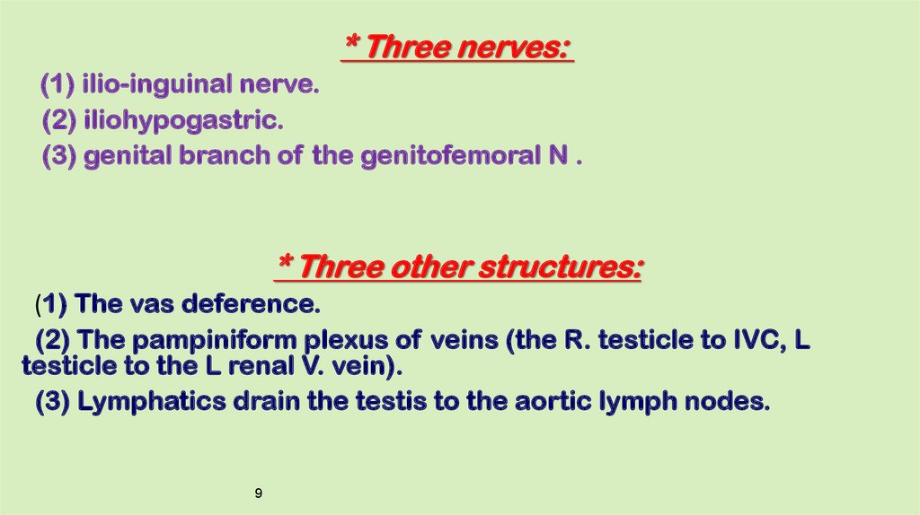
* Three nerves:(1) ilio-inguinal nerve.
(2) iliohypogastric.
(3) genital branch of the genitofemoral N .
* Three other structures:
(1) The vas deference.
(2) The pampiniform plexus of veins (the R. testicle to IVC, L
testicle to the L renal V. vein).
(3) Lymphatics drain the testis to the aortic lymph nodes.
9
10.
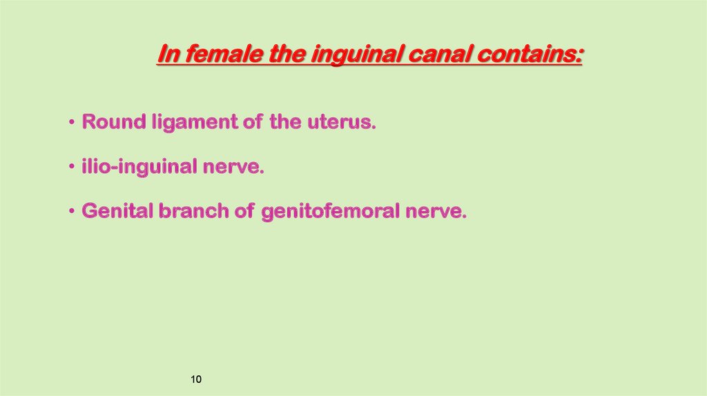
In female the inguinal canal contains:• Round ligament of the uterus.
• ilio-inguinal nerve.
• Genital branch of genitofemoral nerve.
10
11.
Types of Inguinal Hernia:1- Indirect inguinal hernia (LATERAL) (OBLIQUE)
2- Direct inguinal hernia ( MEDIAL) .
3- Sliding hernia.
Occasionally, both lateral & medial hernias are present in the
same patient (pantaloons hernia).
11
12.
Diagnosis Of An Inguinal HerniaIn most cases, the diagnosis of an inguinal hernia is simple & patients
often know their diagnosis as they are so common.
Often the hernia will reduce on lying & reappear on standing.
12
13.
With the patient lying down, Once reduced, surgeon identifies thebony landmarks of the anterior superior iliac spine & pubic tubercle
to landmark the deep inguinal ring at the mid-inguinal point.
Gentle pressure is applied at this point & patient asked to cough.
If hernia is controlled with pressure on the deep inguinal ring then it
is likely to be indirect / lateral & if hernia appears medial to this point
then it is direct / medial.
13
14.
Investigations For Inguinal HerniaMost cases require no diagnostic tests, But:
• US.
• CT scan.
• MRI scan are occasionally used.
• A herniogram involves the injection of contrast into the
peritoneal cavity followed by screening which shows the
presence of a sac or asymmetric bulging of inguinal anatomy.
14
15.
Management of Inguinal HerniaIt is safe to recommend no active treatment in cases of early,
asymptomatic, direct hernia, particularly in elderly patients who
do not wish surgical intervention.
These patients should be warned to seek early advice if the hernia
increases in size or becomes symptomatic.
15
16.
Surgical trusses are not recommended but may be required foroccasional patients who refuse any form of surgical intervention.
Elective surgery for inguinal hernia is a common & simple
operation.
It can be undertaken under local, regional or general anaesthesia.
16
17.
Operations for inguinal herniaA: Herniotomy. & B: Repair (herniorrhaphy):
I: Open repair:
1- Bassini, Shouldice, Desarda.
2- Open mesh repair: Lichtenstein.
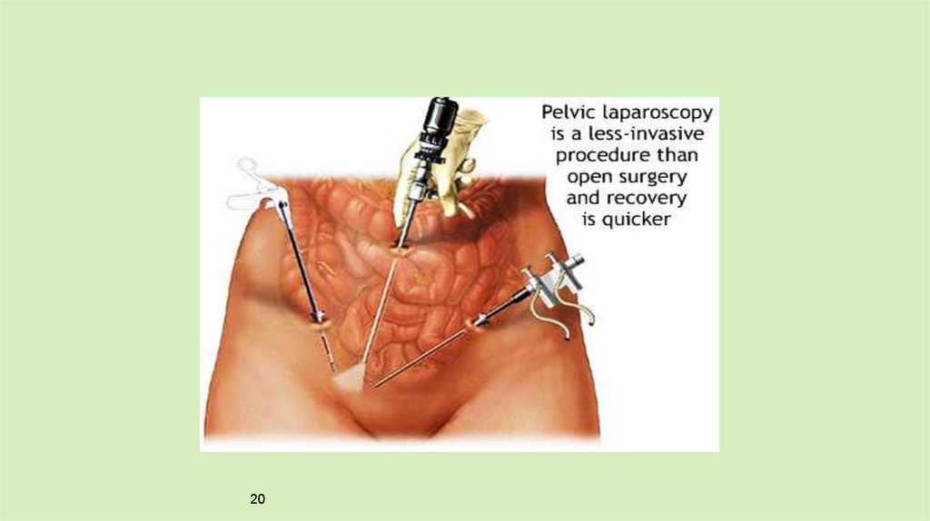
II: Laparoscopic repair:
1- TEP.
2- TAPP.
17
18.
Laparoscopic Herniorrhaphy OfInguinal Hernia
18
19.
Trans-abdominal approach (TAPP):Establishes a pneumoperitoneum & place a synthetic mesh
preperitoneally by dissecting the peritoneum off the hernial
orifices & positioning the mesh beneath the peritoneum before
closing the peritoneum over the mesh.
Preperitoneal approach (TEP):
The preperitoneal plane is opened by either balloon dissection or
direct dissection via paraumbilical incision, the hernial orifices
can be identified bilaterally & any hernial sac reduced & placing a
large mesh over the hernial orifices in preperitoneal plane.
19
20.
2021.
Emergency inguinal hernia surgeryNinety-five per cent of inguinal hernia patients present at clinics &
only 5 per cent present as an emergency with a painful irreducible
hernia which may progress to strangulation & possible bowel
infarction
21
22.
Complications of inguinal hernia surgery• Early: pain, bleeding, urinary retention, anaesthetic related.
• Medium: seroma, wound infection.
• Late: chronic pain, testicular atrophy.
22
23.
Femoral Hernia23
24.
• Less common than inguinal hernia.• It is more common in females than in males.
• Easily missed on examination.
• Fifty per cent of cases present as an emergency with very high
risk of strangulation.
24
25.
2526.
Surgical Anatomy:26
27.
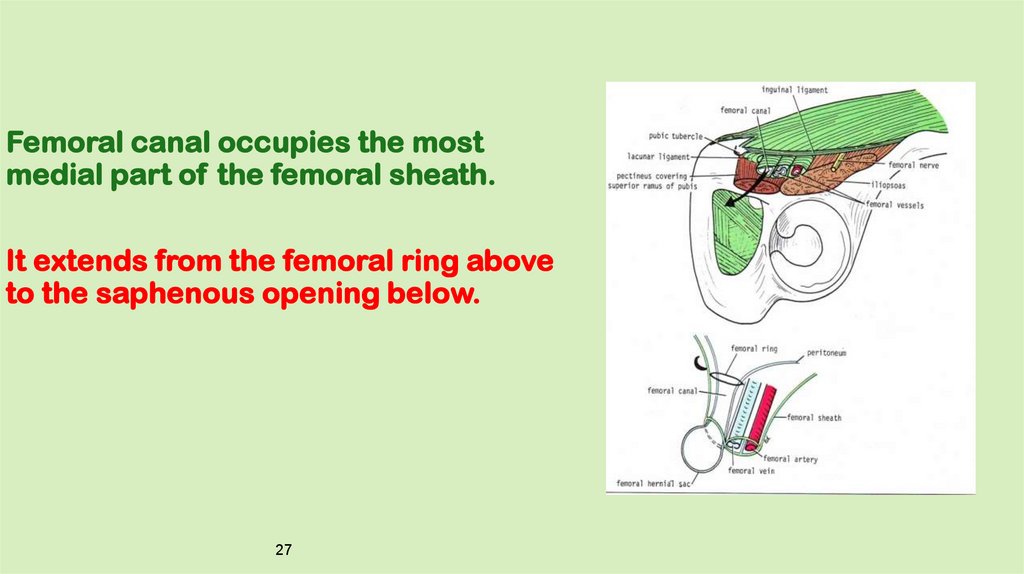
Femoral canal occupies the mostmedial part of the femoral sheath.
It extends from the femoral ring above
to the saphenous opening below.
27
28.
It is 1.25cm long & 1.25cm wide at its base.Femoral canal contains:
• Fat.
• Lymphatic vessels.
• & Cloquet's lymph nodes.
28
29.
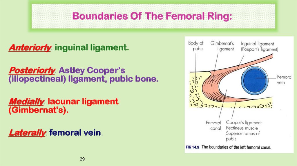
Boundaries Of The Femoral Ring:Anteriorly: inguinal ligament.
Posteriorly: Astley Cooper's
(iliopectineal) ligament, pubic bone.
Medially: lacunar ligament
(Gimbernat's).
Laterally: femoral vein.
29
30.
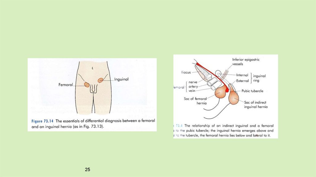
Diagnosis :Diagnostic error is common & often leads to delay in diagnosis &
treatment.
Hernia appears below & lateral to the pubic tubercle & lies in the
upper leg rather than in the lower abd.
Inadequate exposure of this area during routine examination leads
to failure to detect the hernia.
Hernia often rapidly becomes irreducible & loses any cough
impulse due to the30 tightness of the neck.
31.
It may only be 1–2 cm in size & can easily be mistaken for a lymph node.As it increases in size, it is reflected superiorly & becomes difficult to
distinguish from a medial direct hernia which arises only a few
centimetres above the femoral canal.
A direct inguinal hernia leaves the abdominal cavity just above the
inguinal ligament & a femoral hernia just below.
31
32.
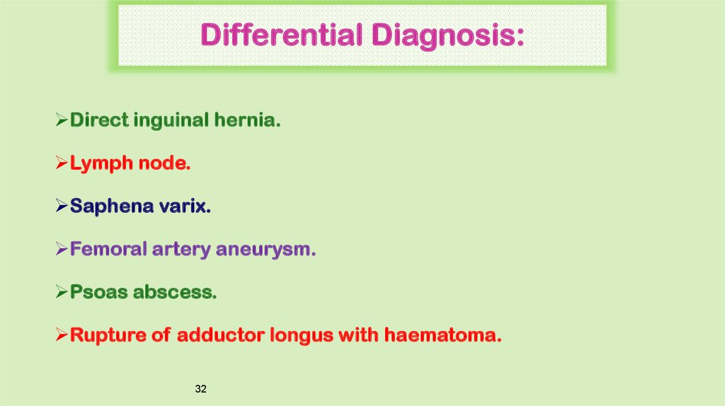
Differential Diagnosis:Direct inguinal hernia.
Lymph node.
Saphena varix.
Femoral artery aneurysm.
Psoas abscess.
Rupture of adductor longus with haematoma.
32
33.
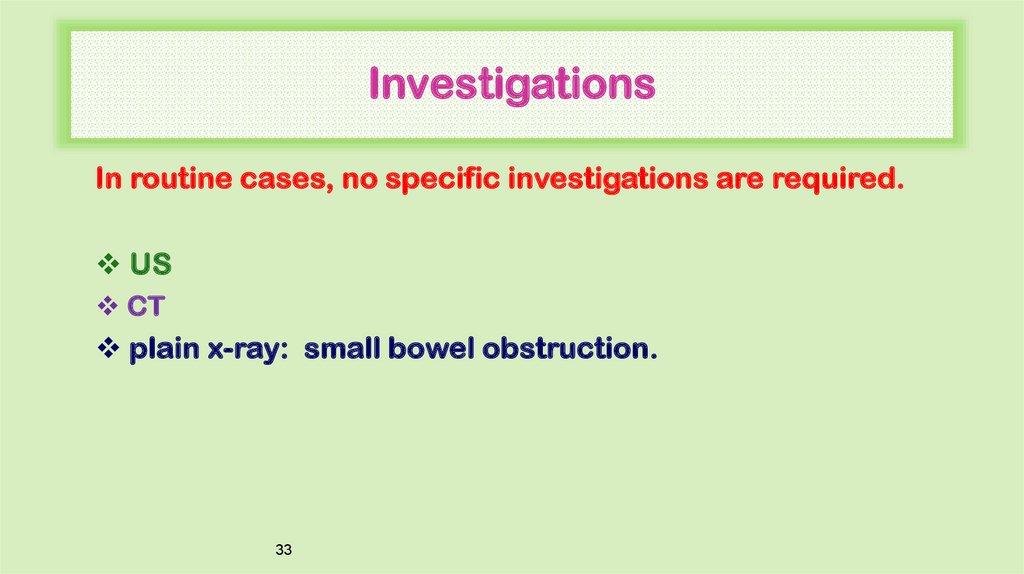
InvestigationsIn routine cases, no specific investigations are required.
US
CT
plain x-ray: small bowel obstruction.
33
34.
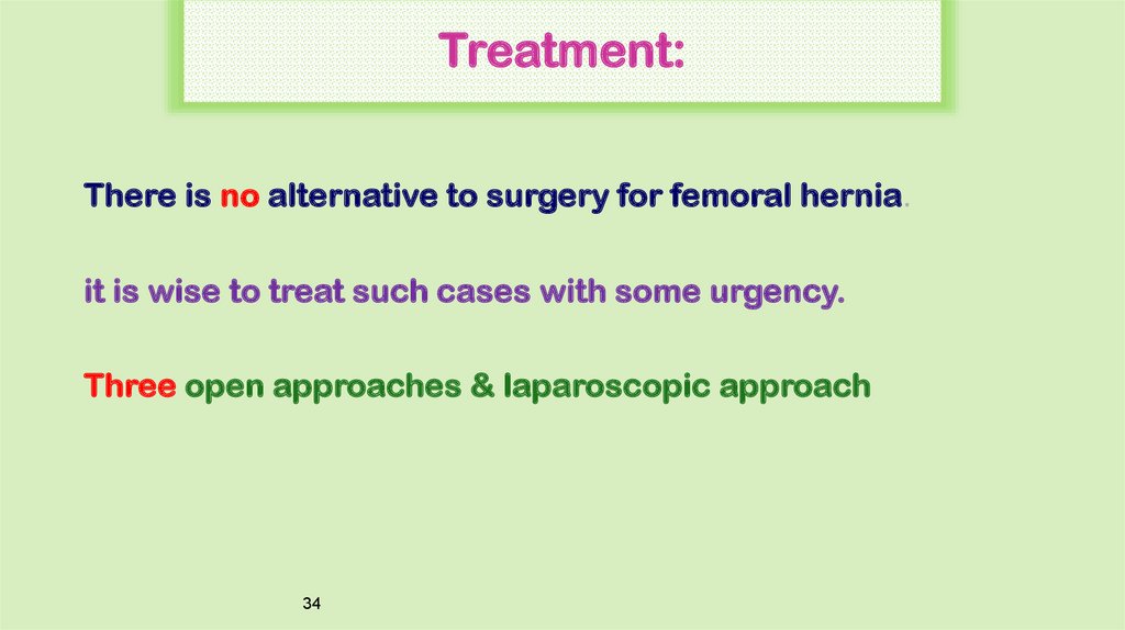
Treatment:There is no alternative to surgery for femoral hernia.
it is wise to treat such cases with some urgency.
Three open approaches & laparoscopic approach
34
35.
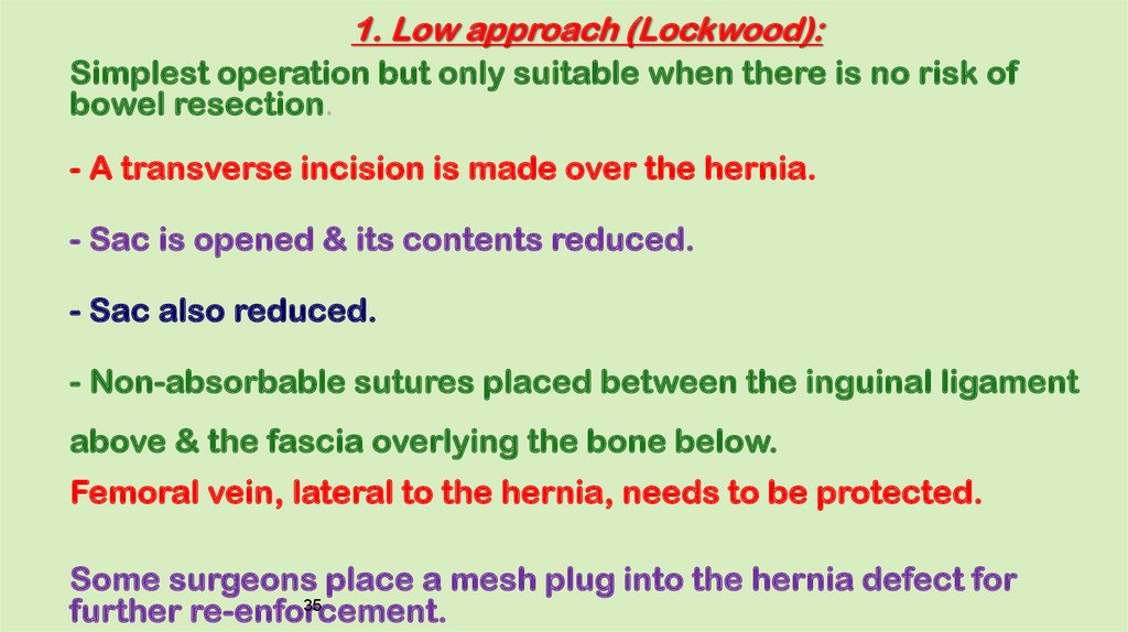
1. Low approach (Lockwood):Simplest operation but only suitable when there is no risk of
bowel resection.
- A transverse incision is made over the hernia.
- Sac is opened & its contents reduced.
- Sac also reduced.
- Non-absorbable sutures placed between the inguinal ligament
above & the fascia overlying the bone below.
Femoral vein, lateral to the hernia, needs to be protected.
Some surgeons place a mesh plug into the hernia defect for
35
further re-enforcement.
36.
2. Inguinal approach (Lotheissen's operation):* Through an inguinal incision.
* A femoral hernia lies immediately below this incision & can be
reduced by a combination of pulling from above and pushing
from below.
* The layers are closed as for inguinal hernia.
36
37.
3. High approach (McEvedy):* This more complex operation is ideal in the emergency
situation where the risk of bowel strangulation is high
* Horizontal incision (classically vertical) is made in the lower
abdomen centred at the lateral edge of the rectus muscle.
* Anterior rectus sheath is incised & the rectus muscle
displaced medially.
37
38.
* Femoral hernia is reduced & sac opened to allow carefulinspection of the bowel.
* Femoral defect is then closed with sutures, mesh or plug.
* This approach allows a generous incision to be made in the
peritoneum which aids inspection of the bowel & facilitates bowel
resection.
38
39.
* Laparoscopic approachBoth the TEP & TAPP approaches can be used for femoral
hernia & a standard mesh inserted.
This is ideal for reducible femoral hernias presenting electively
but not in emergency cases nor for irreducible hernia.
39