


 Медицина
Похожие презентации:


Abdominal Wall Hernias
1.
Crimean state medical university named after S.I.Georgievsky.
Department of surgery № 2.
Head of the department prof. Ilchenko F.N.
Abdominal Wall Hernias
Lecturer - Yuri G. Baranovsky, PhD
2.
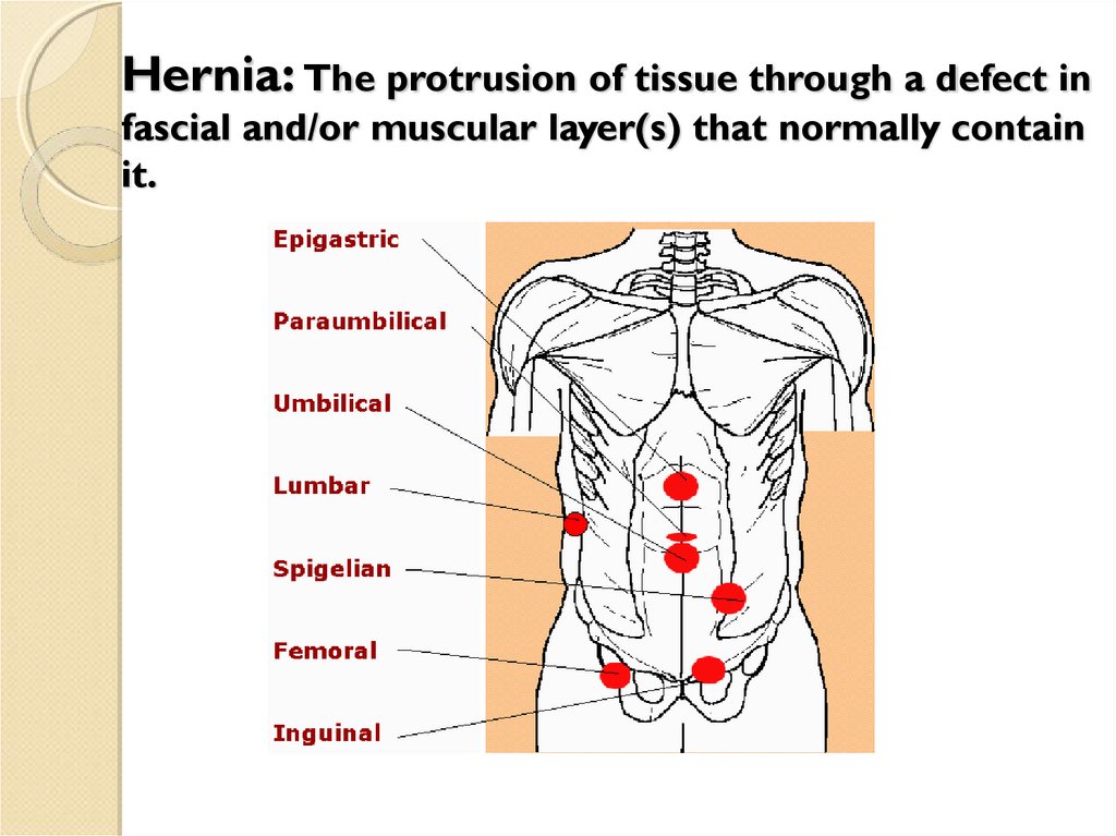
Hernia: The protrusion of tissue through a defect infascial and/or muscular layer(s) that normally contain
it.
3.
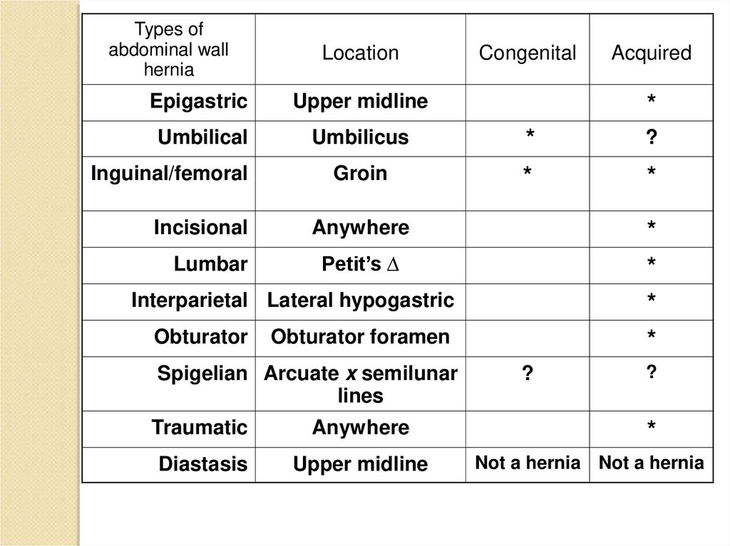
Types ofabdominal wall
hernia
Location
Congenital
Acquired
Epigastric
Upper midline
Umbilical
Umbilicus
*
?
Groin
*
*
Inguinal/femoral
Incisional
Lumbar
*
Anywhere
*
*
*
*
Petit’s ∆
Interparietal Lateral hypogastric
Obturator
Obturator foramen
Spigelian Arcuate x semilunar
lines
Traumatic
Anywhere
Diastasis
Upper midline
?
?
*
Not a hernia Not a hernia
4.
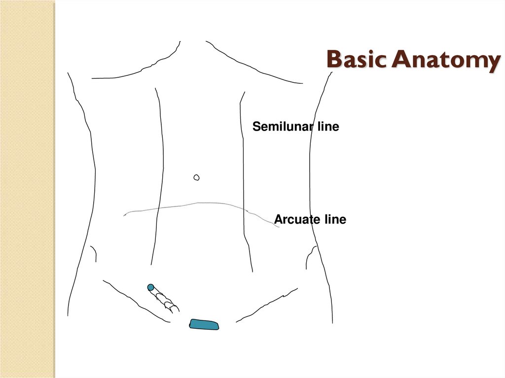
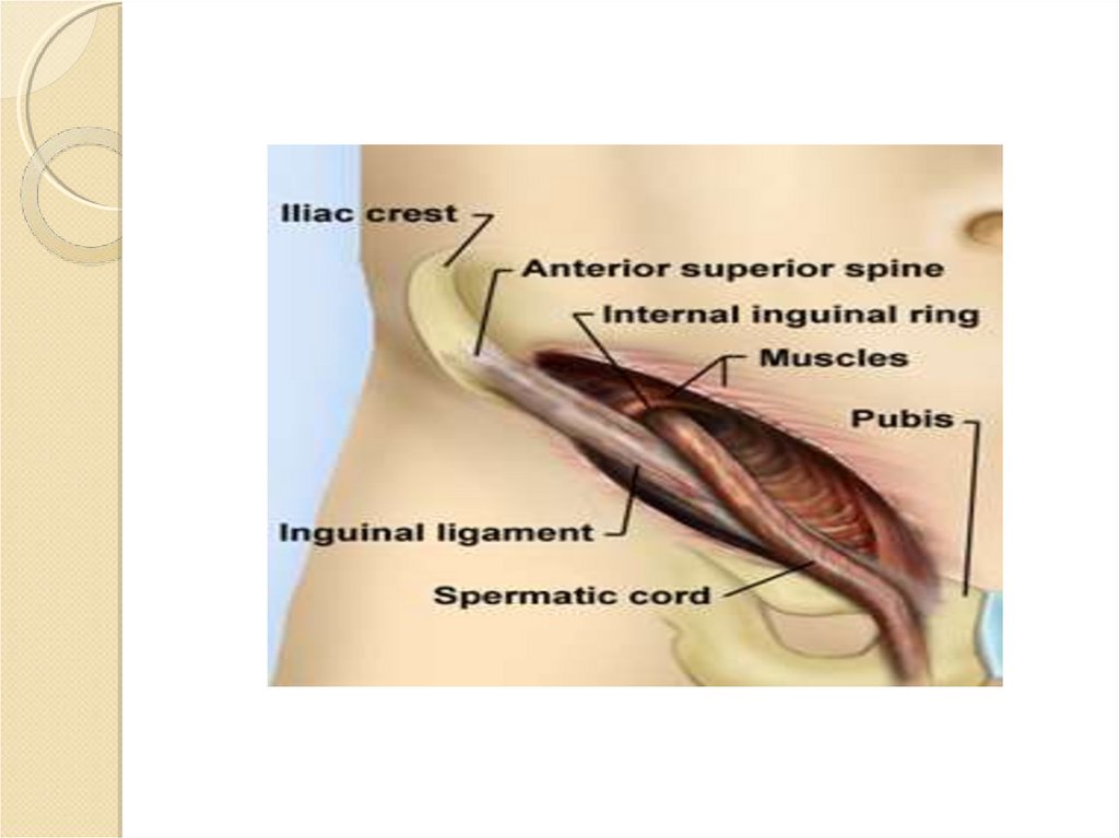
Basic AnatomySemilunar line
Arcuate line
5.
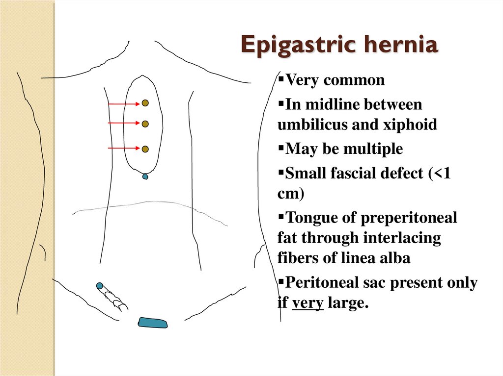
Epigastric herniaVery common
In midline between
umbilicus and xiphoid
May be multiple
Small fascial defect (<1
cm)
Tongue of preperitoneal
fat through interlacing
fibers of linea alba
Peritoneal sac present only
if very large.
6.
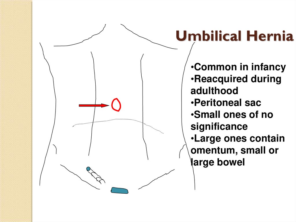
Umbilical Hernia•Common in infancy
•Reacquired during
adulthood
•Peritoneal sac
•Small ones of no
significance
•Large ones contain
omentum, small or
large bowel
7.
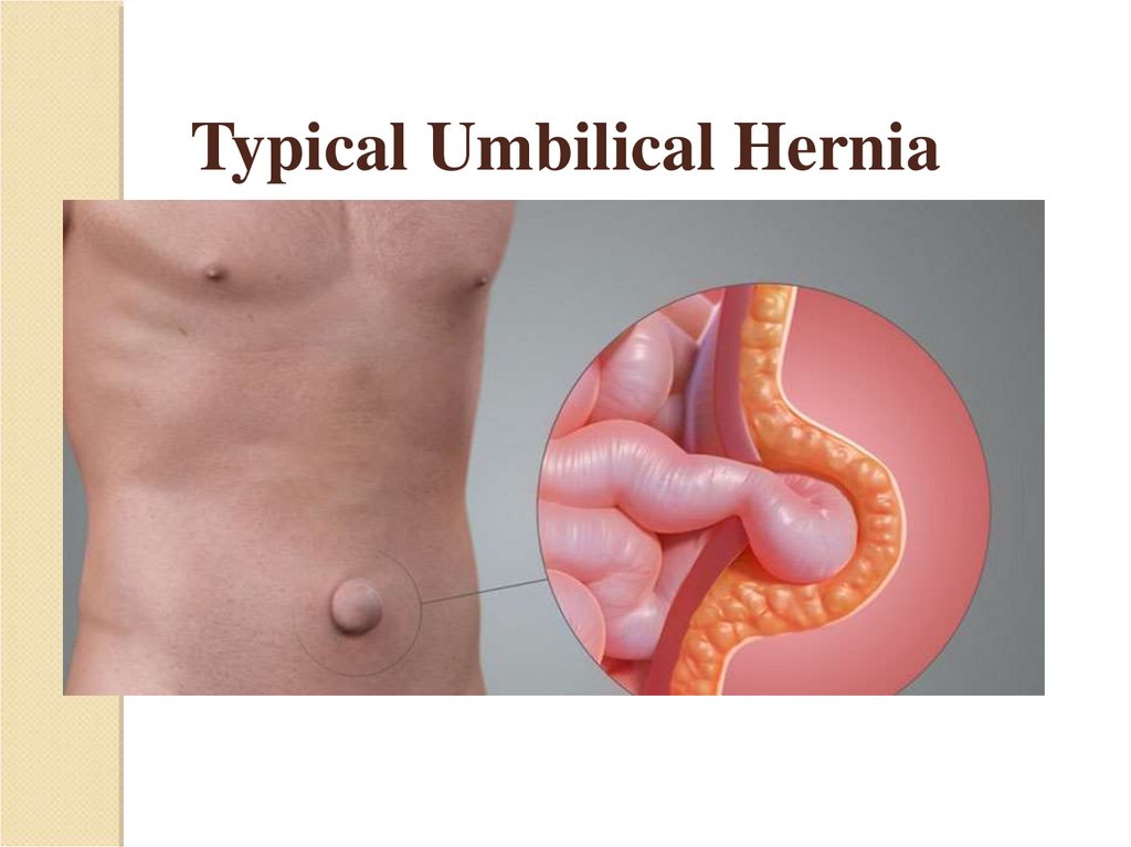
Typical Umbilical Hernia8.
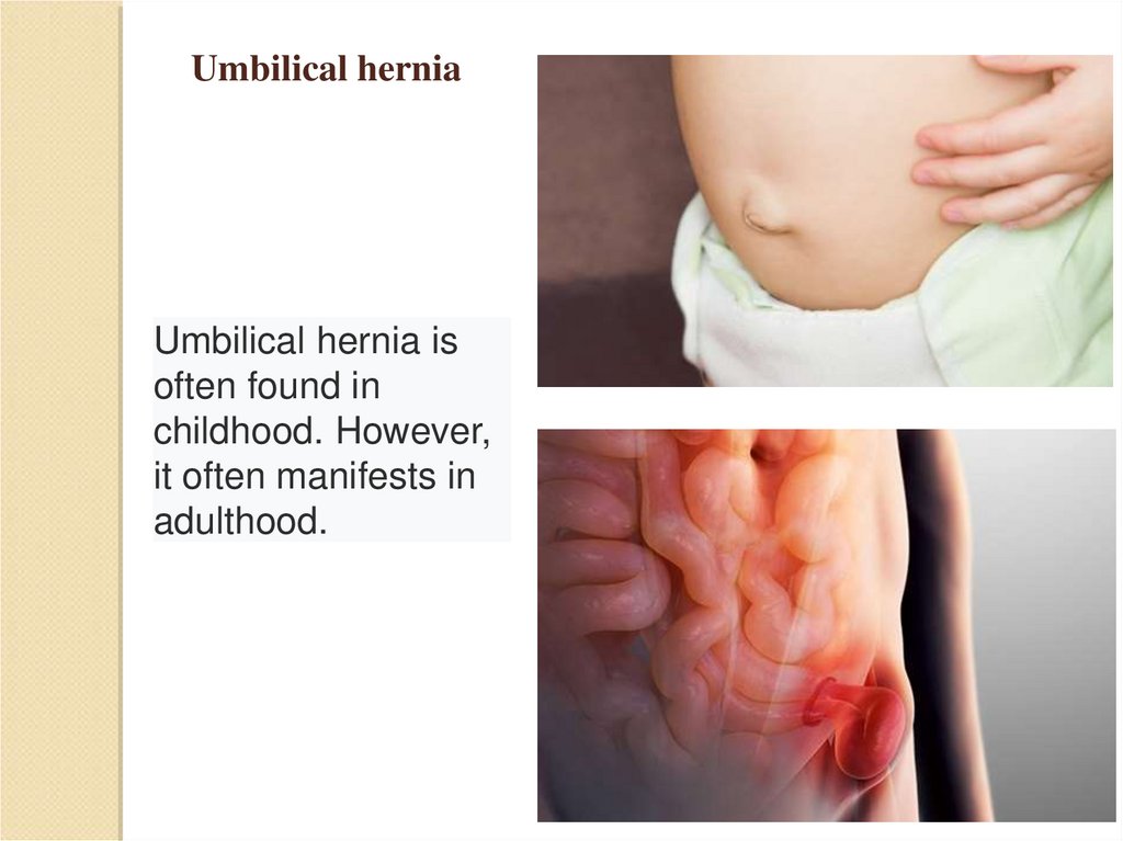
Umbilical herniaUmbilical hernia is
often found in
childhood. However,
it often manifests in
adulthood.
9.
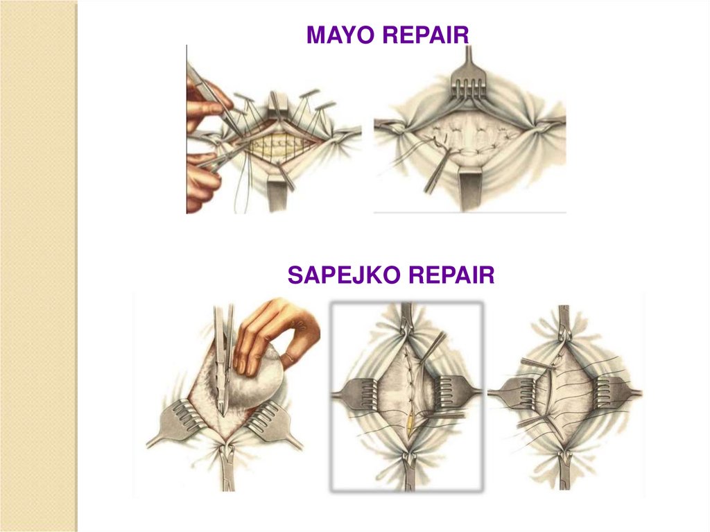
MAYO REPAIRSAPEJKO REPAIR
10.
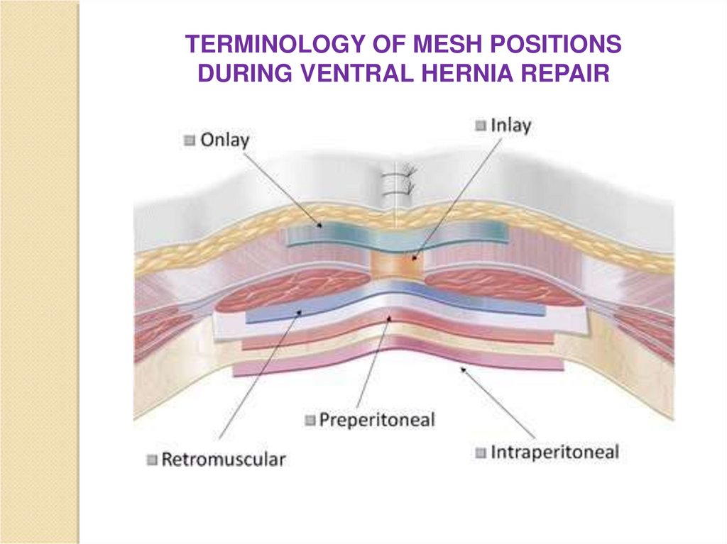
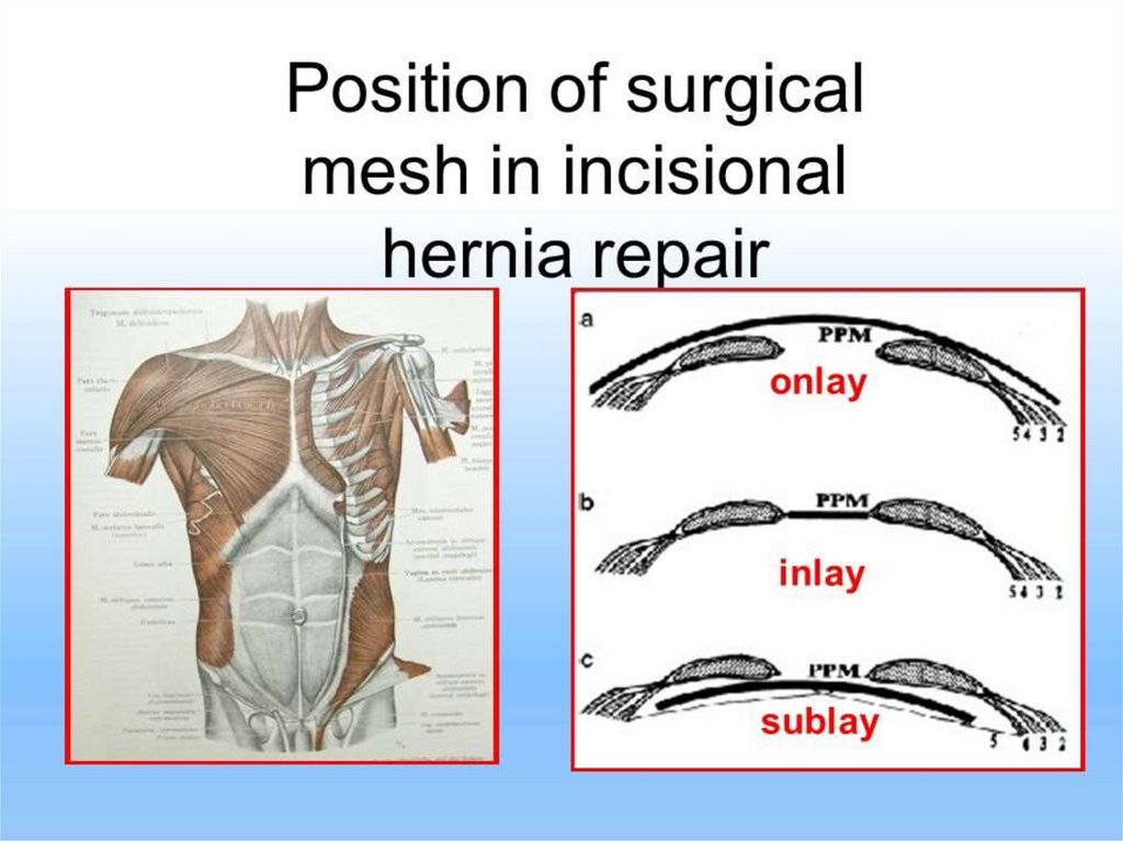
TERMINOLOGY OF MESH POSITIONSDURING VENTRAL HERNIA REPAIR
11.
Umbilical&
Inguinal
Hernias
12.
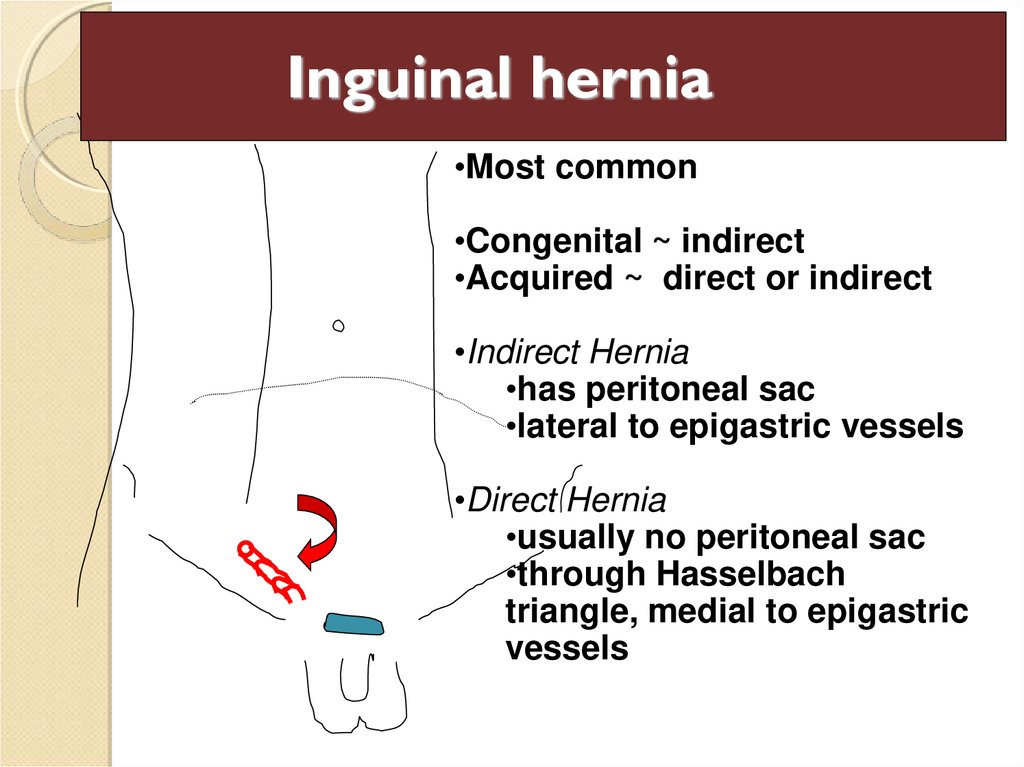
Inguinal hernia•Most common
•Congenital ~ indirect
•Acquired ~ direct or indirect
•Indirect Hernia
•has peritoneal sac
•lateral to epigastric vessels
•Direct Hernia
•usually no peritoneal sac
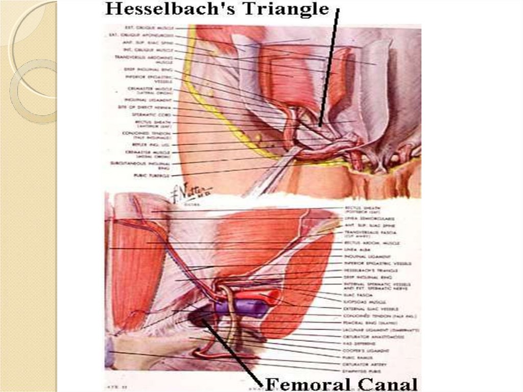
•through Hasselbach
triangle, medial to epigastric
vessels
13.
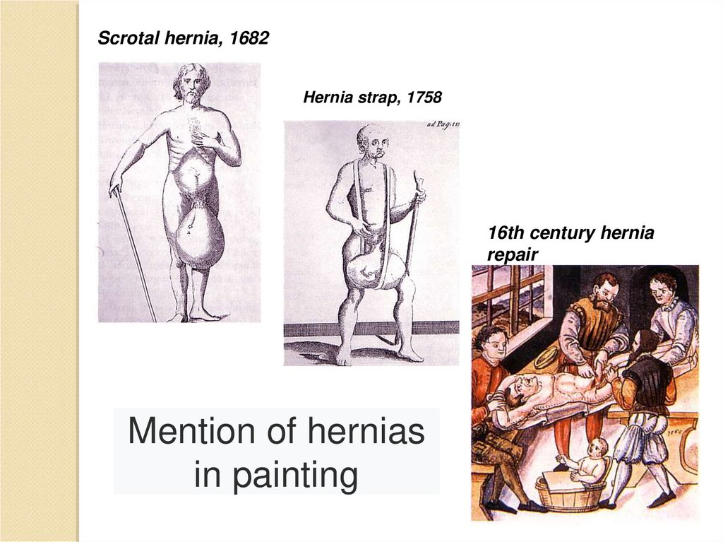
Scrotal hernia, 1682Hernia strap, 1758
16th century hernia
repair
Mention of hernias
in painting
14.
15.
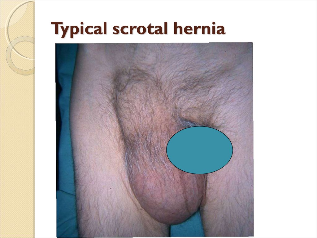
Typical scrotal hernia16.
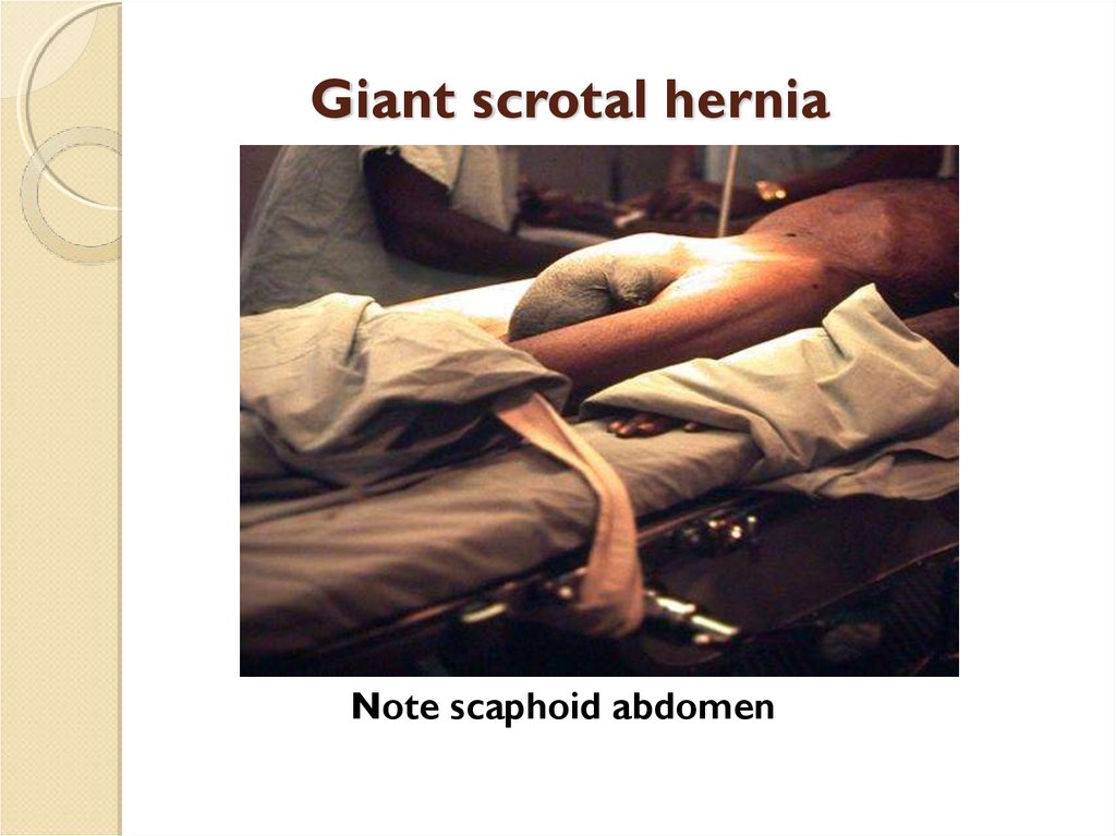
Giant scrotal herniaNote scaphoid abdomen
17. The basic feature of all hernias
Occur at a weak spot .Reduce on lying down ,or with direct pressure.
Have an expansile cough impulse
18. A hernia consist of 3 parts:
1. Sac:consist of a
diverticulum of
peritoneum.
2. Contents:
Omentum, small or
large intestine, urinary
bladder,
Omentum,
ovaries
malignant
nodules or ascetic fluid.
3. Gate:
weak spot of abdominal
wall.
19. Complications of hernias
Irreduciblethe hernia contents cannot be manipulated back into the
abdominal cavity.
Incarcerated
the contents of the sac are literally impression in the sac of
Hernia.
Obstruction
the loop of the bowel become non functioning with normal
blood supply .
Strangulated
cut off the blood supply to the content sac (tender).
20.
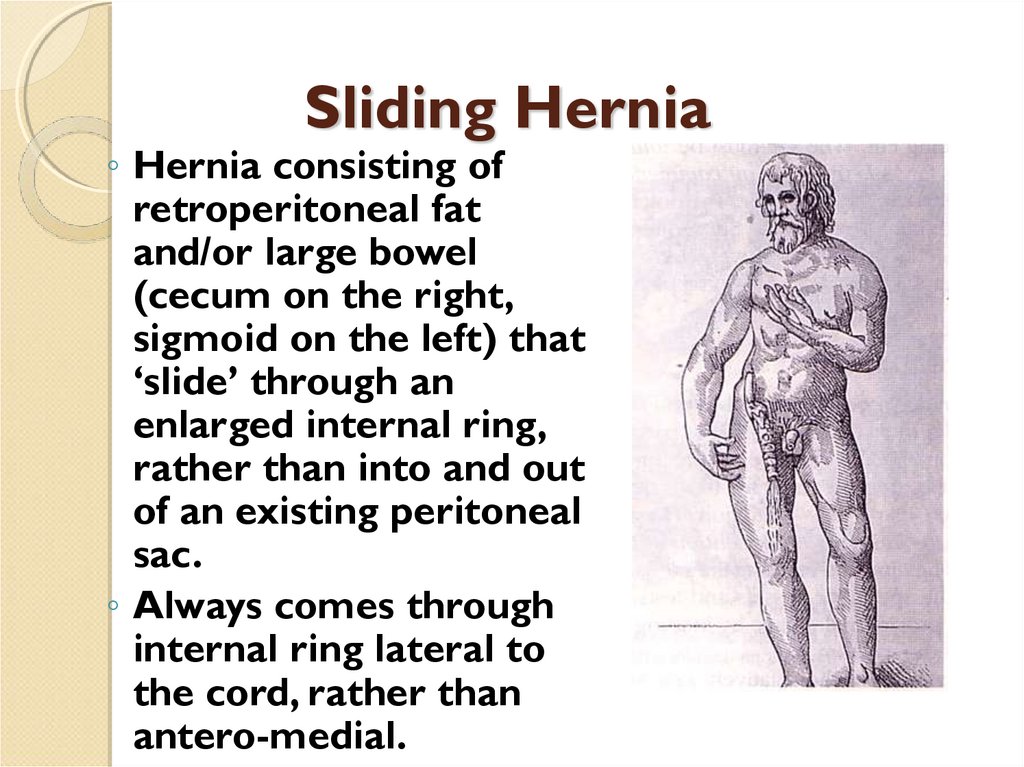
Sliding Hernia◦ Hernia consisting of
retroperitoneal fat
and/or large bowel
(cecum on the right,
sigmoid on the left) that
‘slide’ through an
enlarged internal ring,
rather than into and out
of an existing peritoneal
sac.
◦ Always comes through
internal ring lateral to
the cord, rather than
antero-medial.
21.
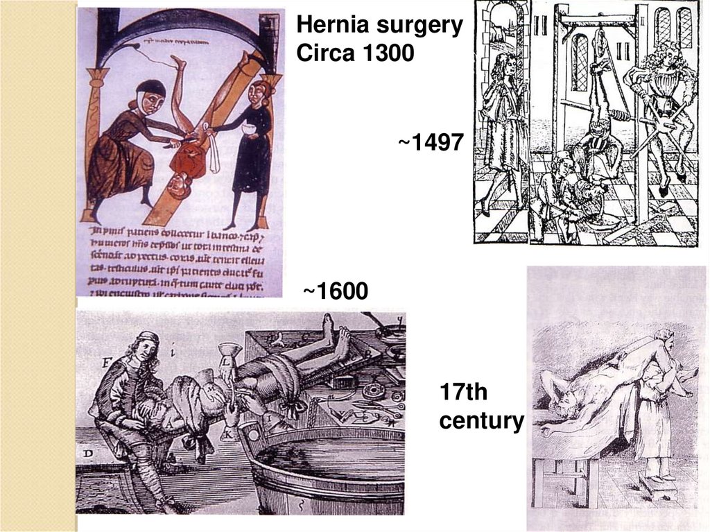
Hernia surgeryCirca 1300
~1497
~1600
17th
century
22.
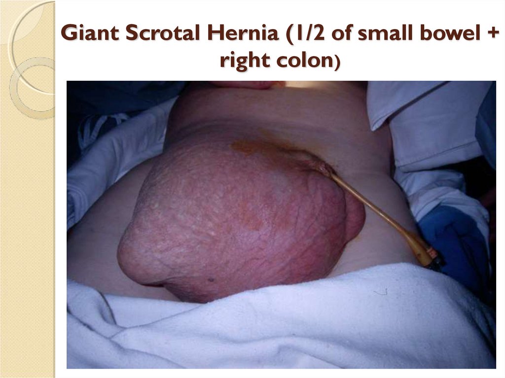
Giant Scrotal Hernia (1/2 of small bowel +right colon)
23.
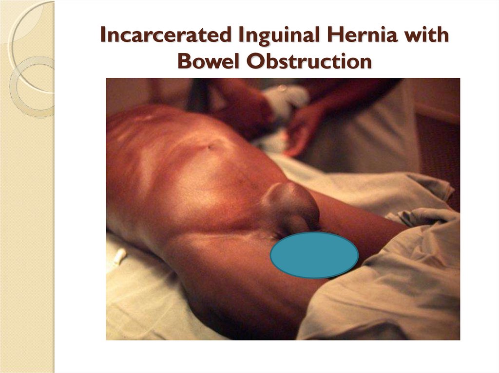
Incarcerated Inguinal Hernia withBowel Obstruction
24.
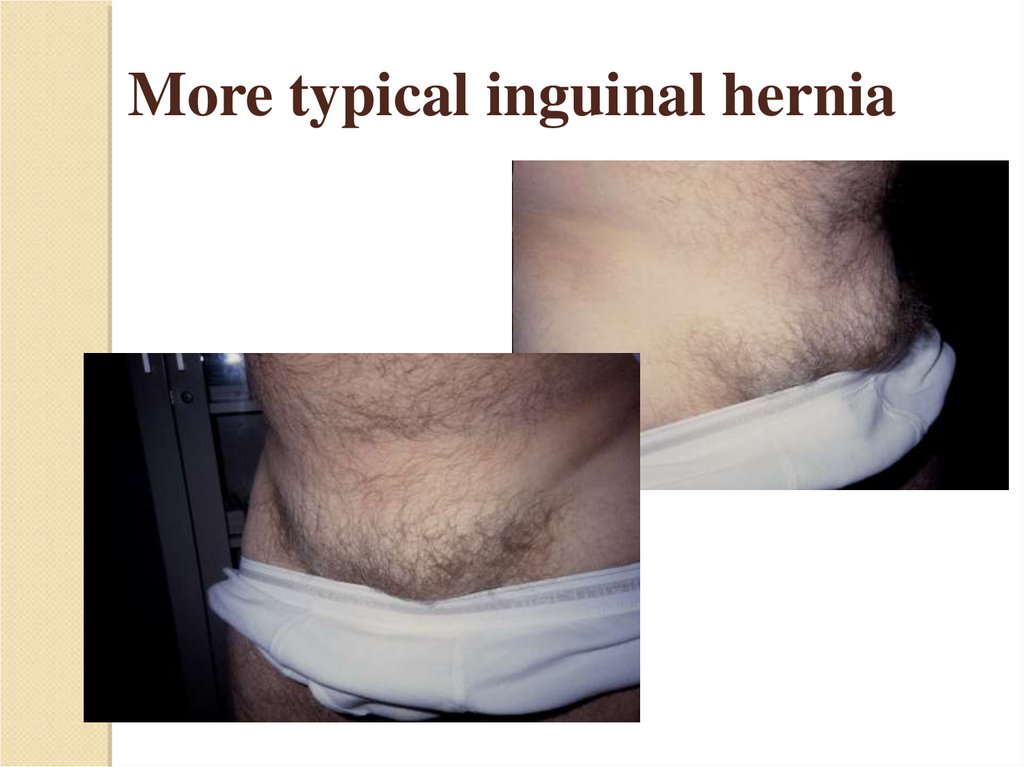
More typical inguinal hernia25.
26.
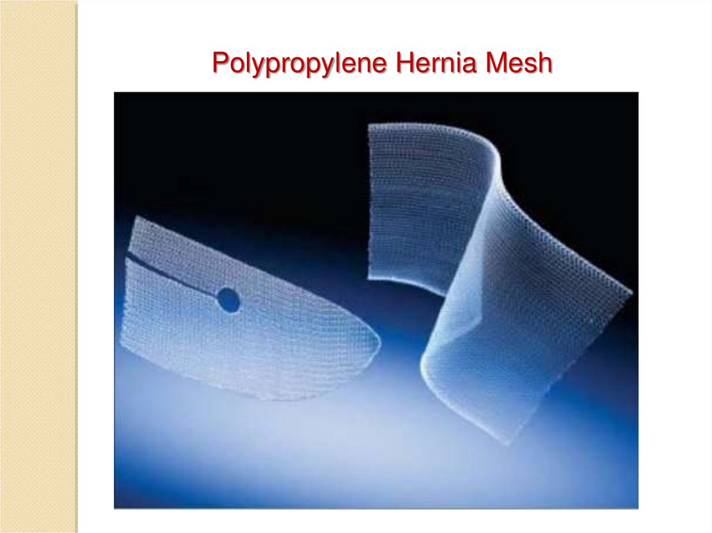
Polypropylene Hernia Mesh27.
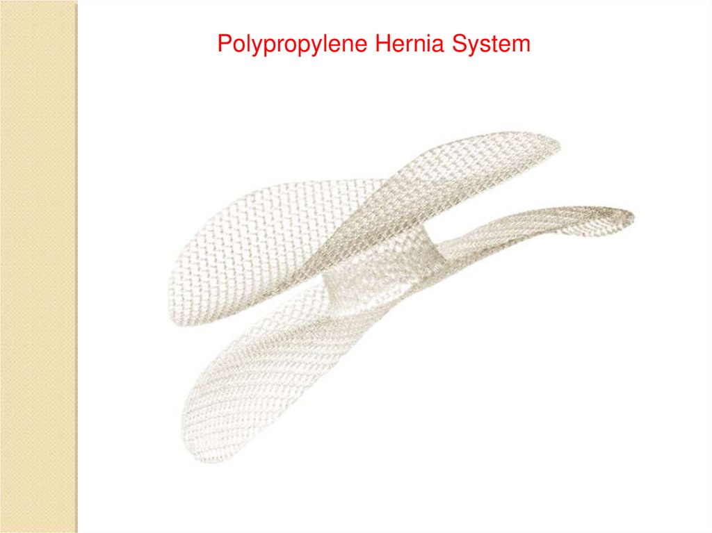
Polypropylene Hernia System28.
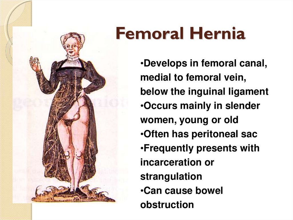
Femoral Hernia•Develops in femoral canal,
medial to femoral vein,
below the inguinal ligament
•Occurs mainly in slender
women, young or old
•Often has peritoneal sac
•Frequently presents with
incarceration or
strangulation
•Can cause bowel
obstruction
29.
30.
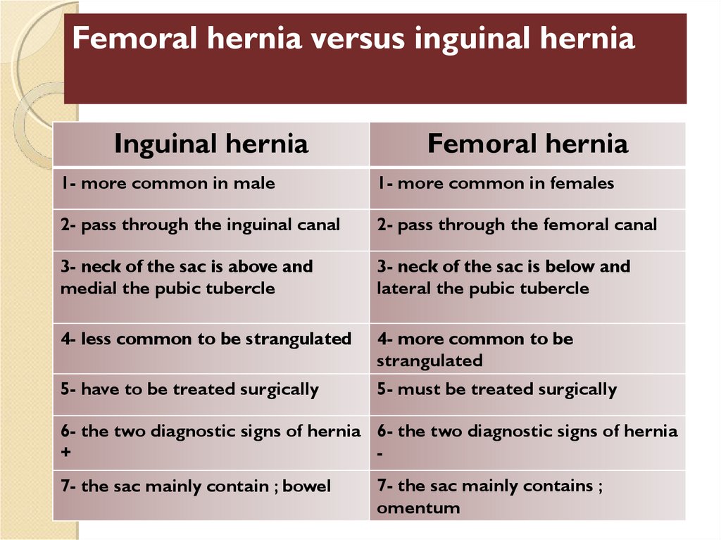
Femoral hernia versus inguinal herniaInguinal hernia
Femoral hernia
1- more common in male
1- more common in females
2- pass through the inguinal canal
2- pass through the femoral canal
3- neck of the sac is above and
medial the pubic tubercle
3- neck of the sac is below and
lateral the pubic tubercle
4- less common to be strangulated
4- more common to be
strangulated
5- have to be treated surgically
5- must be treated surgically
6- the two diagnostic signs of hernia 6- the two diagnostic signs of hernia
+
7- the sac mainly contain ; bowel
7- the sac mainly contains ;
omentum
31.
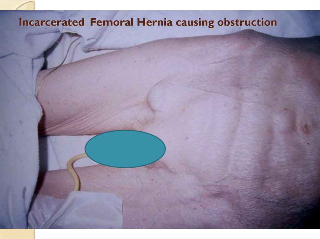
Incarcerated Femoral Hernia causing obstruction32.
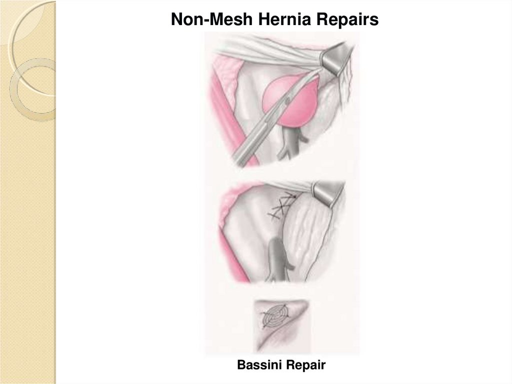
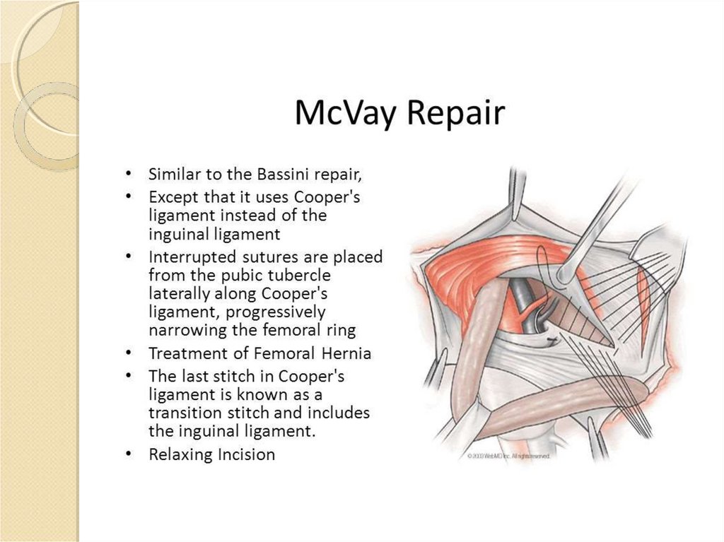
Non-Mesh Hernia RepairsBassini Repair
33.
34.
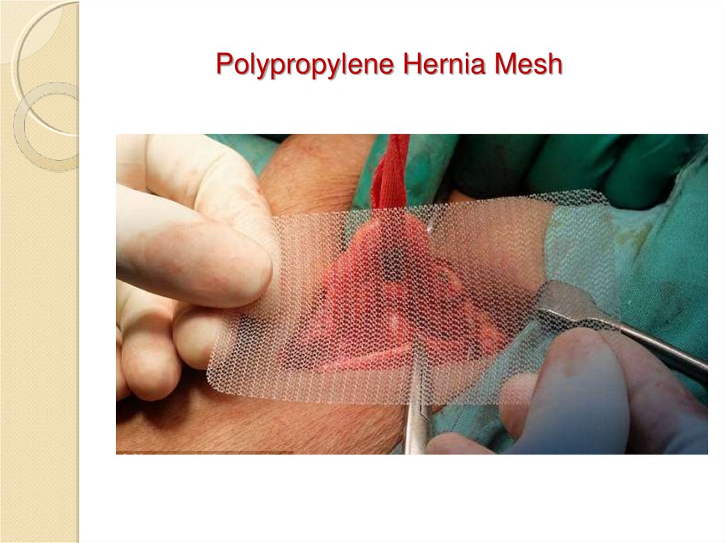
Polypropylene Hernia Mesh35.
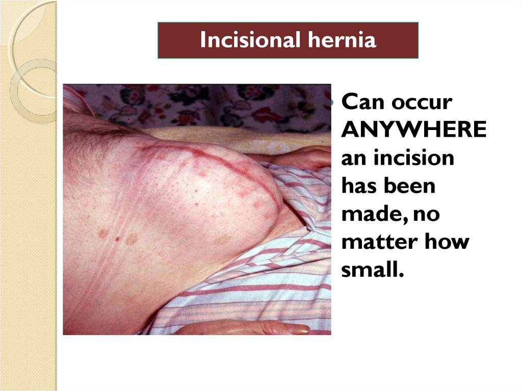
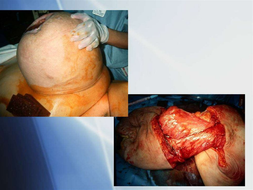
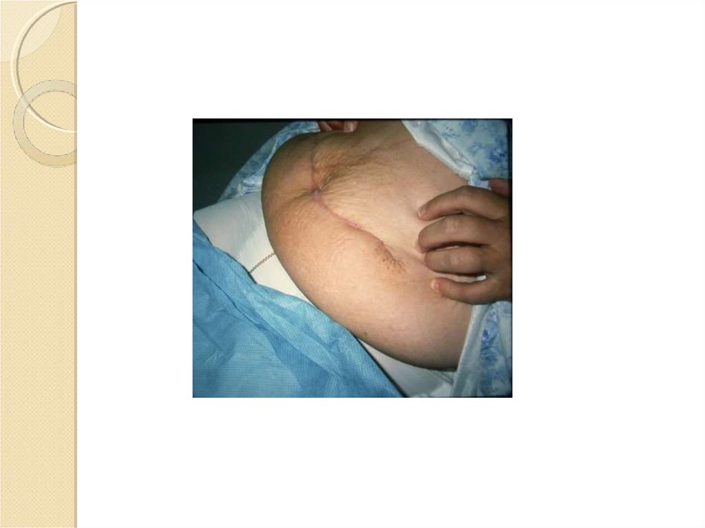
Incisional herniaCan occur
ANYWHERE
an incision
has been
made, no
matter how
small.
36.
Incisional HerniaCan
develop in the original
incision site because of
dehiscence or failure of wound
healing, or can develop at the
sites where sutures are passed
through the tissue during
closure (Swiss cheese-type
hernia), or both.
37.
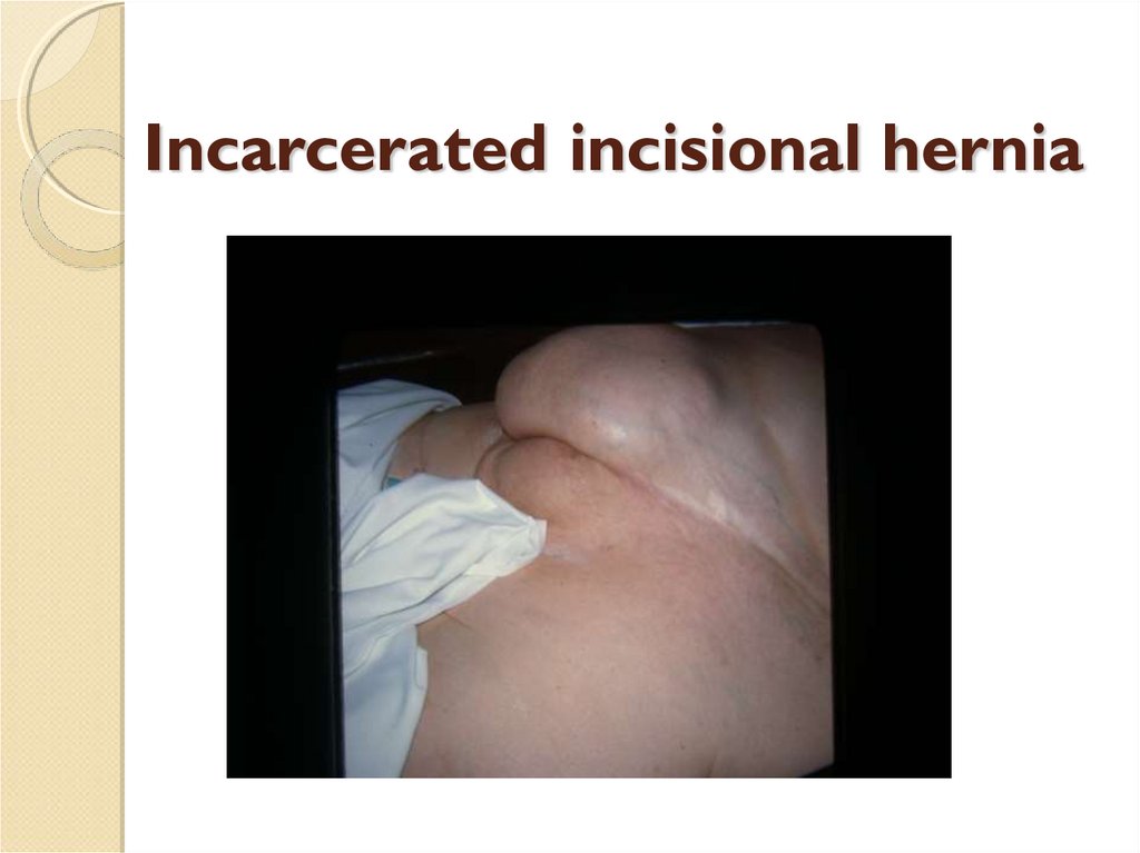
Incarcerated incisional hernia38.
Causes of Incisional HerniaTechnical failure or fascial dehiscence:
◦ Sutures rip through, are placed improperly, or
break
◦ Weak tissue (“ppp”), tension, infection
◦ Occurs within days or weeks after operation
FAILURE OF WOUND HEALING
◦ Most common cause
◦ Seen 6-12 months after operation
39.
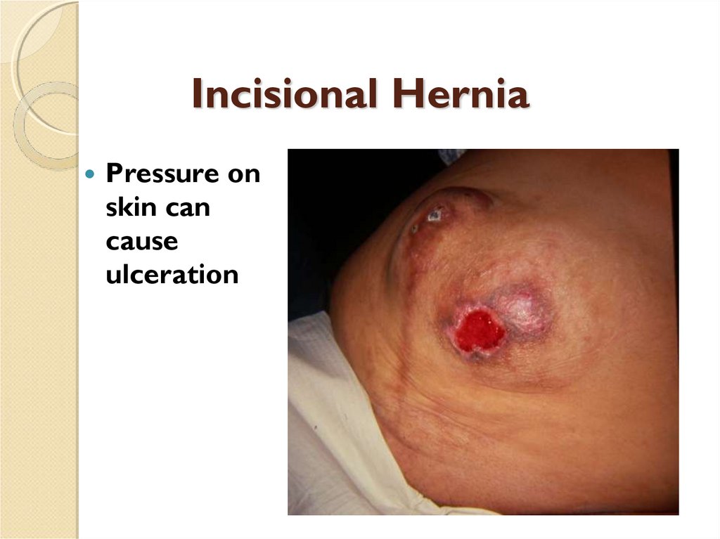
Incisional HerniaPressure on
skin can
cause
ulceration
40.
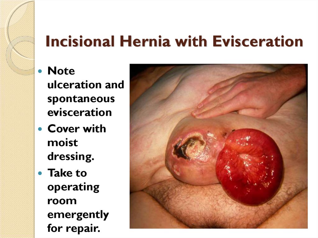
Incisional Hernia with EviscerationNote
ulceration and
spontaneous
evisceration
Cover with
moist
dressing.
Take to
operating
room
emergently
for repair.
41.
42.
43.
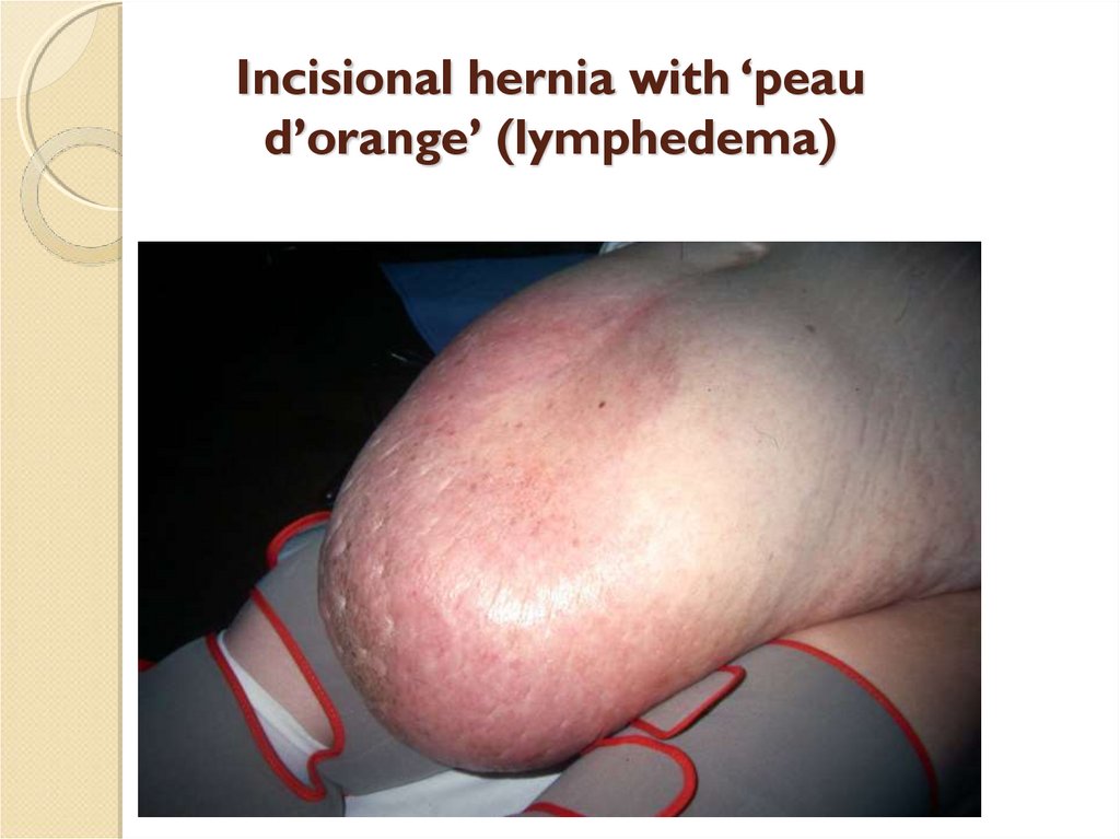
Incisional hernia with ‘peaud’orange’ (lymphedema)
44.
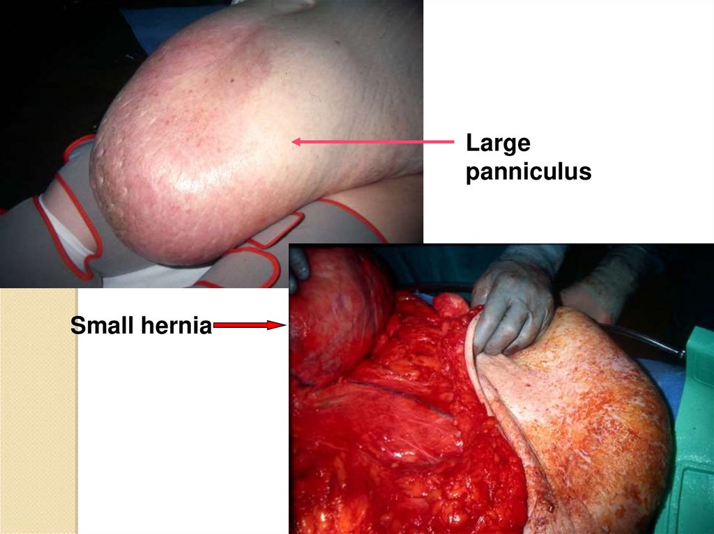
Largepanniculus
Small hernia
45.
46.
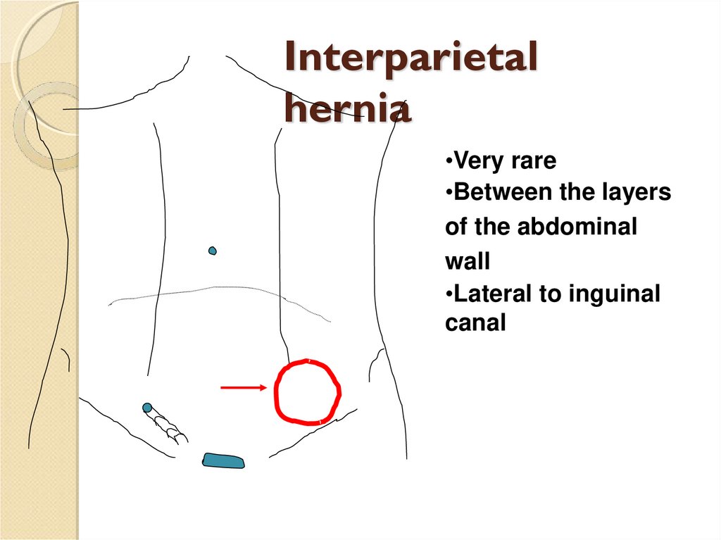
Interparietalhernia
•Very rare
•Between the layers
of the abdominal
wall
•Lateral to inguinal
canal
47.
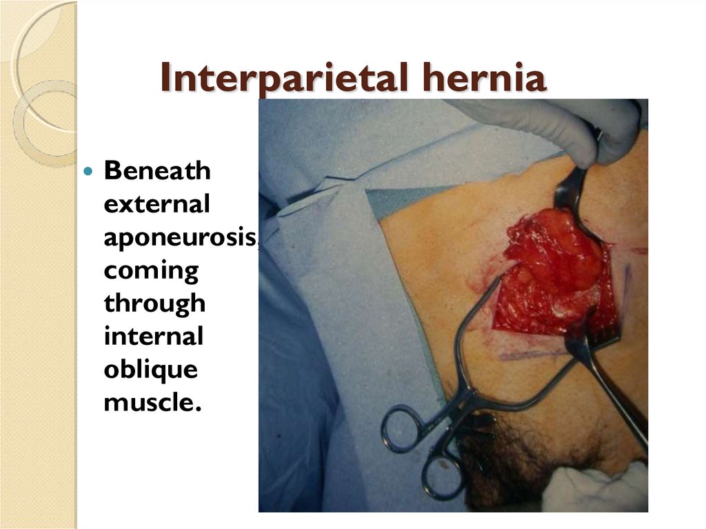
Interparietal herniaBeneath
external
aponeurosis,
coming
through
internal
oblique
muscle.
48.
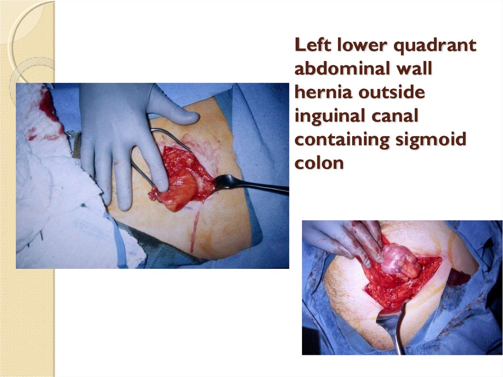
Left lower quadrantabdominal wall
hernia outside
inguinal canal
containing sigmoid
colon
49.
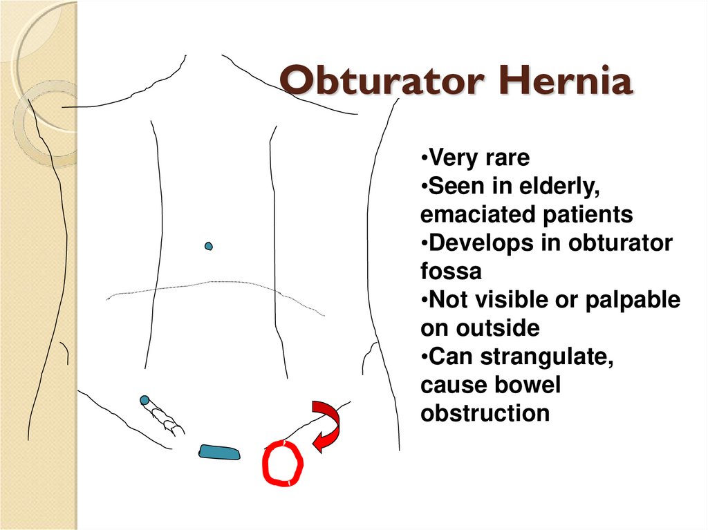
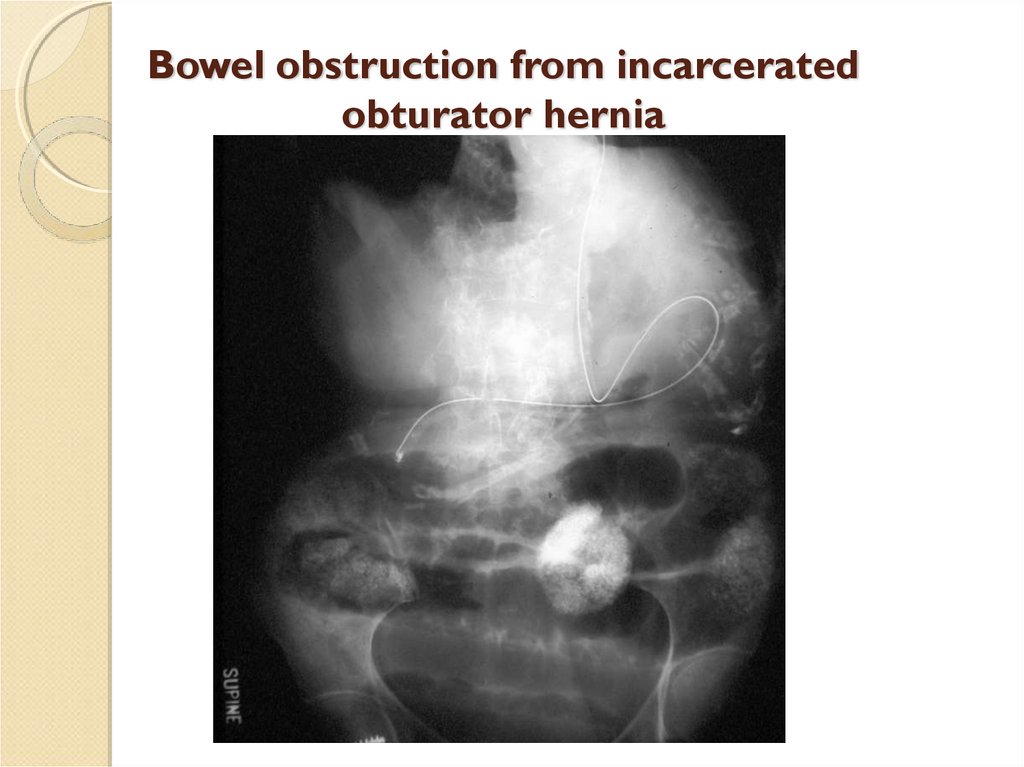
Obturator Hernia•Very rare
•Seen in elderly,
emaciated patients
•Develops in obturator
fossa
•Not visible or palpable
on outside
•Can strangulate,
cause bowel
obstruction
50.
Bowel obstruction from incarceratedobturator hernia
51.
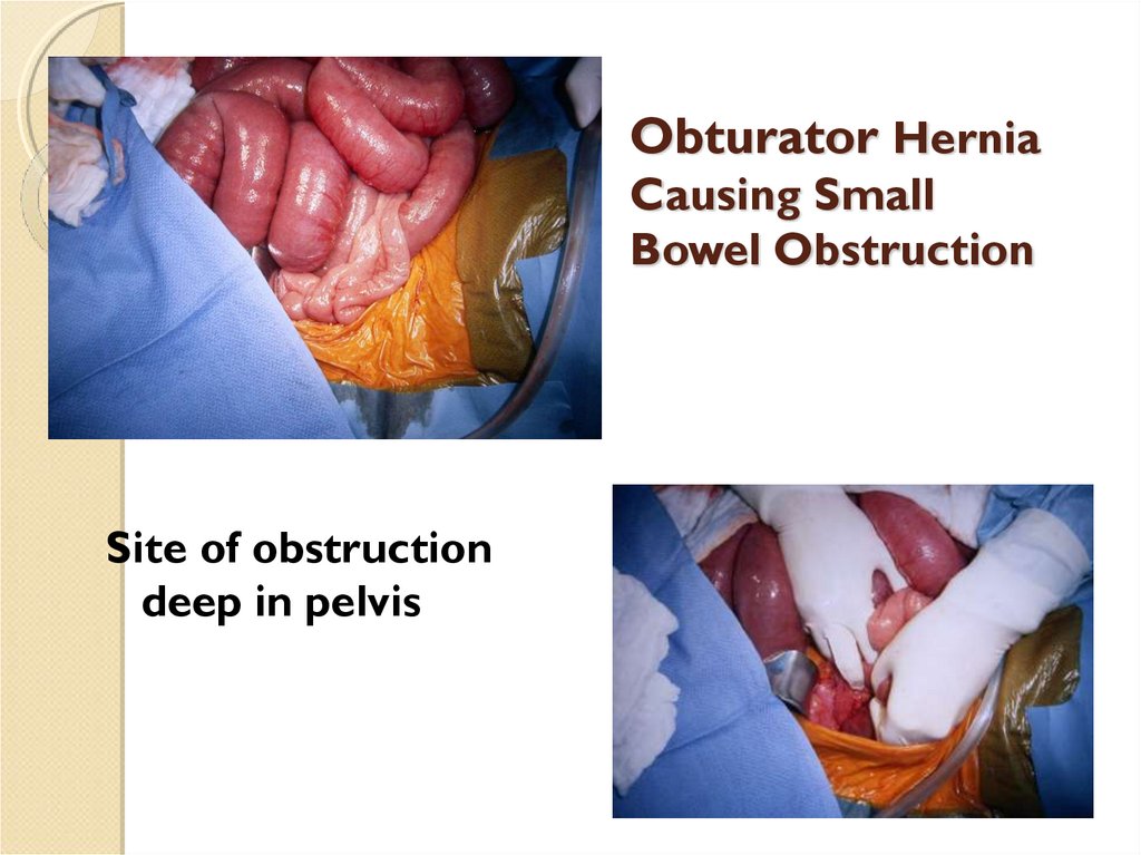
Obturator HerniaCausing Small
Bowel Obstruction
Site of obstruction
deep in pelvis
52.
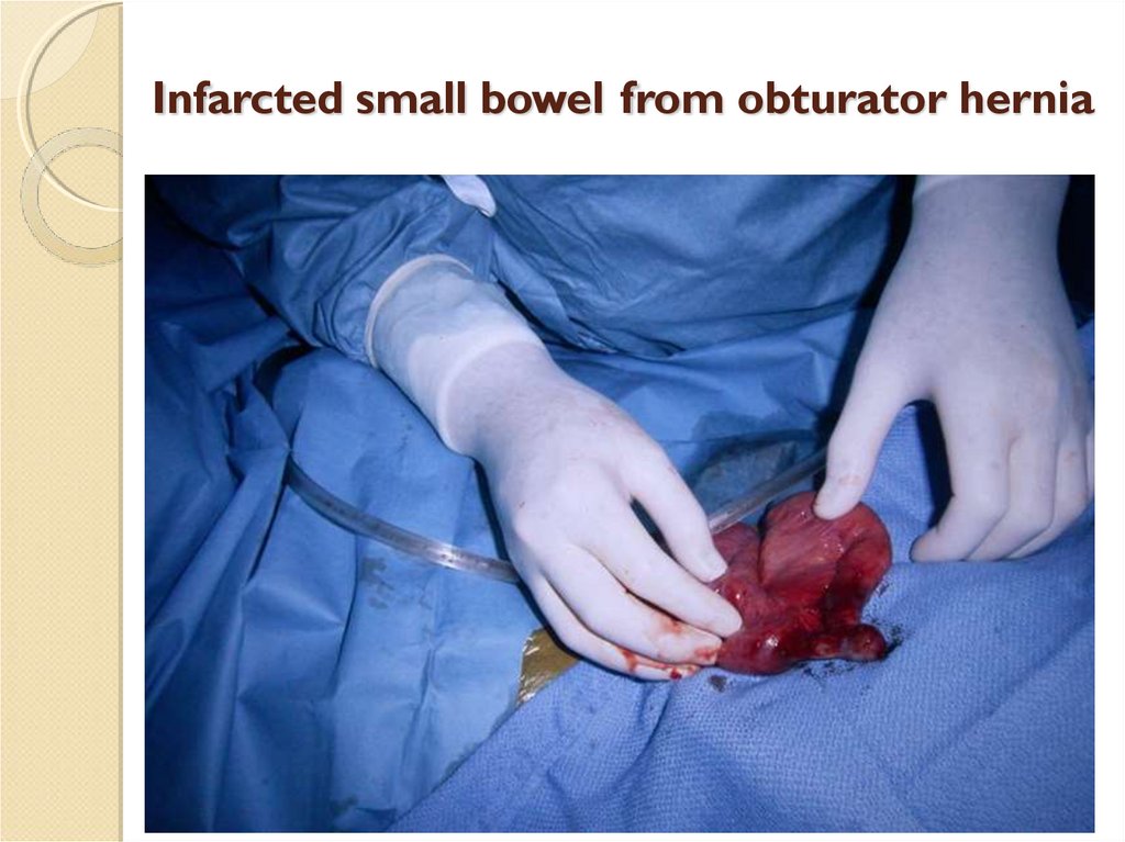
Infarcted small bowel from obturator hernia53.
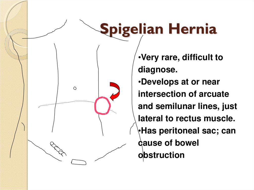
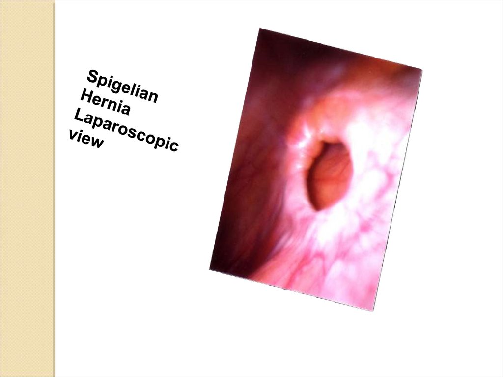
Spigelian Hernia•Very rare, difficult to
diagnose.
•Develops at or near
intersection of arcuate
and semilunar lines, just
lateral to rectus muscle.
•Has peritoneal sac; can
cause of bowel
obstruction
54.
55.
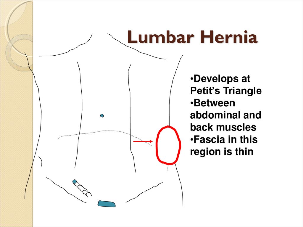
Lumbar Hernia•Develops at
Petit’s Triangle
•Between
abdominal and
back muscles
•Fascia in this
region is thin
56.
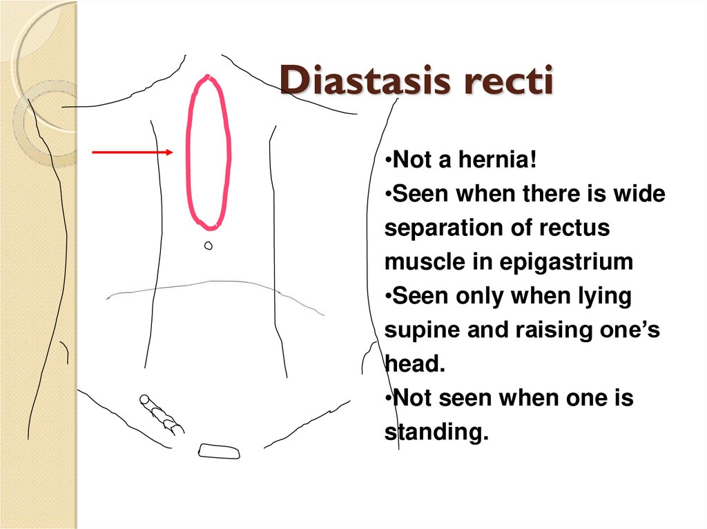
Diastasis recti•Not a hernia!
•Seen when there is wide
separation of rectus
muscle in epigastrium
•Seen only when lying
supine and raising one’s
head.
•Not seen when one is
standing.