
 Медицина
Похожие презентации:


Cardiovascular system
1.
2.
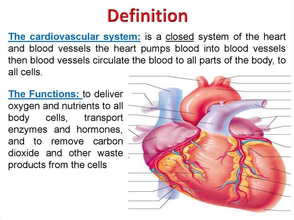
The cardiovascular system: is a closed system of the heartand blood vessels the heart pumps blood into blood vessels
then blood vessels circulate the blood to all parts of the body, to
all cells.
The Functions: to deliver
oxygen and nutrients to all
body
cells,
transport
enzymes and hormones,
and to remove carbon
dioxide and other waste
products from the cells
3.
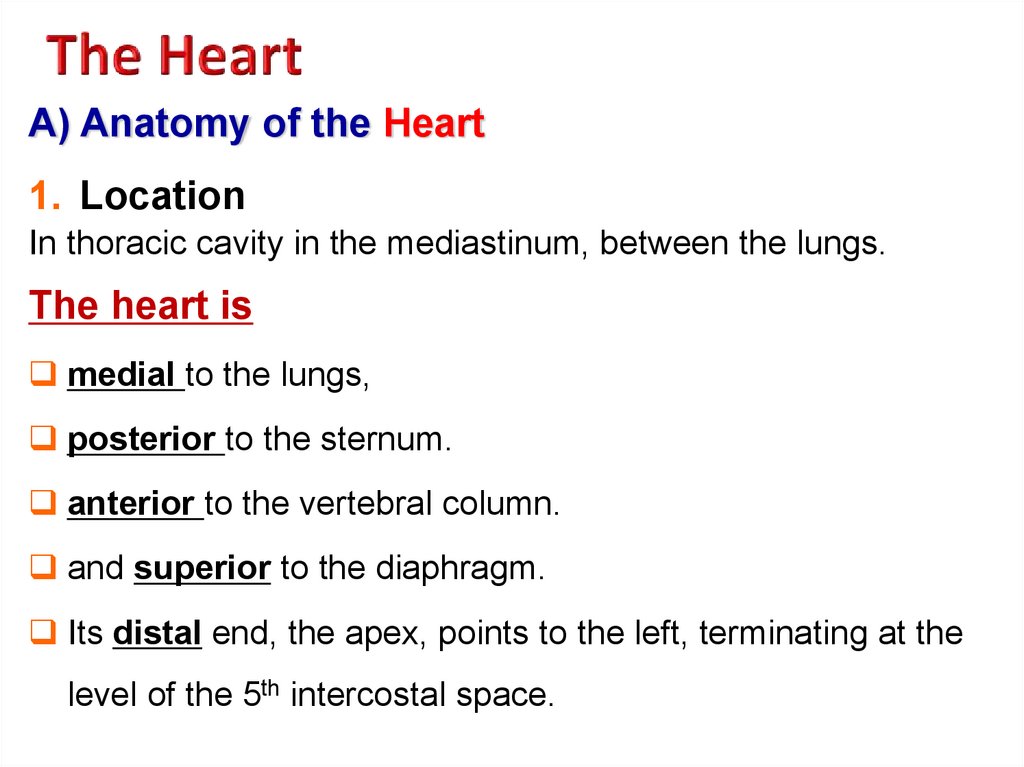
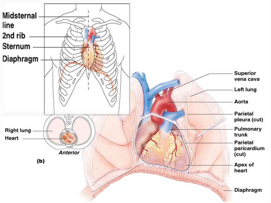
A) Anatomy of the Heart1. Location
In thoracic cavity in the mediastinum, between the lungs.
The heart is
medial to the lungs,
posterior to the sternum.
anterior to the vertebral column.
and superior to the diaphragm.
Its distal end, the apex, points to the left, terminating at the
level of the 5th intercostal space.
4.
5.
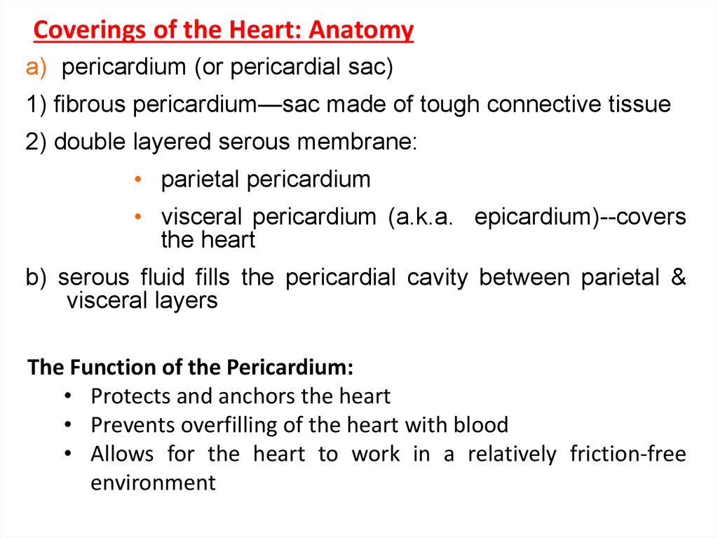
Coverings of the Heart: Anatomya) pericardium (or pericardial sac)
1) fibrous pericardium—sac made of tough connective tissue
2) double layered serous membrane:
• parietal pericardium
• visceral pericardium (a.k.a. epicardium)--covers
the heart
b) serous fluid fills the pericardial cavity between parietal &
visceral layers
The Function of the Pericardium:
• Protects and anchors the heart
• Prevents overfilling of the heart with blood
• Allows for the heart to work in a relatively friction-free
environment
6.
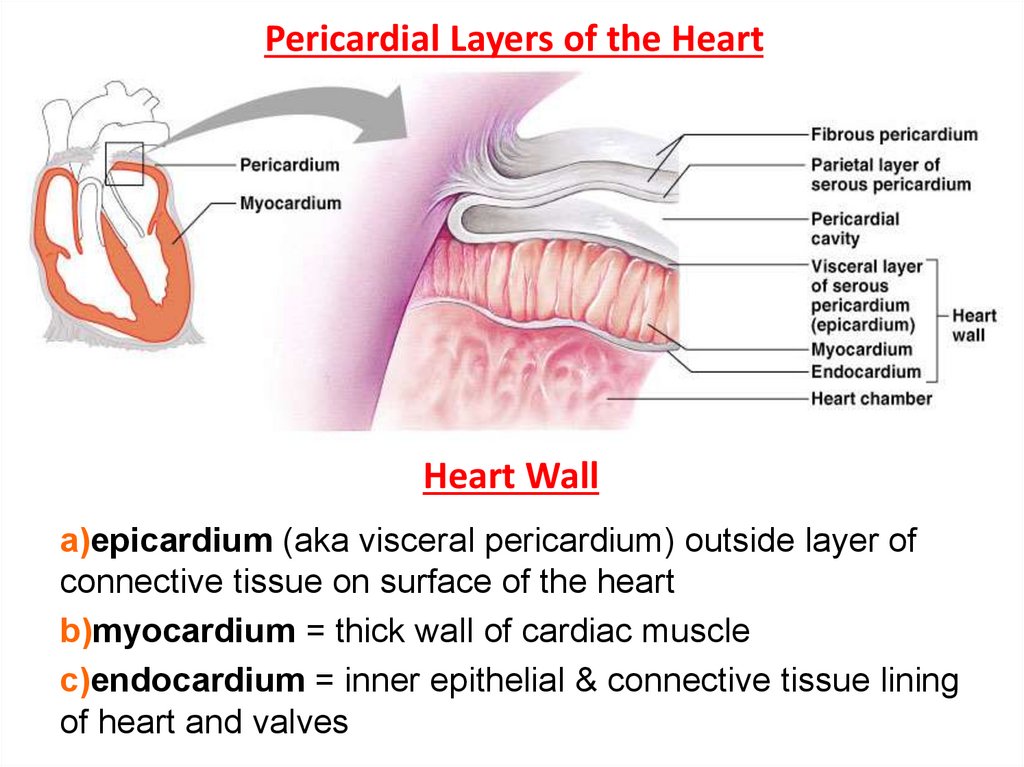
Pericardial Layers of the HeartHeart Wall
a)epicardium (aka visceral pericardium) outside layer of
connective tissue on surface of the heart
b)myocardium = thick wall of cardiac muscle
c)endocardium = inner epithelial & connective tissue lining
of heart and valves
7.
Chambers of the heart (4)• atrium (R & L)—receive blood
each atria extends into a smaller, external chamber called
an auricle
• ventricle (R & L)—inferior to the atria; expel blood out of
the heart
The chambers on the left are separated from the chambers
on the right by a septum (wall of cardiac muscle)
• interatrial septum
• interventricular septum
8.
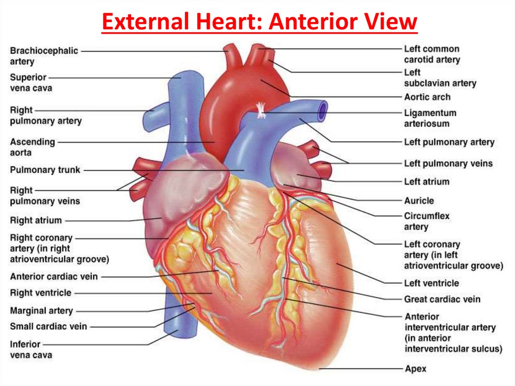
External Heart: Major Vessels of the HeartVessels returning blood to the heart include:
1. Superior and inferior venae cavae
2. Right and left pulmonary veins
Vessels conveying blood away from the heart include:
1. Pulmonary trunk, which splits into right and left pulmonary
arteries
2. Ascending aorta (three branches) –
a. Brachiocephalic
b. Left common carotid
c. Subclavian arteries
9.
External Heart: Anterior View10.
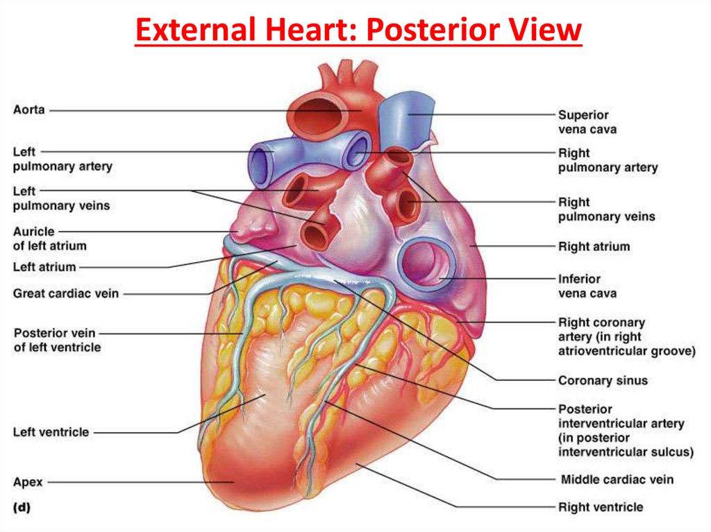
External Heart: Posterior View11.
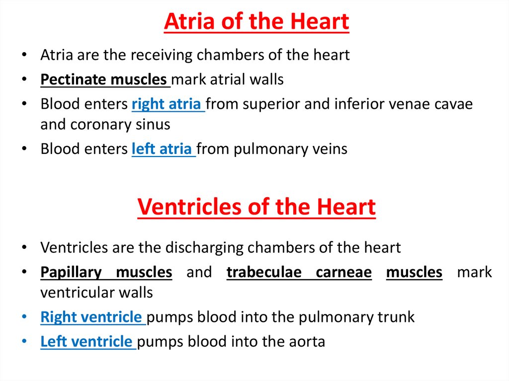
Atria of the Heart• Atria are the receiving chambers of the heart
• Pectinate muscles mark atrial walls
• Blood enters right atria from superior and inferior venae cavae
and coronary sinus
• Blood enters left atria from pulmonary veins
Ventricles of the Heart
• Ventricles are the discharging chambers of the heart
• Papillary muscles and trabeculae carneae muscles mark
ventricular walls
• Right ventricle pumps blood into the pulmonary trunk
• Left ventricle pumps blood into the aorta
12.
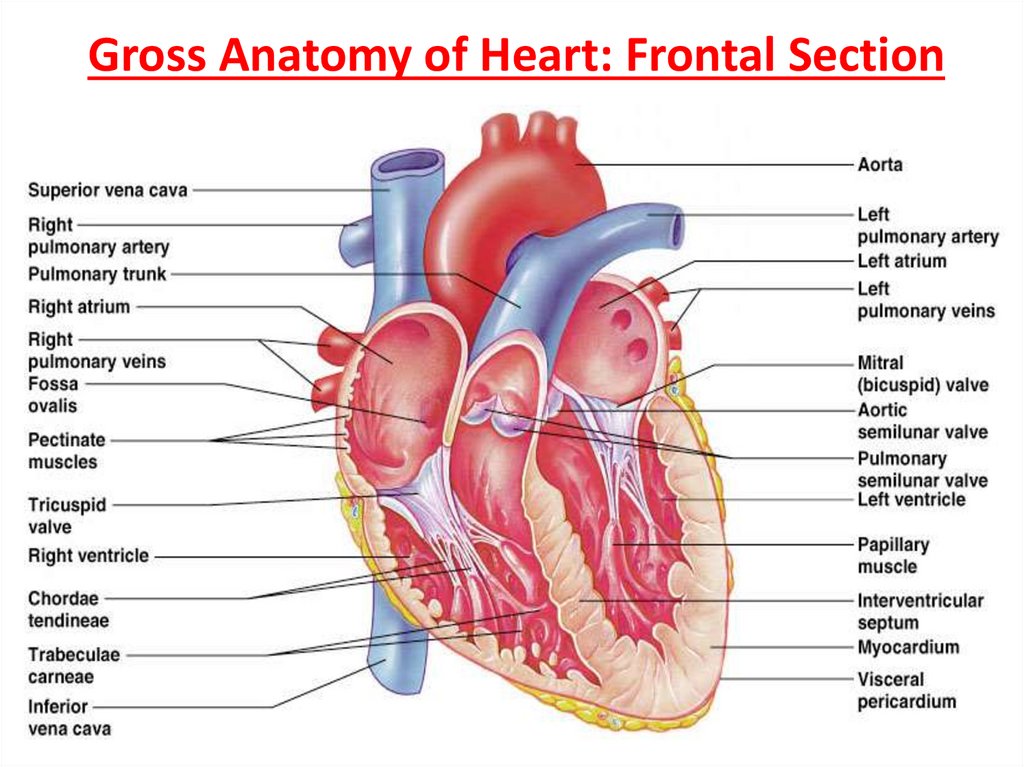
Gross Anatomy of Heart: Frontal Section13.
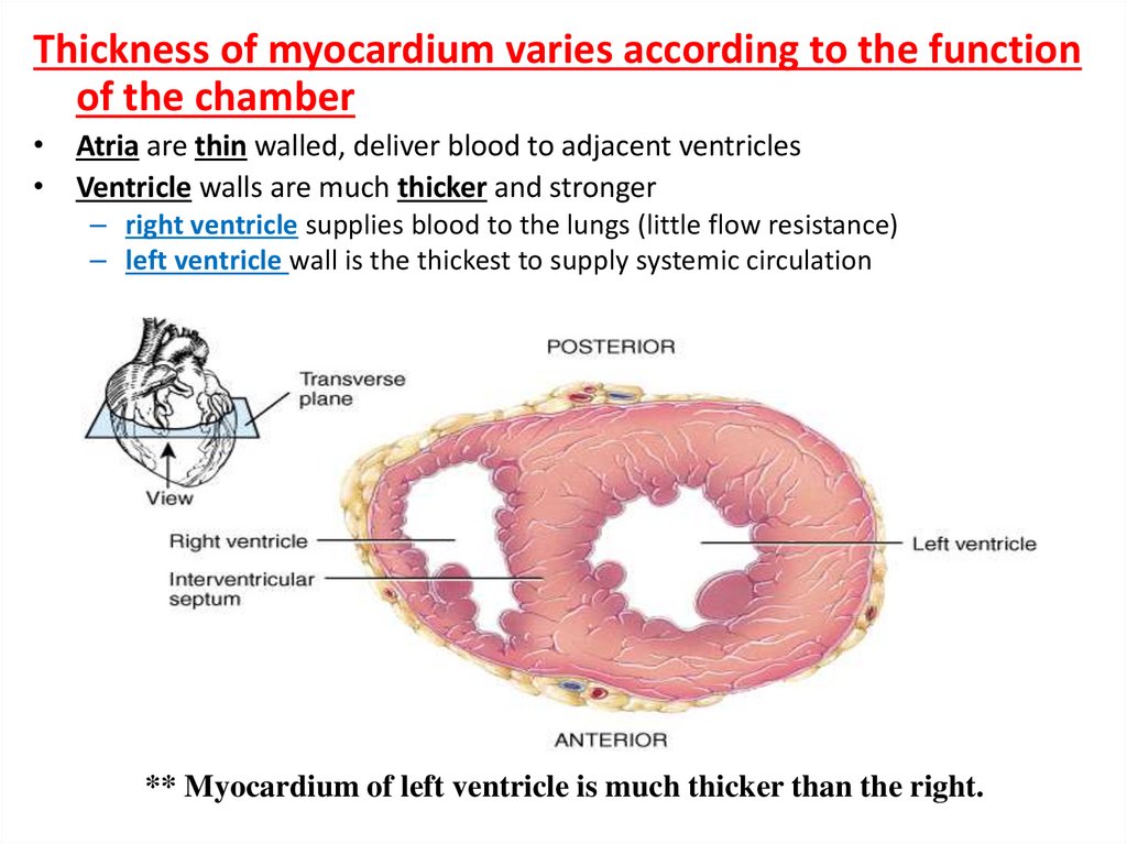
Thickness of myocardium varies according to the functionof the chamber
• Atria are thin walled, deliver blood to adjacent ventricles
• Ventricle walls are much thicker and stronger
– right ventricle supplies blood to the lungs (little flow resistance)
– left ventricle wall is the thickest to supply systemic circulation
** Myocardium of left ventricle is much thicker than the right.
14.
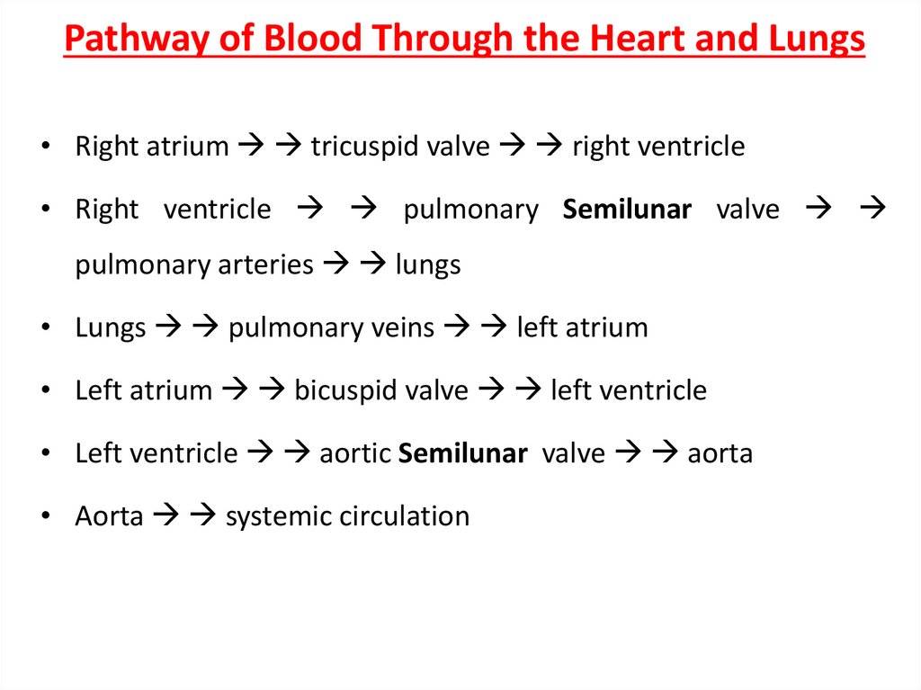
Pathway of Blood Through the Heart and Lungs• Right atrium tricuspid valve right ventricle
• Right ventricle pulmonary Semilunar valve
pulmonary arteries lungs
• Lungs pulmonary veins left atrium
• Left atrium bicuspid valve left ventricle
• Left ventricle aortic Semilunar valve aorta
• Aorta systemic circulation
15.
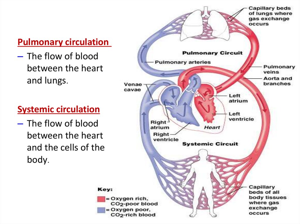
Pulmonary circulation– The flow of blood
between the heart
and lungs.
Systemic circulation
– The flow of blood
between the heart
and the cells of the
body.
16.
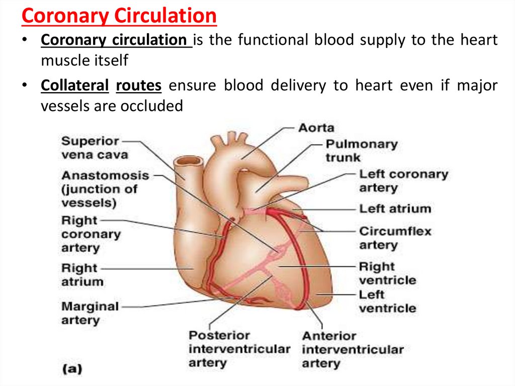
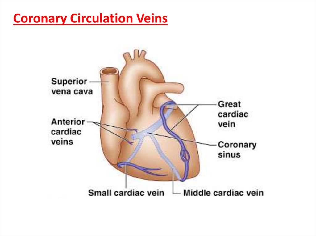
Coronary Circulation• Coronary circulation is the functional blood supply to the heart
muscle itself
• Collateral routes ensure blood delivery to heart even if major
vessels are occluded
17.
Coronary Circulation Veins18.
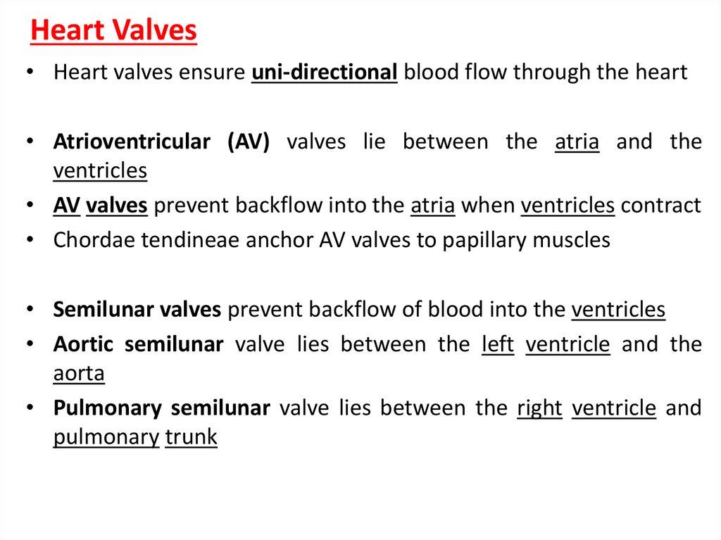
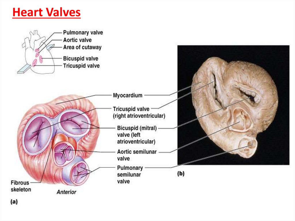
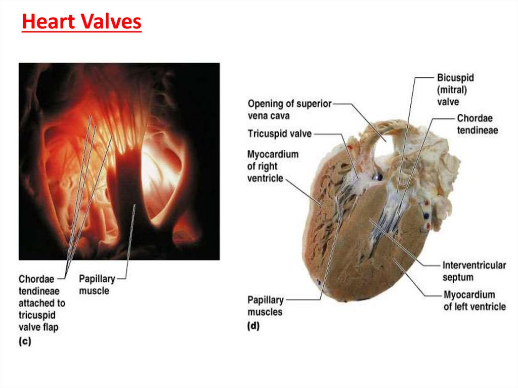
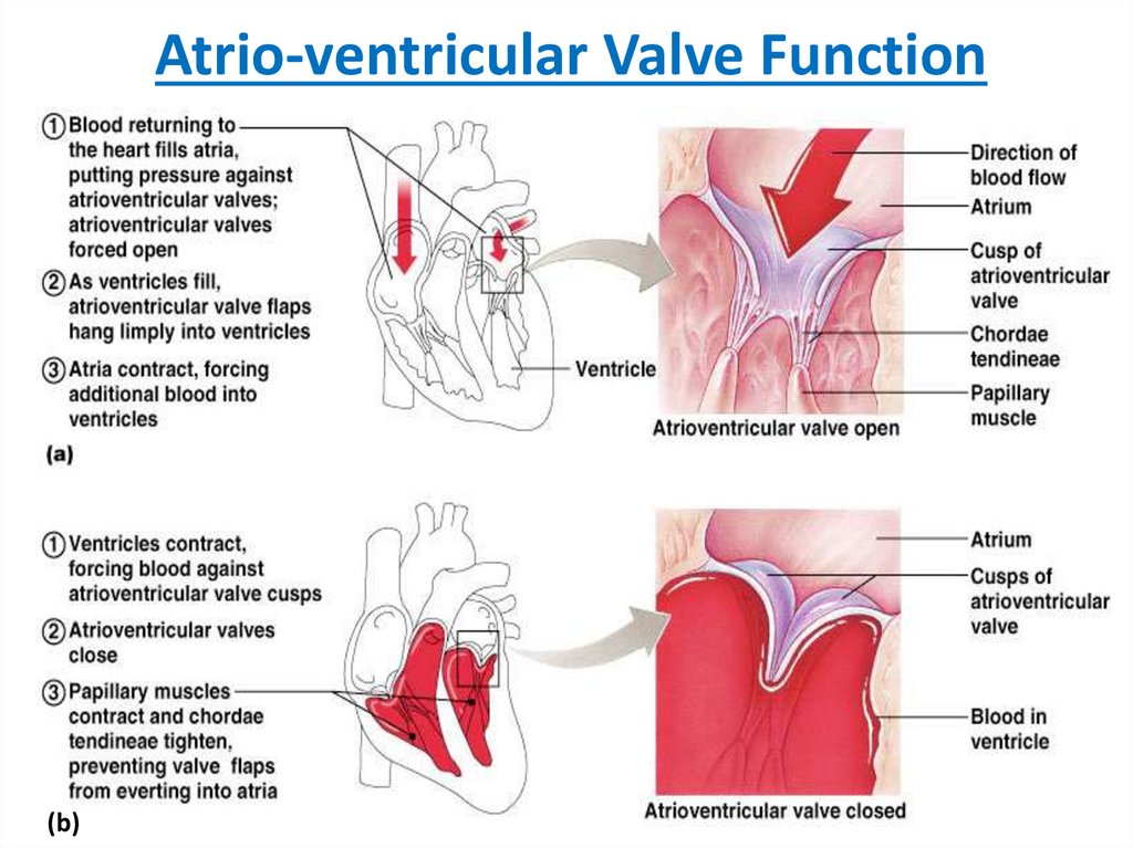
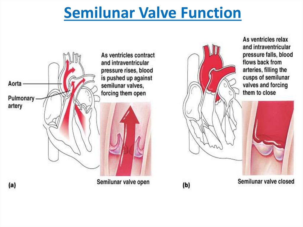
Heart Valves• Heart valves ensure uni-directional blood flow through the heart
• Atrioventricular (AV) valves lie between the atria and the
ventricles
• AV valves prevent backflow into the atria when ventricles contract
• Chordae tendineae anchor AV valves to papillary muscles
• Semilunar valves prevent backflow of blood into the ventricles
• Aortic semilunar valve lies between the left ventricle and the
aorta
• Pulmonary semilunar valve lies between the right ventricle and
pulmonary trunk
19.
Heart Valves20.
Heart Valves21.
Atrio-ventricular Valve Function(b)