


 Медицина
Похожие презентации:


Department of "Visual diagnostics" Radiological research methods and radiological semiotics of acquired diseases
1.
Department of "Visual diagnostics"Radiological research methods and
radiological semiotics of acquired
diseases of the mitral valve
Almaty 2021
2.
Anatomyof theheartThe wall of the heart consists of three
membranes
⦿endocardium (endocardium) –connective fabric with a large number
of elastic fibers and smooth muscle cells, endothelial layer
All heart valves - endocardial folds mitral,
aortic, pulmonary,tricuspid
⦿ myocardium (miocardium)
⦿ pericardium (pericardium) - serous
membrane
Heart
⦿ left atrium - atrium s.
⦿ right atrium - atrium d.
⦿ left ventricle - ventriculus s.
⦿ right ventricle - ventriculus d.
3.
Anatomyof the heartRight atrium
the veins of the great circle flow blood
circulation superior vena cava - v. cava s. collects blood from the head, neck, upper
limbs and chest wall inferior vena cava - v.
cava i. - carrying blood from the lower
extremities, organs and walls of the
abdominal cavity
Left atrium
4 pulmonary veins flow (2 from each lung)
that carry arterial blood from the lungs
4.
Anatomyof the heartRight ventricle - pulmonary
artery exits
The left ventricle exits the
aorta
5.
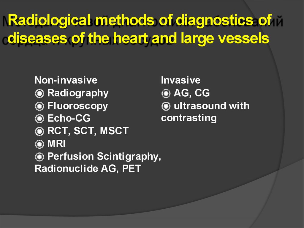
Radiological methods of diagnostics ofdiseases of the heart and large vessels
Non-invasive
Invasive
⦿ Radiography
⦿ AG, CG
⦿ Fluoroscopy
⦿ ultrasound with
contrasting
⦿ Echo-CG
⦿ RCT, SСT, MSCT
⦿ MRI
⦿ Perfusion Scintigraphy,
Radionuclide AG, PET
6.
X-ray, fluoroscopy⦿ Condition of lung tissue and pulmonary pattern (pulmonary
circulation)
⦿ Position, shape, size of the heart, diameter of large vessels
⦿ Study of the shape of the cardiovascular shadow -the ratio of
the cavities of the heart and large vessels
⦿ Study of heart function – contraction myocardium - direct
observation of the screen -fluoroscopy with contrasting the
esophagus
7.
Anterior radiograph of the heart and a diagram to ita - right transverse dimension of the heart shadow (MR)
b - left transverse dimension (ML)
в - oblique size
g - longitudinal heart
8.
Chart of basic measurements of the heart•Of
the proposed options for measuring the shadow of the
heart, the most important is the determination of the
dimensions of the length and diameter
•The length is a line running from the apex of the
cardiovascular angle on the right to the apex of the heart
•The cross section is the sum of two linear measurements horizontal lines running perpendicular to the median sagittal
plane from the most distant points of the right and left
curvature of the heart
•The length of the heart in men is 12-13 cm
•Diameter - 11 - 12 cm
•The length and diameter of the heart in women is 1 cm less
9.
Variants of the location of the heart are normalA - oblique location of the heart (normostenic);
B - horizontal position of the heart (hypersthenic);
C- vertical arrangement of the heart (asthenic)
10.
Chart of basicmeasurementsoftheheart
⦿ Normostenic -
the angle of
inclination of the longitudinal
axis of the heart shadow to the
diameter - 43-48 °MR / ML - 1:
2
⦿ Asthenic - angle - 49-56 °MR /
ML - 1: 1.8
⦿ Hypersthenic - 39-42 °MR /
ML - 1: 2.3
10
11.
Radiometric dimensions that can be determinedon aradiograph
12.
X-ray of the heart withcontrastingesophagus
Barium sulfate - per os
⦿3 standard projections - straight,
right (first) oblique, left (second)
oblique
⦿ Optional - left lateral projection
⦿
12
13.
X-ray silhouette of the heart in frontalprojection
Arcs on the right contour
1 Superior vena cava or
ascending aorta
2 Right atrium
Left contour
1 Aorta
2 Pulmonary cone
3 Left atrial appendage
4 Left ventricle
14.
Radiographs of the heart in direct projectionSigns of a normal heart cardiovascular
shadow occupies two thirds of the
height of the chest cavity
-along the right contour, the upper (A)
and lower arch (PP) are equal to each
other, the atriovasal angle divides the
cardiovascular shadow
-half
-on the left side four arcs are
differentiated - as a rule, the arch of the
left ventricular appendage does not
normally protrude one third of the
shadow of the heart in diameter lies on
the right, two-thirds on the left,
-acute cardio-diaphragm angles
15.
X-ray examination of the heartDiagramoftheimageandradiographoftheheartindirectprojection
Left 4 arcs -aorta, pulmonarycone,left atrial appendage,leftventricle,on theright 2arcs right atrium, aorta
16.
X-ray examination of the heartDiagram of the image and radiograph of the heart in the left lateral projection
Toassess thesize oftheright andleft ventricle -theirratio is1: 1
16
17.
Radiograph of the heart in the right oblique projection- the posterior contour is formed by the atria (left
at the top), the anterior contour by the ventricles
(right at the top - conus pulmonalis)
Signs of the norm-Cardiovascular
shadow is rectangular in shape
triangle
- between the posterior contour
of the heart and the shadow of
the spine, rectangular
enlightenment
- - the anterior contour of the
heart is not reaches the chest
wall, including in the area of the
left ventricular arch
- - no significant swelling of
individual chambers and
vessels
- - the contrasting esophagus is
not deviates
18.
Radiograph of the heart in the left oblique projection – the right partsof the heart are located anteriorly, theleft ones areposteriorly, above are the
atria, below are the ventricles
Signs of the norm
-between the posterior contour of
the left atrium and the shadow
spine enlightenment triangular
shape
- the anterior contour of the
heart is not reaches the chest
wall, including in the area of
the arch of the right ventricle
- - no significant bulging
separate departments and
vessels
19.
X-ray examination of the heartDiagram of the image and radiograph of the heart in the right oblique projection
the posterior contour is formed by the atria (left top), the anterior
contourventricles (right above - conus pulmonalis)
19
20.
X-ray examination of the heartDiagram of t he image and radiograph of the heart in the left oblique
projection
the right heart is located anteriorly, the left is posterior above - atria,
below - ventricles
21.
Heart in childhood x-ray of a 4year-old child22.
Various forms of cardiovascular shadow - normal, mitralconfiguration, aortic configuration, trapezoidal shadow
23.
Radiation methods of researchof theheartFirst choice methods
-Echocardiography with doppler
echocardiography(CDM, energy and tissue
DG)
-Doppler ultrasound - measurement of blood
flow velocity using ultrasound based on the
Doppler effect.
-Doppler effect - a physical phenomenon,
according to which the frequency the
perception of a sound emitted by a moving
object changes when it is perceived by a
stationary object
24.
Features of radiation examinationof the heart
1. 1.The
heart is examined together with the pulmonary circulation -hemodynamic
research principle
2. 2. Study of heart function is mandatory
3. 3. Study of blood flow is mandatory
4. 4. the heart is examined with contrasting the esophagus
The state of the pulmonary circulation can be examined on a plain chest x-ray
Plain radiography and detailed ultrasound are sufficient to make a diagnosis in
most medical situations
25.
Left atrioventricular foramen stenosis (mitralstenosis)
⦿Smoothness of the waist of the heart
⦿Bulging of 3 arcs along the left contour of the heart
⦿In oblique projections, the contrasted esophagus is deflected by the
left atrium along a small radius
⦿Atriomegaly
⦿In some cases, calcification of the leaflets of the mitral valve is visible
⦿Enlargement of the right ventricle and right atrium
⦿The shadow of the heart is enlarged in diameter, sometimes to the
left, if the enlarged right ventricle becomes edge-forming, and the
unchanged left ventricle rotates posteriorly
⦿Signs of venous stasis are especially pronounced with defect
decompensation
26.
Insufficiency of the mitral valve (mitralregurgitation)
⦿Left atrial enlargement
⦿Deviation of the contrasted esophagus along an arc of a large radius
(more6 cm)
⦿The waist of the heart is smoothed, the 3rd arch bulges (enlarged left
atrium)
⦿Increased left ventricular arch - signs of its hypertrophy and dilatation
⦿In the presence of hemodynamic disorders, increased vascular, and in
further and interstitial component of the pulmonary pattern
⦿With prolonged pulmonary hypertension - an increase in the right heart
⦿Difference from mitral stenosis - the pulmonary cone of the right
ventricle and the arch of the pulmonary trunk bulge weakly
27.
Mitral heart disease - mitral valveinsufficiency
28.
Mitral insufficiency29.
Mitral stenosis32
30.
Hemosiderosis withmitral stenosis
33
31.
Mitral stenosis32.
Еchocardiography
⦿Subtlemorphologicalfeaturesof thestructureof the
endocardium
⦿Valvefunction
⦿Myocardialthickness, dimensions ofheartcavities
⦿Myocardialcontractilefunction
⦿Blood flow
33.
Ultrasound examination of theheartStandard
projections
for ultrasound
imaging
-Parasternal
-Subcostal
-Apical
-Suprasternal
34.
4335.
Principle of image acquisition in M-modeUltrasound examination is carried out in standard positions
The ability to obtain all the necessary standard positions and
analyze them forms the basis of knowledge echocardiography
The study is carried out in one-dimensional mode, twodimensional mode and using dopplerography.
36.
EchocardiographyM-mode - detailed information about the dynamics of the
behavior of reflecting structures located along the
ultrasound beam; the form of a group of curves, each of
which corresponds to certain structure
Violations of local contractility of the left ventricle
normal
akinesia
hypokinesia
dyskinesia
37.
Principle of acquiring an image in two-dimensionalmode
Sonography (B - mode) - two-dimensional examination of the
heart Provides an opportunity to observe the movements of the
walls of the heart and valves in real time on the display screen
In standard projections, the sensor is positioned along the long
or short axis of the heart Microconvex probe, 3.5 MHz
The diagram shows a study at the level of the mitral valves.
Descending sector scanning shows skin, right ventricle,
interventricular wall, left ventricular cavity, mitral valve, left
atrium
38.
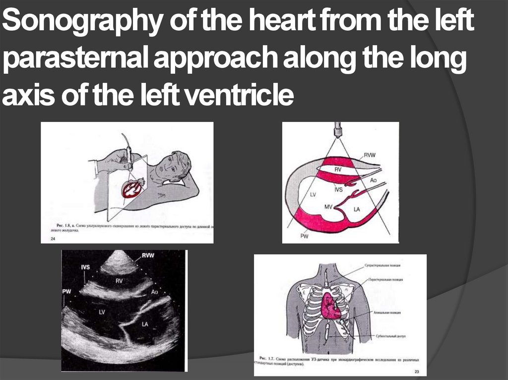
Sonography of the heart from the leftparasternal approach along the long
axis of the left ventricle
39.
Parasternal access along theshort axis at the level of the aortic
valve
40.
Scanning at the level of theaortic valve
41.
Scanning from parasternal access atthe mitral level valve
42.
Scanning by subcostal approachalong the long axis of the heart
43.
Ultrasound signs vegetations on theleaflets of the mitral valve
44.
Diastolic blood flow in normalconditions and with stenosis of the
mitral foramen
45.
Insufficiency of the mitral valve46.
Reduction of diastolic opening of the mitral valveand narrowing of the mitral opening with stenosis
(right), left -norm. Parasternal access along the
short axis.
47.
48.
Mitral stenosis (B-mode)49.
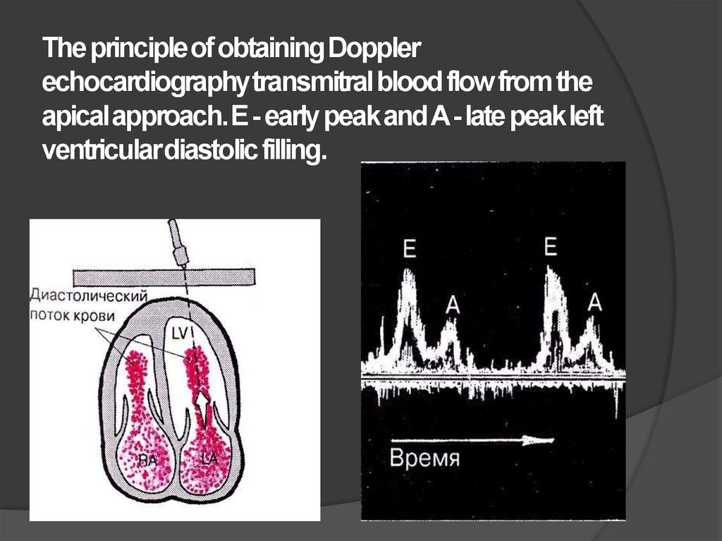
The principle of obtaining Dopplerechocardiography transmitralblood flow from the
apical approach. E - early peak and A -late peak left
ventricular diastolic filling.
50.
Transmitral blood flow Doppler for mitralstenosis
51.
Quantitative assessment of blood flow velocitywith Doppler ultrasonography.
52.
RCT, SCTIndications
⦿Clarification of the nature of changes in
the lung tissue
⦿Localization of pathological education
(intra-, paracardial)
⦿Diseases of the pericardium
⦿Aortic aneurysm of any location
53.
limitations⦿Ionizing radiation
⦿Insufficient image acquisition
speed
54.
Multispiral computed tomographyadvantages
⦿Increase research speed
⦿Enhanced resolution
⦿High quality reconstructions in various planes
⦿CT angiography
55.
magnetic resonance imagingIndications
⦿Arrhythmogenic pancreatic dysplasia
⦿Diagnosis of heart tumors and blood
clots
⦿Complicated CHD
56.
Cardiometry, MRI69
57.
MRI aortic stenosis70
58.
MRI of theheart
71
59.
Aortic aneurysmthrombosis
72
60.
Radionuclide methodsIndications
⦿Assessment ofmyocardial perfusion
⦿Assessment ofthereserve capacity oftheheartmuscleEquilibrium VH
⦿Pumping function ofthemyocardium
⦿Thenature ofthemovement ofthewallsofthe heart
PET
⦿ Assessment of myocardial
perfusion and metabolism
61.
Coronary angiographyIndications
⦿Assessment of the condition of the
coronary arteries
⦿Study of blood flows in the chambers
of the heart during
-complex CHD
-arrhythmogenic dysplasia of right
ventricle
Contraindication: Contrast intolerance
62.
MRI of theheart