


")

")


")


")
")


 Медицина
Похожие презентации:


Management of patients with cardiomegaly and heart failure
1. MANAGEMENT OF PATIENTS WITH CARDIOMEGALY AND HEART FAILURE
2.
Cardiomegaly is a considerableenlargement of the heart from its
dilatation and/or hypertrophy,
accumulation of waste products due to
impaired metabolism or development of
neoplastic processes.
It is usually manifested by symptoms of
heart failure, rhythm and conduction
disorders.
3. COMMON SIGNS OF CARDIOMEGALY
Enlargement of the heartRhythm and conduction disturbances
Physical findings: widened borders of the heart,
dull sounds, weakened 1st sound over the
apex, presystolic gallop, additional 3rd and 4th
sounds, murmurs of regurgitation
Signs of the underlying disease which has
caused cardiomegaly
4. MAIN CAUSES OF CARDIOMEGALY
IHD: atherosclerotic cardiosclerosis, postinfarction cardiosclerosis, ischemiccardiomyopathy, cardiac aneurysm
Arterial hypertension
Heart defects (congenital, acquired)
Diffuse myocarditis
Primary and secondary cardiomyopathy
Pericarditis
Cardiac tumours
Athlete’s heart
5. DIAGNOSIS OF CARDIOMEGALY
Interviewing the patient to find out the maincomplaints: dyspnoea, fatigability, weakness,
less tolerance to physical exertion; feeling of
heaviness in the right hypochondrium,
peripheral oedemas, pains in the chest (angina
or cardialgia), heart palpitations
6. DIAGNOSIS OF CARDIOMEGALY
History. We should specify: consequence ofdevelopment of heart failure symptoms (left or
right ventricular failure separately or total heart
failure at once), episodes of BP elevation,
history of acute rheumatic fever, heart
murmurs revealed in childhood, history of
myocardial infarction, aches in the heart in the
past, severe diabetes, connection with past
infections, vaccination, alcohol consumption,
medications, family history.
7. DIAGNOSIS OF CARDIOMEGALY
PHYSICAL EXAMINATION:Inspection: cyanosis, acrocyanosis, paleness,
ruddiness of cheeks, swollen veins in the neck,
pulsating vessels or precardial area, widened
venous network, enlarged abdomen,
orthopnoea;
Palpation: assessing the pulse rate (slow, of
small amplitude or full, rapid; pulse deficit),
chest tremors, apical pulse shifted to the left;
Taking BP: normal, hypertension, high pulse
pressure
8. DIAGNOSIS OF CARDIOMEGALY
PHYSICAL EXAMINATION:Percussion: wider vascular bundle, wider
borders of heart dullness
Auscultation: dull sounds, weakened 1st sound,
protodiastolic or presystolic gallop rhythm,
extra 3rd and 4th sounds, murmurs of
regurgitation, cardiac rhythm disorders;
9. DIAGNOSIS OF CARDIOMEGALY
LABORATORY FINDINGS:CDC: diagnosis of anaemia, polycythemia (COPD,
cyanotic congenital heart defects), leucocytosis and
elevated ESR (infectious endo-, myocarditis, exudative
pericarditis)
Blood and urine glucose: diabetes mellitus
Lipid profile: CHD, aortic atherosclerosis
CRP test: endo-, myocarditis, systemic connective
tissue diseases (SCTD)
Antinuclear and rheumatoid factors: SCTD
Bacterial blood culture: infectious endocarditis
Т3, Т4, TSH: hypo-, hyperthyroidism
Urea, electrolytes, creatinine: CKD
10. DIAGNOSIS OF CARDIOMEGALY
INSTRUMENTAL INVESTIGATIONS:Chest X-ray (shape of the heart, enlargement
of certain chambers, vessels): ‘mitral’, ‘aortic’ or
spherical shape of the heart;
ECG: the changes are non-specific and
manifold (hypertrophy of chambers of the
heart, rhythm and conduction disorders,
changes due to scarring of the heart)
11. DIAGNOSIS OF CARDIOMEGALY
INSTRUMENTAL INVESTIGATIONS:Echocardiography is the most valuable noninvasive methods of diagnosis assesses
thoroughly morphological changes in the
chambers and valves of the heart, peculiarities
of movement of valves, thickened areas and
calcinosis, impaired movement of blood in the
heart, signs of pulmonary hypertension,
elevated LVEDP, width of heart walls,
asymmetry due to hypertrophy or symmetry,
areas of hypo- or akinesia.
12. MANAGEMENT OF PATIENTS WITH CARDIOMEGALY
To confirm cardiomegaly (to determineenlargement of the chambers, dilation or
hypertrophy, to estimate the degree of
enlargement of the chambers)
To reveal the cause of cardiomegaly
To assess its functional significance
To plan management of the patient
13. MANAGEMENT OF PATIENTS WITH CARDIOMEGALY
ASSESSMENT OF FUNCTIONALSIGNIFICANCE OF CARDIOMEGALY:
Symptoms of dyspnoea, weakness, fatigability
Cardiac ventricular function (EF), congestive
heart failure
Determination of NYHA functional class of heart
failure
14. MANAGEMENT OF PATIENTS WITH CARDIOMEGALY
PLANNING MANAGEMENT OF THEPATIENT:
Prevention: changing lifestyle, treatment of
hypertension, CHD or any other underlying
disease
Medical treatment: diuretics, ACE inhibitors (or
sartans), beta-blockers, nitrates,
antiaggregants, anticoagulants, cardiac
glycosides, antiarrhythmic drugs
Surgical treatment
15. CARDIOMYOPATHIES (CM)
European Society of Cardiology (ESC),2008
“Cardiomyopathies are structural and
functional myocardial diseases in the
absence of systemic hypertension, coronary
atherosclerosis, valvulopathies, or
congenital heart disease”.
16. CARDIOMYOPATHIES
CM phenotypesHCM (hypertrophic CM)
2. DCM (dilated CM)
3. ARVD (arrhythmogenic right ventricular
dysplasia)
4. RCM (restrictive CM)
5. Non-classified:
Non compacted myocardium
Stress CM (Takotsubo CM)
1.
17. ESC RECOMMENDATIONS (2008)
All CM phenotypes are divided into:1. Familial (inherited, genetic)
Non-identified genetic disorder
A disease subgroup (including those with
known gene mutation, metabolic disorders,
glycogen storage disease, impaired fatty acid
metabolism, lysosome storage disorders)
18.
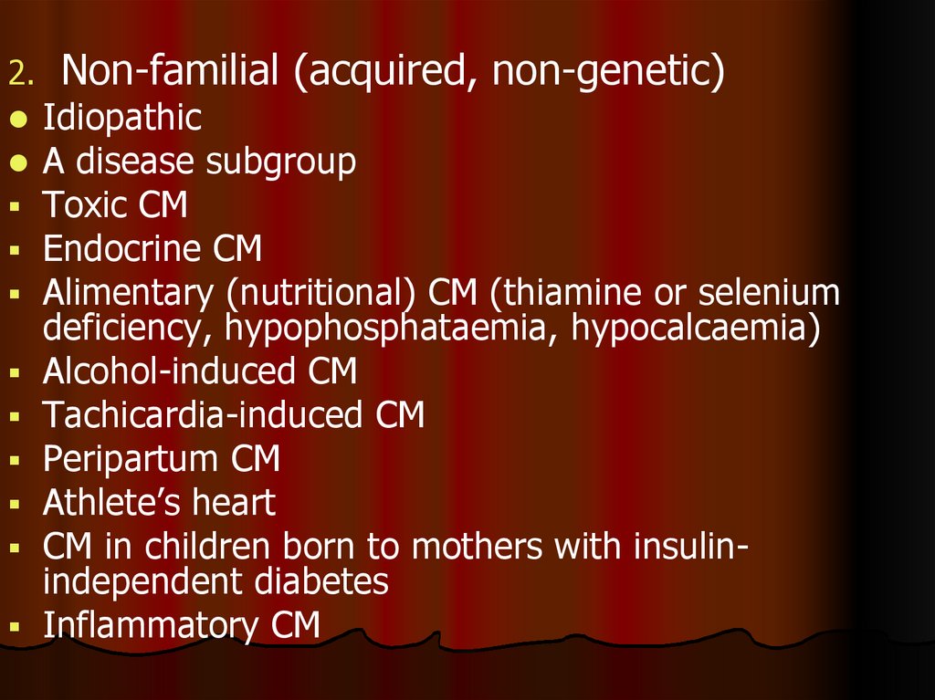
2.Non-familial (acquired, non-genetic)
Idiopathic
A disease subgroup
Toxic CM
Endocrine CM
Alimentary (nutritional) CM (thiamine or selenium
deficiency, hypophosphataemia, hypocalcaemia)
Alcohol-induced CM
Tachicardia-induced CM
Peripartum CM
Athlete’s heart
CM in children born to mothers with insulinindependent diabetes
Inflammatory CM
19. HYPERTROPHIC CARDIOMYOPATHY
Hypertrophic cardiomyopathy is defined by thepresence of increased left ventricular (LV) wall
thickness that is not solely explained by abnormal
loading conditions.
HCMP occurrence is 0.02-0.23% in adults and
unknown in children, incidence being equal
approximately to 0.3-0.5 per 100000 of population
(0.005-0.07%). Most studies note that men develop
the disease more often than women; CM prevalence
rate in different races is similar.
20. HYPERTROPHIC CARDIOMYOPATHY
2014 ESC guidelines on diagnosis andmanagement of hypertrophic cardiomyopathy
HCM is prevalently a genetic disease of the muscle of
the heart characterised by a set of specific
morphological and functional changes and progressing
steadily with a high risk of development of severe lifethreatening arrhythmia and sudden cardiac death.
HCM synonyms
Idiopathic hypertrophic subaortic stenosis
Muscular subaortic stenosis
Hypertrophic obstructive cardiomayopathy
21. HYPERTROPHIC CARDIOMYOPATHY
HCM is the main cause ofsudden cardiac death (SCD)
in the young, in sportsmen
in particular
The findings of autopsies of sportsmen who died of
SCD show that death may have been caused by nonrevealed or clinically silent HCM in 36% of cases.
22. HYPERTROPHIC CARDIOMYOPATHY
HCM is characterised by considerable(more than 15 mm) hypertrophy of
myocardium of the left and/or in rare
cases right ventricle, often asymmetric
due to thickened IVS, with frequent
development of left ventricular outflow
tract obstruction (LVOTO) in the absence
of known causes (hypertension, heart
defects and specific heart diseases).
23. HYPERTROPHIC CARDIOMYOPATHY
24. HYPERTROPHIC CARDIOMYOPATHY
25. HYPERTROPHIC CARDIOMYOPATHY
26. HYPERTROPHIC CARDIOMYOPATHY
27. HYPERTROPHIC CARDIOMYOPATHY
Pathogenesis of HCM includes 4 interrelatedprocesses:
1. Left ventricular outflow tract obstruction
(LVOTO)
2. Diastolic dysfunction
3. Myocardial ischaemia
4. Mitral regurgitation
28. HYPERTROPHIC CARDIOMYOPATHY
CLINICAL MANIFESTATION:Asymptomatic course in 25% cases
Dyspnoea on exertion (90%), orthopnoea;
Angina (70-80%);
Syncope (20%), presyncope (50%)
Greater
obstruction in augmentation of cardiac
contractility due to exertion;
Cardiac arrhythmias (90%)
Thromboembolic risks of atrial fibrillation
Sudden cardiac death
29. HYPERTROPHIC CARDIOMYOPATHY
ON EXAMINATION:intense, raised cardiac impulse shifted slightly
to the left
double, triple or even quadruple impulse over
the apex of the heart
alternating pulse
An ejection systolic murmur over the apex or in
the 3rd-4th intercostal space at the left sternal
edge, ‘rhomboid’ character of systolic murmur
30. HYPERTROPHIC CARDIOMYOPATHY
DIAGNOSIS:DNA-diagnosis using polymerase chain
reaction (PSR)
Genetic testing of relations in the first degree
to assess the risk of development of HCM
ECG and daily monitoring of ECG
Chest X-ray, EchoCG, MRI
Cardiac stress tests
Coronary angiography
31. HYPERTROPHIC CARDIOMYOPATHY
32. HYPERTROPHIC CARDIOMYOPATHY
Left ventricular wall or IVS thickness >15 mm33. HYPERTROPHIC CARDIOMYOPATHY
MEDICAL TREATMENT:ß-blockers
Verapamil (480 mg)
Improve diastolic function of the LV
Relieve symptoms (especially pain behind the breastbone)
Disopyramide
Increase diastolic filling/relaxation of the LV
Are first choice in obstructive and non-obstructive forms
Decrease myocardial oxygen demand
Have shown no effect on SCD risk
Is used in combination with ß-blockers
Negative inotropic effect
Diuretics
Amiodarone, Sotalol (to treat arrhythmia)
34. HYPERTROPHIC CARDIOMYOPATHY
Invasive methods of HCM managementTransaortic septal myectomy (Morrow’s procedure)
is a ‘gold’ standard to
decrease left ventricular
outflow tract obstruction
(LVOTO) for both children
and adults.
35. HYPERTROPHIC CARDIOMYOPATHY
Percutaneous transluminal septal alcohol ablationMay be chosen for highly
symptomatic adult patients
with LVOTO, resistent to
medical treatment and if
other methods are
undesirable for them
36. DILATED CARDIOMYOPATHY (DCM)
Is a disease of the cardiac musclecharacterised by dilation and impaired
contractility of the left ventricle or both
ventricles of the heart (WHO).
37. DILATED CARDIOMYOPATHY
Dilated cardiomyopathy is responsible for 9%of all cases of heart failure. Incidence of dilated
cardiomyopathy is 3 to 10 cases per 100 000
people.
It affects men more often, occurring mostly in
adults 20 to 50
ESC (2008) DCM diagnosis is confirmed if
there is dilation and impaired contractility of
the left ventricle in the absence of CHD,
valvular pathology or hypertension.
38. CLINICAL MANIFESTATIONS OF DCM
Symptoms: palpitation, syncopes, weakness,dyspnoea, reduced exercise tolerance and sudden
cardiac death.
Most often DCM symptoms occur in adults from 30 to
40
DCM clinical manifestations are connected with:
Progressing CHF
Reduced heart output
Ventricular and supraventricular arrhythmia
Conduction disorders
Thromboembolism, including pulmonary embolism and
acute impaired cerebral circulation
Sudden death or death caused by heart failure
Sudden death may occur before Class III HF develops
39. CLINICAL MANIFESTATIONS OF DCM
Physical changesInspection, palpation:
Swollen, pulsating jugular veins
Diffuse apical pulse shifted to the left
Tachypnoea, orthopnoea
Oedemas, anasarca
Percussion: widened borders of the heart to the left, to the
right
Auscultation:
The heart – regurgitation murmurs:
– mitral or mitral-tricuspid
Gallop rhythm
(Often) arrhythmia (tachycardia, extrasystolia, atrial
fibrillation)
The lungs – rales: moist, stagnant
40. DIAGNOSIS OF DCM
ECG: no specific changes- Ventricular arrhythmia
- Atrial fibrillation
- Impaired contractility
- Complete left bundle
branch block (LBBB)
- Non-specific ST – T
changes
- increase in the
amplitude of the Rwave between leads
V1-V4
41. DIAGNOSIS OF DCM
Cardiomegaly(cardiothoracic
ratio > 50%)
Pulmonary
congestion
42. DIAGNOSIS OF DCM
Dilation of heartcavities
EF < 40%
Sings of pulmonary
hypertension
Hypokinesis of walls
No findings to support
IHD, defects and
other cardiac
problems
Sings of dyssynchrony
of myocardium
43. DIAGNOSIS OF DCM
Radionuclide methodsCan be used to assess the size of heart chambers, contractility
of the left and right ventricles, dyssynchrony, focal changes.
Differential diagnosis with IHD. Allow to make an early
diagnosis of impaired areas and take a biopsy from these areas
MRI and MSCT
1. Differential diagnosis with other cardiomyopathies: ARVD,
endocardial fibroelastosis (EFE), amyloidosis, sarcoidosis,
myocarditis, between infiltrative and inflammatory CM.
2. Identifying patients with a high risk of sudden cardiac death
(with vast areas of fibrosis).
Coronary ventriculography
To reveal intact arteries. Invasive measurement of parameters
Endomyocardial biopsy
44. EXCLUSION CRITERIA FOR DCM
Systemic arterial hypertension (> 160/100 mm Hg)Ischaemic heart diseases (50% coronary stenosisin
one or several vessels on coronary ventriculography)
Alcohol abuse (> 40 g/day for females ,> 80 g/day
for males)
Systemic diseases of the connective tissue
Specific diseases of pericardium
Congenital heart defects
Acquired heart defects
Pulmonary heart disease
45. MANAGEMENT OF DCM
To exclude factors which may worsendysfunction of myocardium
2. Medical treatment:
Management of heart failure
Treatment and prevention of arrhythmias/
sudden cardiac death
Prevention of thromboembolism
3. Surgical treatment
1.
46. MYOCARDITIS
Inflammatory impairment of the heart muscle due toinfluence (direct or indirect through immune mechanisms)
of a number of factors; associated with damage to
mechanical and electric functions of the heart.
The true incidence of myocarditis in population is
unknown.
A cause of sudden death.
Clinical symptoms vary from subclinical disease to sudden
death in newly developed atrial or ventricular arrhythmia,
complete heart block or acute symptoms resembling
myocardial infarction.
Most researches mention prevalence of the disease in
males.
47. ETIOLOGY OF MYOCARDITIS
BacteriaRickettsiae and Spirochaete
Viruses
Protozoa
Fungi
Parasitic diseases
Deficiencies (hypophosphataemia, hypomagnesemia,
hypocalcaemia, carnitine or selenium deficiency)
Allergic and toxic reactions
Action of some medications and cardiotoxic factors
Autoimmune diseases
Sequelae of burns, corrosions and frostbite
Post-transplantation conditions
48. VIRAL INFECTION IN MYOCARDITIS
Coxsackie of A and B groups, ЕСНО, A and B flu,herpes (herpes virus type 6), cytomegalovirus,
Epstein-Barr virus , parvovirus В 19, coronavirus,
arbovirus, hepatitis В, С, D viruses, HIV, epidemic
parotitis, polio.
The most common viral genome identified in biopsy of
myocardium in European population is parvovirus В 19
and human herpes virus type 6.
Among pathogenic bacteria, intracellular pathogens
(of Chlamidia genus) have been the most significant
recently.
49. MYOCARDITIS
THE COURSE OF THE DISEASEMild: mostly focal, without cavity dilation,
systolic dysfunction, potentially dangerous
arrhythmias, heart failure stages 0-1.
Moderate: focal or diffuse with initial dilation,
moderate impairment of LV contractility,
without malignant arrhythmia
Severe: diffuse myocarditis with cardiomegaly,
systolic dysfunction, life-threatening rhythm
and conduction disorders
50. DIAGNOSIS OF MYOCARDITIS
1 CRITERIA OF INFLAMMATION, INFECTION:Fatigue, hyperthermia, accelerated ESR,
leucocytosis, elevation of C-reactive protein
Routine microbiologic and serologic reactions
(positive neutralisation reaction, complementbinding reaction, haemagglutination) are of
significance to make a diagnosis of non-viral
myocarditis only
Immune, histochemical study of biopsy
material, PCR-guided diagnosis to confirm viral
myocarditis
51. DIAGNOSIS OF MYOCARDITIS
2 CRITERIA OF MYOCARDIAL INVOLVEMENT:Clinical: cardialgia, heart palpitations, irregular heart work, HF
symptoms associated with infection, allergy or other underlying
disease, weakened I(II) sound, systolic murmur at the apex,
widened borders of the heart
ECG: tachycardia, bradicardia, arrhythmias, blockades,
decreased voltage, repolarisiation disorders, long QT
EchoCG: cavity dilation,< EF, hypokinesis of myocardium,
thicker walls, fluid accumulation in a pericardial cavity, valve
regurgitation, blood clots in the cavities
Biochemical: elevated cardiac troponin levels, CPK-MB, LDH,
level of antimyocardial antibodies (to sarcolemmal and
microfibrillar proteins of cardiomyocytes)
52. DIAGNOSIS OF MYOCARDITIS
New York Heart Association (NYHA)History of infection confirmed clinically and
biochemically or another cause (allergy, toxins,
medications, burns, etc.)
Sinus tachycardia
Weakened S1
Gallop rhythm
Enlarged heart
Congestive heart failure
Pathological changes on an ECG
Elevated serum enzyme or isoenzyme activity
53. MANAGEMENT OF MYOCARDITIS
1 Etiotropic treatmentAntibacterial, antiviral, antiparasitic drugs
2 Pathogenic treatment
Non-steroidal anti-inflammatory drugs (NSAIDs)
Glucocorticoids (GCs) (if severe)
Immunosuppressive drugs (second-line therapy)
3 Symptomatic treatment
Management of HF
Treatment of rhythm and conduction disorders
Prevention and treatment of thromboembolism
54.
HF is a clinical syndrome characterized bytypical symptoms (e.g. breathlessness, ankle
swelling and fatigue) that may be accompanied
by signs (e.g. elevated jugular venous pressure,
pulmonary crackles and peripheral oedema)
caused by a structural and/or functional cardiac
abnormality, resulting in a reduced cardiac
output and/or elevated intracardiac pressures at
rest or during stress.
55.
A state in which the heart cannot providesufficient cardiac output to satisfy the metabolic
needs of the body
It is commonly termed congestive heart failure
(CHF) since symptoms of increase venous pressure
are often prominent
56.
HF – is an imprecise term used to describe thepathological state that develops when the heart
cannot maintain an adequate cardiac output or can
do so only at the expense of an elevated filling
pressure.
In practice,HF may be diagnosed whenever a
patient with significant heart disease develops the
signs or symptoms of a low cardiac
output,pulmonary congestion or systemic venous
congestion.
57. CLASSIFICATION
Heart failure can be classified in severalways
1 - Acute and chronic HF
2 – Left , right and biventricular HF
3 - Systolic and diastolic dysfunction
4 - Forward and backward HF
5 - High-output HF
6 - Functional classes (NYHA)
58. ACCF/AHA stages of HF
Stage A: At high risk for HF but withoutstructural heart disease or symtoms of HF
Stage B: Structural heart disease but
without signs or symptoms of HF
Stage C: Structural heart disease with prior
or current symptoms of HF
Stage D: Refractory HF Requiring
specialized interventions
ACCF/AHA guidelines, 2001
59. ESC Guidelines for diagnostic and treatment of acute and chronic HF (2016)
Definition of heart failure with:preserved (HFpEF), mid-range (HFmrEF)
and reduced ejection fraction (HFrEF)
1) LVEF < 40% with reduced EF
2) LVEF – 40-49% with mid-range EF
3) LVEF > 50 % with preserved EF
60.
I class. Patients with cardiac disease but without resultinglimitations of physical activity. Ordinary physical activity does
not cause undue fatigue, palpitation, dyspnoea or anginal
pain.
II class. Patients with cardiac disease resulting in slight
limitation of physical activity. They are comfortable at rest.
Ordinary physical activity results in fatigue, palpitation,
dysponea, or anginal pain.
III class. Patients with cardiac disease resulting in marked
limitation of physical activity. They are comfortable at rest.
Less than ordinary physical activity causes fatigue, palpitation,
dysponea or anginal pain.
IV class. Patients with cardiac disease resulting in inability to
сап on any physical activity without discomfort. Symptoms of
cardiac insufficiency or of the anginal syndrome may be
present even at rest. If any physical activity is undertaken,
discomfort is increased.
61. MANAGEMENT OF HEART FAILURE (HF)
The main purposes:1.
2.
3.
4.
5.
6.
To
To
To
To
To
To
reduce mortality !!!
relieve HF symptoms
slow down HF progress
improve the quality of life (QOL)
reduce duration of hospital treatment
improve prognosis
62. THE MAIN PRINCIPLES OF HF MANAGEMENT
To reveal and exclude triggering factorsTo normalise cardiac output
To eliminate fluid retention in the body
To reduce peripheral tension
To reduce sympathoadrenal effects
To improve blood supply and metabolism of
myocardium
63. METHODS OF HF MANAGEMENT
Non-medical (changing lifestyle)Pharmacotherapy (ACE inhibitors or ARBs, betablockers, aldosterone antagonists, diuretics, cardiac
glycosides, ivabradine, anticoagulants, antiarrhythmic
drugs, statins, cardiometabolic drugs)
Mechanical (thoracocentesis, paracentesis, dialysis,
ultrafiltration)
Surgical (pace-makers, ICD (implantable cardioverter
defibrillator), coronary revascularisation, heart
transplantation)
64. Pharmacotherapy for HF
1 DRUGS PROVED TO BE ABLE TO REDUCE MORBIDITYAND MORTALITY RATES IN CASE OF CHF EXACTLY
a)
b)
used for all patients (ACE inhibitors or ARBs, beta-blockers,
aldosterone antagonists);
used under certain clinical conditions (diuretics, cardiac
glycosides, ivabradine, anticoagulants);
2 DRUGS NOT INFLUENCING PROGNOSIS FOR CHF BUT
RELIEVING SYMPTOMS IN CERTAIN CLINICAL
SITUATIONS
(antiarrhythmic drugs, statins, calcium channel blockers (CCB),
antiaggregants, cytoprotectants, vasodilators)
65. ACE inhibitors recommended by Russian Cardiology Society
EnalaprilCaptopril
Fosinopril
Perindopril
Lisinopril
Ramipril
Spirapril
Trandolapril
Chinapryl
Zofenopril
2,5×2 - 20×2
6,25×3 - 50×3
5×1 - 20×1
2×1 - 8×1
2,5×1 - 20×1
2,5×2 - 5×2
3×1 - 6×1
1×1 - 4×1
5×1 - 40×1
7,5×1 - 30×1
66. RULES FOR ADMINISTRATION OF ACE INHIBITORS
1.2.
3.
4.
5.
To discontinue active diuretic therapy or to
reduce the dosage of diuretics within 24 h.
To discontinue or to reduce the dosage of
systemic vasodilators.
Not to start treatment if BP is < 90 mm Hg,
plasma К > 5.0 mmol/L, creatinine > 220
mcmol/L.
To monitor BP, plasma K, creatinine after
each dose titration, then once in three
months.
If GFR is 15-30% reduced the dose may
remain the same; in 30-50% the dose should
be decreased by two times, in >50% the drug
should be discontinued
67. ESC recommendations. ARBs II with proved influence on prognosis
Candesartan from 4-8 mg daily to 32 mg dailyValsartan from 20-40 mg twice a day to 160 mg twice a day
Losartan from 50 mg daily to 150 mg daily
68. ESC recommendations. β-blockers with proved influence on prognosis
Bisoprololfrom 1.25 mg
Carvedilol
from 3.125 mg
Metoprolol succinate
from 12.5 mg
Nebivolol
from 1.25 mg
daily
to 10 mg
daily
twice a day to 25-50 mg twice a day
daily
to 200 mg
daily
daily
to 10 mg
daily
69. Peculiarities of taking ß-blockers
To all patients with manifestations of CHFdue to IHD or DCM, if the level of EF <45%.
If the level of EF <25%, carvedilol is
preferable.
Treatment should start after correction of
hypervolemia.
Individual titration, aiming at reaching target
doses.
70. ESC recommendations. Aldosterone antagonists
Eplerenonefrom 25 mg daily to 50 mg daily
Spironolactone
from 25 mg daily to 25-50 mg daily
71. ESC recommendations. Aldosterone antagonists
Contraindicated:K level >5.0 mmol/L, creatinine >220 mcmol/L,
While taking other sparing diuretics, concomitant
use of ACE inhibitors and ARBs II.
Treatment only monitoring potassium and creatinine
levels
Initial dosage is 12.5-25 mg (50 mg for patients not
taking ACE inhibitors or ARBs).
Therapeutic dosage is 25-75 mg (100-150 mg for
patients not taking ACE inhibitors or ARBs).
72. IVABRADIN, a standard medication for CHF management
Reviewing European recommendations on HF(2012):
Ivabradin should be included into medical treatment
for CHF of every patient with CHF Class II-IV and
LVEF <35%, heart rate 72≥70 bpm, sinus rhythm.
Heart rate has been recognised as a routine
parameter determining the further management
Heart rate <70 bpm is included into the algorithm of
management of patients with CHF
Ivabradin improves outcomes in patients with CHF
73. Indications for administration of diuretics:
To eliminate clinical symptoms of fluidretention. They contribute to better
exercise tolerance.
Preventive consumption for
haemodynamically stable patients
disposed to hypervolemia.
74. Doses of diuretics during active stage of HF treatment
Furosemidefrom 20–40 mg to 40-240 mg
Torasemide
from 5–10 mg to 10–100 mg
Hydrochlorothiazide
from 12.5-25 mg to 25-100 mg
Indapamide
Spironaloctone
from 2.5 mg to 2.5 - 5 mg
from 12.5-25 mg in combination with
ACE inhibitors or ARBs II to 25-75 mg in combination with
ACE inhibitors or ARBs II
from 50 mg without ACE inhibitors or
ARBs II to 100-150 mg without ACE inhibitors or ARBs II