


 Медицина
Похожие презентации:


Purulent surgical infection
1. Purulent surgical infection
Lection2. Overall manifestations
Signs of sepsis or other systemic disease arenonspecific and include disturbances of
thermoregulation or evidence of dysfunction of
multiple organ systems.
1.Disturbances of thermoregulation - fever
(temperature >38°C), hypothermia (temperature
<36°C), or temperature instability.
2. Cardiovascular disturbances - tachycardia (pulse
>180 beats per minute ), hypotension (systolic
blood pressure <60 mm Hg in full-term infants),
or delayed capillary refill (<2-3 s).
3.
3. Respiratory disturbances - apnea,tachypnea (respirations >60/min),
grunting, flaring of the alae nasi,
intercostal or subcostal retractions, or
hypoxemia.
4. Gastrointestinal tract disturbances - rigid
or distended abdomen or absent bowel
sounds.
5. Cutaneous abnormalities - jaundice,
petechiae, or cyanosis.
6. Neurologic abnormalities - irritability,
lethargy, hypotonia, or hypertonia.
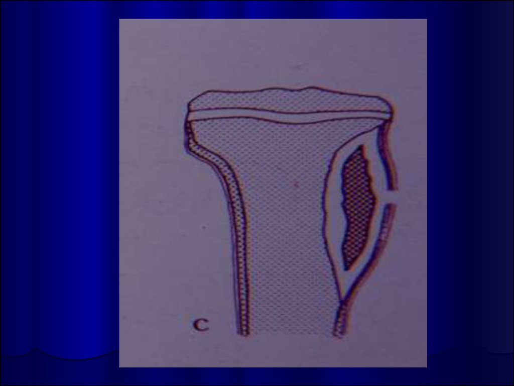
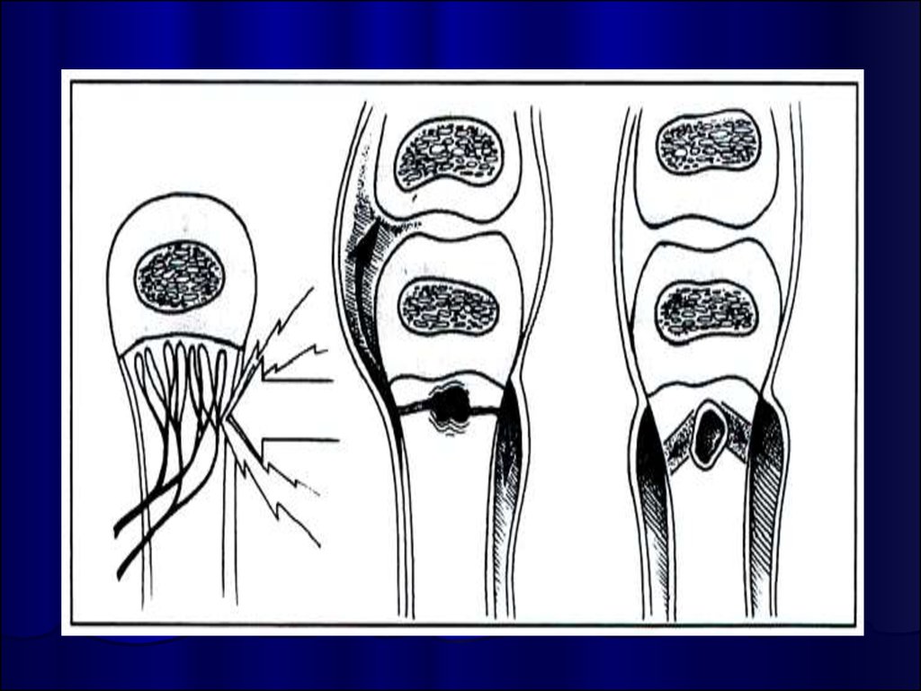
4. Hematogenous Osteomyelitis
Hematogenous infection begins in the medullarycavity of bones, is encased in a rigid structure,
which does not allow for the expansion of the
inflammatory process. . Progression of the
infection restricts medullary blood supply.
Passage of pus through the cortex elevates the
periosteum and the resulting sub-periosteal
abscess causes bony infarction as the cortical
bone is supplied by end-arteries from the
periosteum.
5.
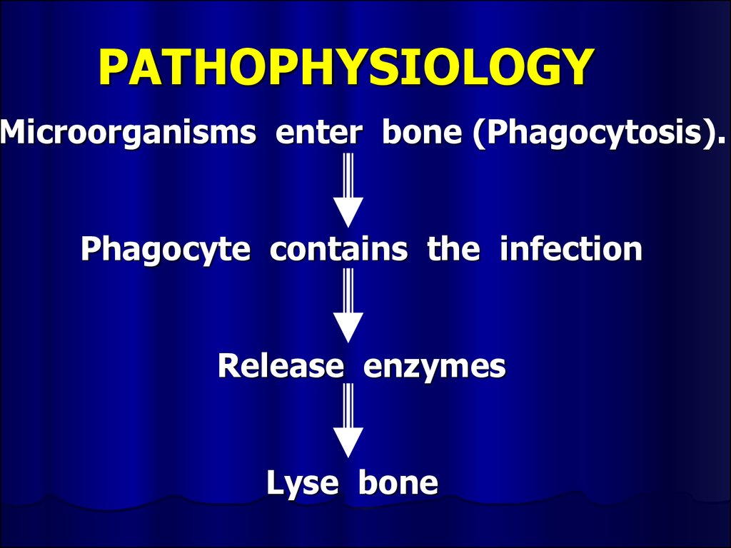
PATHOPHYSIOLOGYMicroorganisms enter bone (Phagocytosis).
Phagocyte contains the infection
Release enzymes
Lyse bone
6.
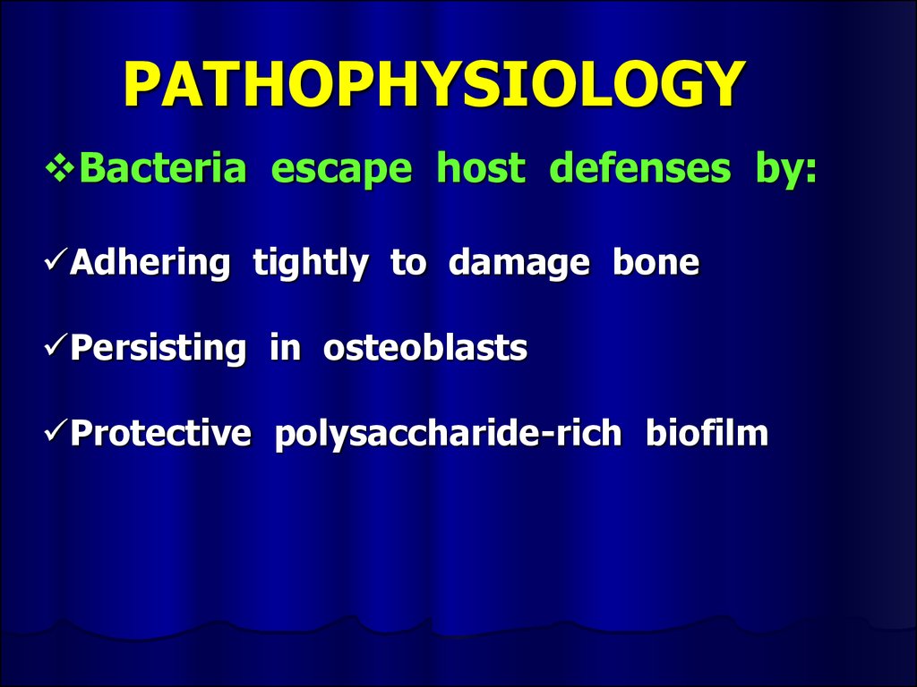
PATHOPHYSIOLOGYBacteria escape host defenses by:
Adhering tightly to damage bone
Persisting in osteoblasts
Protective polysaccharide-rich biofilm
7.
PATHOLOGYAcute Congested or thrombosed vessels
Chronic Necrotic bone
Absence of living osteocyte
Mononuclear cells predominate
Granulation & fibrous tissue
8. Stages
Toxic(adynamic) stage
Septicopyemic stage
Local stage
9. Forms
AcuteOsteomyelitis
Sub-acute Osteomyelitis
Chronic Osteomyelitis
10. Symptoms in newborn
Clinicalof septicemia : fever (36
- 74 %) irritable, refuses to feed,
rapid pulse
Joint swelling
Tenderness and resistance to
movement of the joint
Look for umbilical infection
11. Symptoms in infant
DrowsyIrritable
History
of birth difficulties
History of umbilical artery
catheterization
Metaphyseal tenderness and
resistance to joint movement
12. Symptoms in child
Severepain
Malaise
Fever
Toxemia
History of recent infection
Local inflammation
pus
escape from bone
Lymphadenopathy
13. Outcomes
Suppuration:4-5
days
Pus formation
Subperiosteal abscess
Pus spreading
epiphysis
joint
medullary cavity
soft tissue
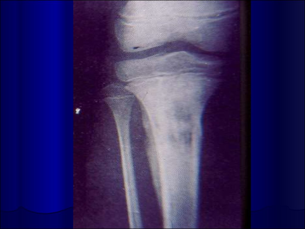
14. Necrosis
Bonedeath by the end of a week
Bone destruction ← toxin
← ischemia
Epiphyseal plate injury
Sequestrum formation
small removed by
macrophage,osteoclast.
large remained
15.
16. New bone formation
the end of 2nd week(10 – 14 days)
New bone formation from deep layer of
periosteum.
If infection persist- pus discharge through
sinus to skin surface Chronic
osteomyelitis
By
17.
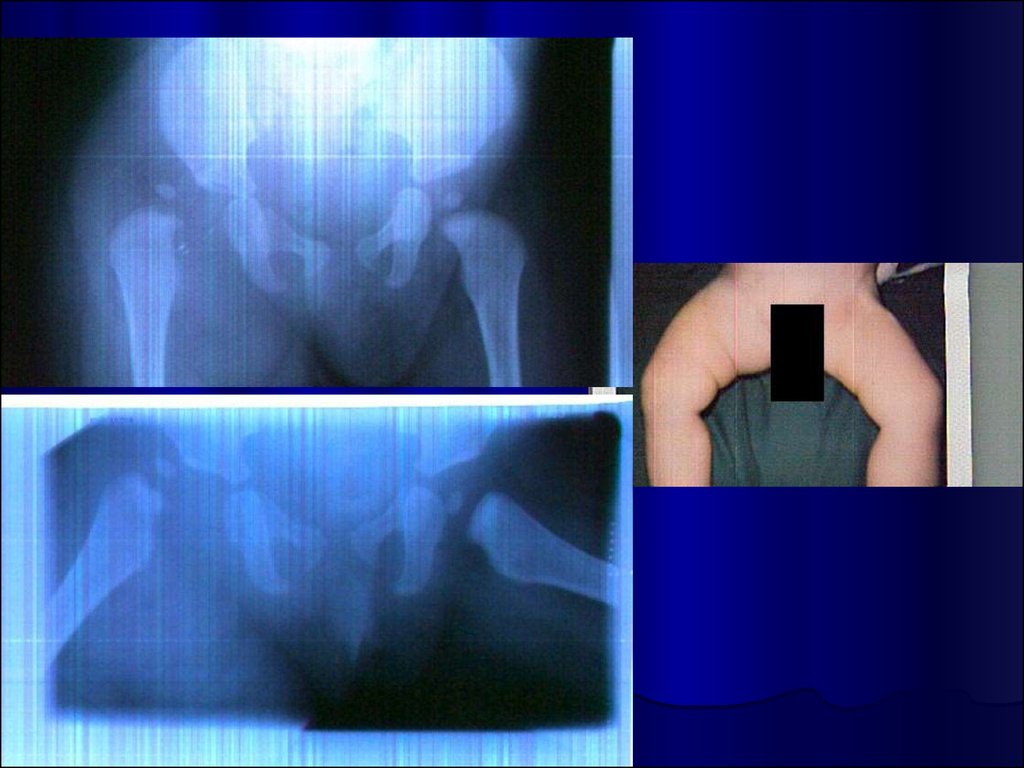
18. Joint capsule of 4 metaphysis cause of osteomyelitis
Femoral head and neck ( hip )Humeral head ( shoulder )
lateral side of distal tibia ( ankle joint )
radial head and neck ( elbow joint )
19.
20. Septic Arthritis
21. Differential diagnosis
Toxicsynovitis
Juvenile rheumatoid arthritis
Cellulitis
Pyomyositis
Psoas abscess
22. Investigation
Laboratory testsPlain
film
Ultrasonic diagnosis
Aspirate bone liquid
CT-scan
23.
24.
25.
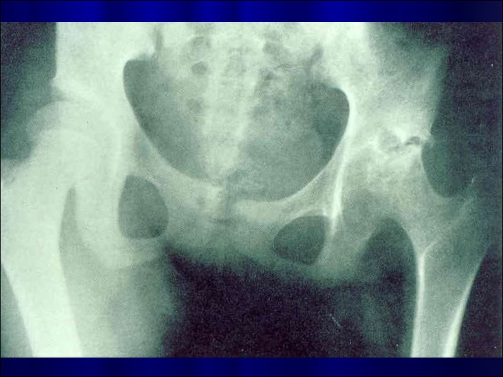
26.
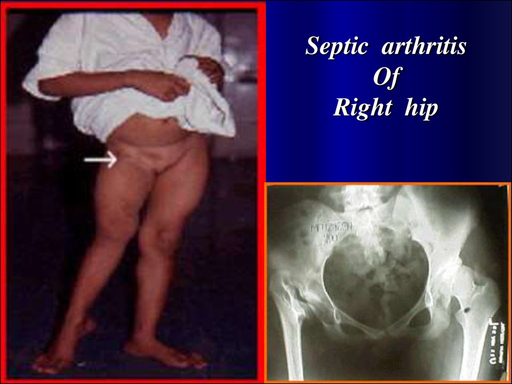
Septic arthritisOf
Right hip
27. Investigation : Aspiration
confirmdiagnosis
smear for cell and organism
culture and sensitivity test
28.
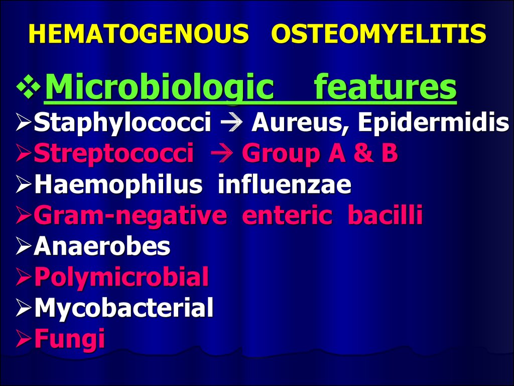
HEMATOGENOUS OSTEOMYELITISMicrobiologic
features
Staphylococci Aureus, Epidermidis
Streptococci Group A & B
Haemophilus influenzae
Gram-negative enteric bacilli
Anaerobes
Polymicrobial
Mycobacterial
Fungi
29.
TREATMENTInitial treatment shoud be aggressive.
Inadequate therapy Chronic disease
Antibiotic use:
Parenteral
High doses
Good penetration in bone
Full course
Empiric therapy
Surgery
30. Antibiotic treatment
AgePathogen
Drugs
1.Healthy Neonate
(< 1 mo)
-Staphylococcal Gr.
B infection
- cloxcillin 50
mg/kg/day
2. Infant and
children
-Staph. Aureus
-Gram neg.
infection
-Haemophilus
infection
-2nd generation
Cephalosporins or
Amoxycillin with
clavulanic acid
3. Adolescent (11 –
15 years)
-Staph. Aureus
150 – 200 mg/kg/day
IV divide q 4 – 6 hr.
max 12 gm./day
-Neisseria
gonorrhea
4.Sickle-cell patient
-Salmonella
infection
- Co-trimoxazole
- Amoxycillin with
clavulanic acid
31.
TREATMENTIndication for Surgery
Diagnostic
Hip joint involvement
Neurologic complication
Poor
Sequestration
32.
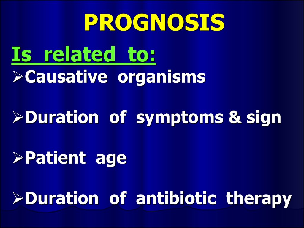
PROGNOSISIs related to:
Causative organisms
Duration of symptoms & sign
Patient age
Duration of antibiotic therapy
33.
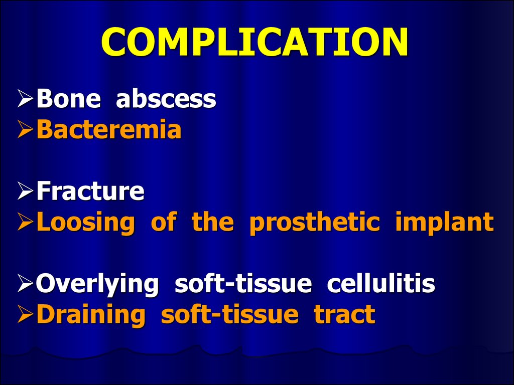
COMPLICATIONBone abscess
Bacteremia
Fracture
Loosing of the prosthetic implant
Overlying soft-tissue cellulitis
Draining soft-tissue tract
34.
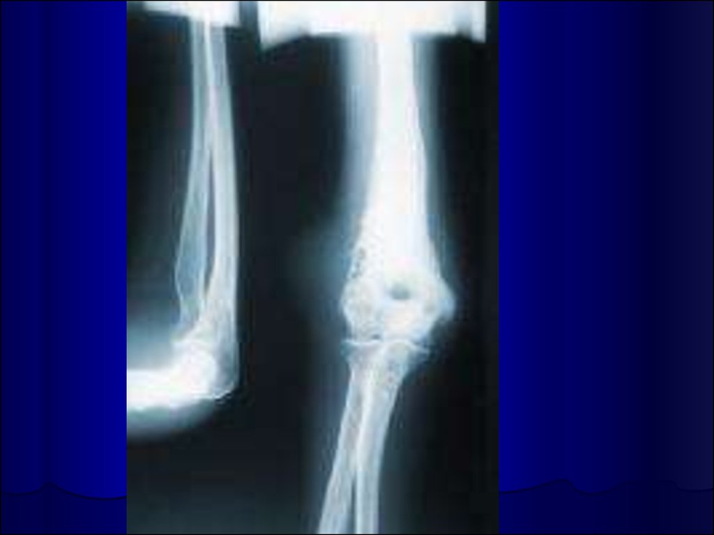
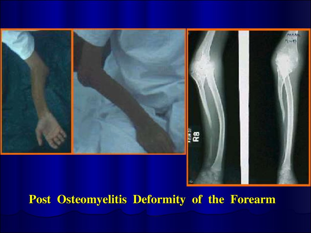
Post Osteomyelitis Treatment35.
Septic OsteomyelitisPost Osteomyelitis Scar
36.
Post Osteomyelitis Deformity of the Forearm37. Necrotizing pneumonia
Necrotizing pneumonia is characterized byinflammation of the alveoli and terminal
airspaces in response to invasion by an
infectious agent introduced into the lungs
through hematogenous spread or
inhalation.
38. Pathophysiology
The alveoli fill with proteinaceous fluid, which triggers abrisk influx and polymorphonuclear cells followed by the
deposition of fibrin and the degradation of inflammatory
cells.
Intra-alveolar debris is ingested and removed by the
alveolar macrophages.
This consolidation leads to decreased air entry and
dullness to percussion.
Inflammation in the small airways leads to crackles.
The patient must increase his or her respiratory
rate to maintain adequate ventilation.
39. Physical examination
Newborns:1.
2.
3.
rarely cough
they more commonly present with tachypnea,
retractions, grunting, and hypoxemia
grunting suggests a lower respiratory tract disease
Older infants:
1.
2.
3.
grunting may be less common
tachypnea, retractions, and hypoxemia are common
may be accompanied by a persistent cough,
congestion, fever, irritability, and decreased
feeding
40.
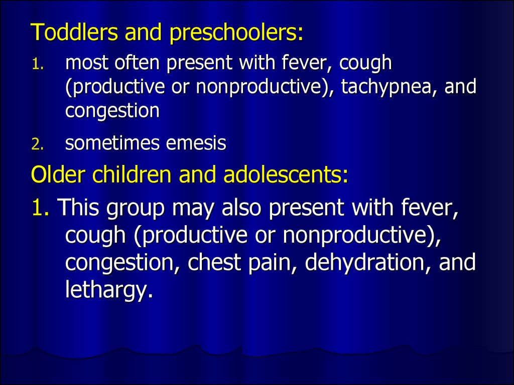
Toddlers and preschoolers:1.
most often present with fever, cough
(productive or nonproductive), tachypnea, and
congestion
2.
sometimes emesis
Older children and adolescents:
1. This group may also present with fever,
cough (productive or nonproductive),
congestion, chest pain, dehydration, and
lethargy.
41. Generalized symptoms
Intoxication sundromeNasal flaring
Auscultation: dry or bubbling rales,
wheezing, diminished breath sounds,
tubular breath sounds, pleural friction rub.
The affected lung field may be dull to
percussion.
Decreased tactile and vocal fremitus.
42. Extrapulmonary symptoms
1.2.
3.
Abdominal pain or an ileus accompanied
by emesis in patients with lower lobe
pneumonia.
Nuchal rigidity in patients with right
upper lobe pneumonia.
Rub caused by pericardial effusion in
patients with lower lobe pneumonia due
to Haemophilus influenzae infection.
43. Diagnosis
Laboratory tests (inflammation signs).Radiography
Lung aspirate
Sputum culture
Blood culture
Polymerase chain reaction
Skin tests (TB pneumonia BCG)
Bronchoscopy
CT - scan
44.
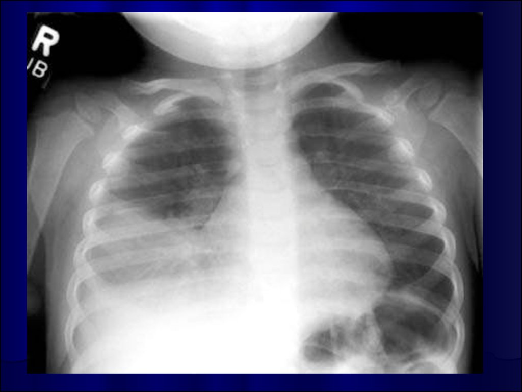
45. Segmental-lobar opacification
46. Segmental-lobar opacification with pleural effusion
47.
48. Differential diagnosis
Afebrile Pneumonia SyndromeAirway Foreign Body
Aspiration Syndromes
Bronchiectasis
Bronchiolitis
Bronchitis, Acute and Chronic
Chronic Granulomatous Disease
Congenital Pneumonia
Cystic Adenomatoid Malformation
Cystic Fibrosis
Empyema
Gastroesophageal Reflux
Pulmonary Sequestration
49. Antibacterial therapy
Cephalosporins (III-IV gen.): Ceftriaxone(Rocephin), Cefotaxime (Claforan),
Cefuroxime (Zinacef, Ceftin, Kefurox).
Macrolide antibiotics: Azithromycin
(Zithromax), Clarithromycin (Biaxin),
Erythromycin (E.E.S., E-Mycin, Ery-Tab),
50. Tube Thoracostomy
51.
52.
53. Necrotic phlegmon
Purulent lesions in the skin and hypodermictissue, usually this process localisations in
the scapular and sacrcococcygeal regions.
Necrotic phlegmon is predominantly a
disease of the neonate.
54.
55. Causes
Vulnerability epidermisA lot of intrecellular liquid
Progress vasculature
Congenital hypoplasia subjacent tissues
56. Clinical stages
Intoxication syndromeHyperaemia
Compression soft tissues
Edema
Fluctuation
Exfolation skin
57. Differential diagnosis
Aseptic necrosisErythematous erysipelas
Idiopathic erysipelas
Phlegmonous erysipelas
58. Treatment
Fluid therapyAntibacterial therapy (cephalosporinis IIIIV gen.)
General health-improving therapy
Surgical treatment – chess incisions in the
lesion region, irrigation aspiration.
59.
60.
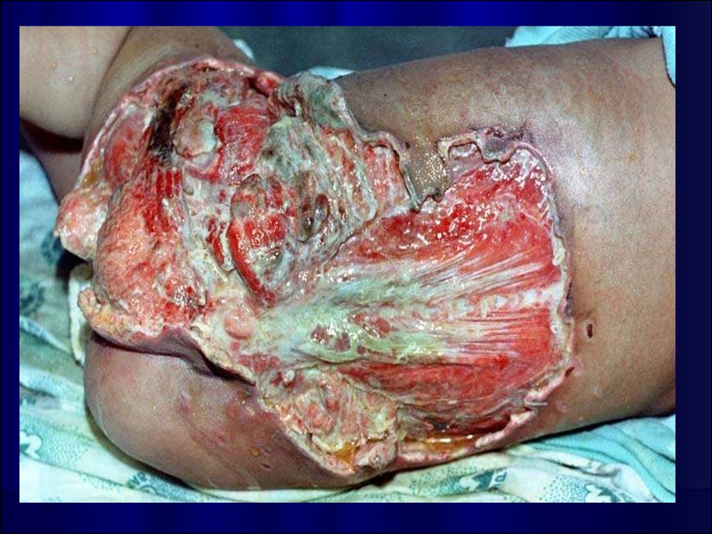
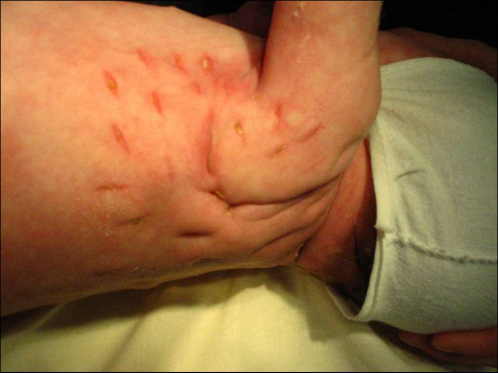
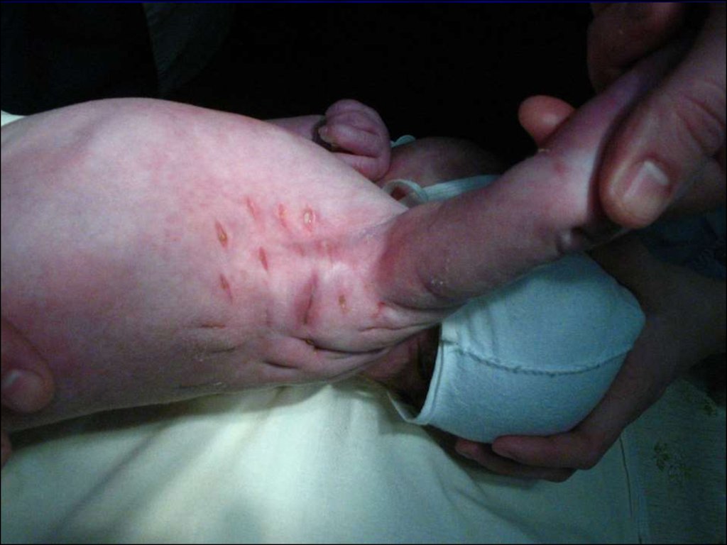
61. Omphalitis
Omphalitis is an infection of the umbilicalstump. Omphalitis typically presents as a
superficial cellulitis that may spread to
involve the entire abdominal wall and may
progress to necrotizing fasciitis,
myonecrosis, or systemic disease. Aerobic
bacteria are present in approximately
85% of infections, predominated by
Staphylococcus aureus, group A
Streptococcus, Escherichia coli, Klebsiella
pneumoniae, and Proteus mirabilis
62.
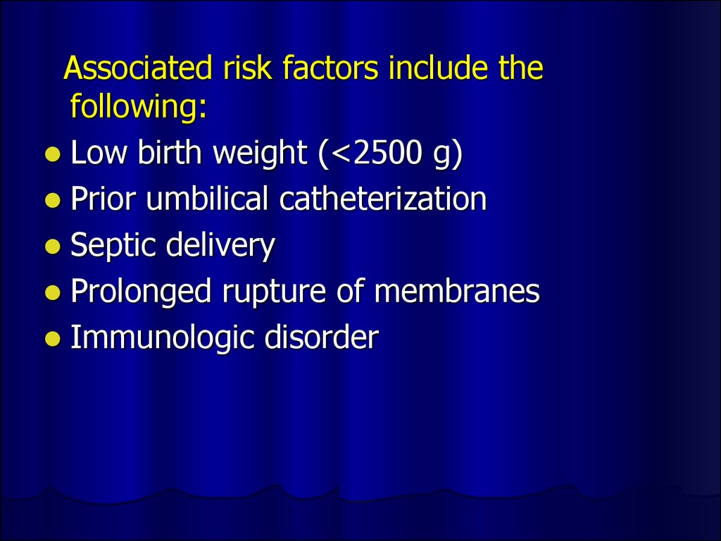
Associated risk factors include thefollowing:
Low birth weight (<2500 g)
Prior umbilical catheterization
Septic delivery
Prolonged rupture of membranes
Immunologic disorder
63. Clinic
Purulent or malodorous discharge fromthe umbilical stump
Periumbilical erythema
Edema
Tenderness
Ecchymoses
Progression of cellulitis despite
antimicrobial therapy
64. Differential diagnosis
Umbilical fistulaSoaking umbilical
Enterocystoma
65. Complications
Necrotizing fasciitisMyonecrosis
Sepsis
Septic embolization
Particularly endocarditis and liver abscess
formation
Abdominal complications
66. Treatment
Fluid therapyAntibacterial therapy (cephalosporinis IIIIV gen.)
Surgical care: management of necrotizing
fasciitis and myonecrosis involves early
and complete surgical debridement of the
affected tissue and muscle
67. Neonatal Sepsis
Clinical syndrome of systemic illnessaccompanied by bacteremia occurring in
the first month of life
Incidence
1-8/1000
live births
13-27/1000 live births for infants < 1500g
Mortality rate is 13-25%
Higher
rates in premature infants and those
with early fulminant disease
68. Early Onset
First 5-7 days of lifeUsually multisystem fulminant illness with
prominent respiratory symptoms (probably
due to aspiration of infected amniotic fluid)
High mortality rate
5-20%
Typically acquired during intrapartum
period from maternal genital tract
Associated
with maternal chorioamnionitis
69. Late Onset
May occur as early as 5 days but is mostcommon after the first week of life
Less association with obstetric
complications
Usually have an identifiable focus
Most
often meningitis or sepsis
Acquired from maternal genital tract or
human contact
70. Causative organisms
Primary sepsisGroup
B streptococcus
Gram-negative enterics (esp. E. coli)
Listeria
monocytogenes, Staphylococcus, other
streptococci (entercocci), anaerobes, H. flu
Nosocomial sepsis
Varies
by nursery
Staphylococcus
epidermidis, Pseudomonas,
Klebsiella, Serratia, Proteus, and yeast are
most common
71. Risk factors
Prematurity and low birth weightPremature and prolonged rupture of membranes
Maternal peripartum fever
Amniotic fluid problems (i.e. mec, chorio)
Resuscitation at birth, fetal distress
Multiple gestation
Invasive procedures
Galactosemia
Other factors: sex, race, variations in immune
function, hand washing in the NICU
72.
Clinical presentationClinical signs and symptoms are
nonspecific
Differential diagnosis
RDS
Metabolic
disease
Hematologic disease
CNS disease
Cardiac disease
Other infectious processes (i.e. TORCH)
73.
Temperature irregularity (high or low)Change in behavior
Skin changes
Intolerance, vomiting, diarrhea, abdominal distension
Cardiopulmonary
Poor perfusion, mottling, cyanosis, pallor, petechiae, rashes,
jaundice
Feeding problems
Lethargy, irritability, changes in tone
Tachypnea, grunting, flaring, retractions, apnea, tachycardia,
hypotension
Metabolic
Hypo or hyperglycemia, metabolic acidosis
74. Diagnosis
CulturesBlood
Confirms
sepsis
94% grow by 48 hours of age
Urine
Don’t
need in infants <24 hours old because UTIs
are exceedingly rare in this age group
CSF
Controversial
May
be useful in clinically ill newborns or those
with positive blood cultures
75. Treatment
AntibioticsPrimary
sepsis: ampicillin and gentamicin
Nosocomial sepsis: vancomycin and
gentamicin or cefotaxime
Change based on culture sensitivities
Don’t forget to check levels
76. Supportive therapy
RespiratoryCardiovascular
Treat DIC with FFP and/or cryo
CNS
Support blood pressure with volume expanders and/or
pressors
Hematologic
Oxygen and ventilation as necessary
Treat seizures with phenobarbital
Watch for signs of SIADH (decreased UOP, hyponatremia)
and treat with fluid restriction
Metabolic
Treat hypoglycemia/hyperglycemia and metabolic acidosis
77.
Thank you forattention!