


 Медицина
Похожие презентации:


The treatment of Lobular Pneumonia
1. The treatment of Lobular Pneumonia
2.
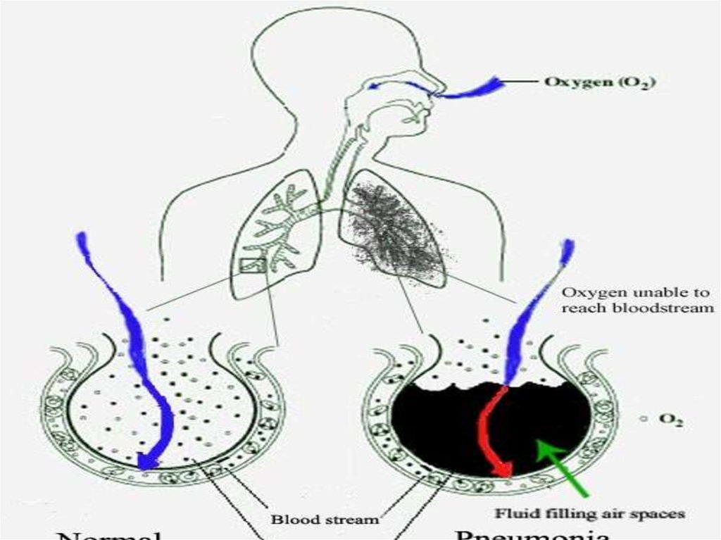
• Pneumonia is an inflammation of the lung, usuallycaused by bacteria, viruses or protozoa. If the
infection is localized to one or two lobes of a lung it
is referred to as «lobar pneumonia» and if the
infection is more generalized and involves primarily
the bronchi it is known as «bronchopneumonia». A
wide range of infecting organisms has been
implicated.
3.
4.
5. Pneumonia: infecting organisms in approximate descending order of frequency
Community acquired
Streptococcus pneumoniae
Mycoplasma pneumoniae
Influenza virus A
Haemophilus influenzae
Legionella pneumophila
Staphylococcus aureus
Coxiella burneti
Chlamydia psittaci
Hospital acquired
Gram-negative bacilli
Staphylococcus aureus
Streptococcus pneumoniae
Legionella pneumophila
Haemophilus influenzae
Pseudomonas spp
Immunocompromised patients
Pneumocystis carinii
Cytomegalovirus
Mycobacterium avium-intracellulare
Mycobacterium tuberculosis
Streptococcus pneumoniae
Haemophilus influenzae
Legionella pneumophila
Actinomyces israelii
Aspergillus fumigatus
Nocardia asteroides
6.
7.
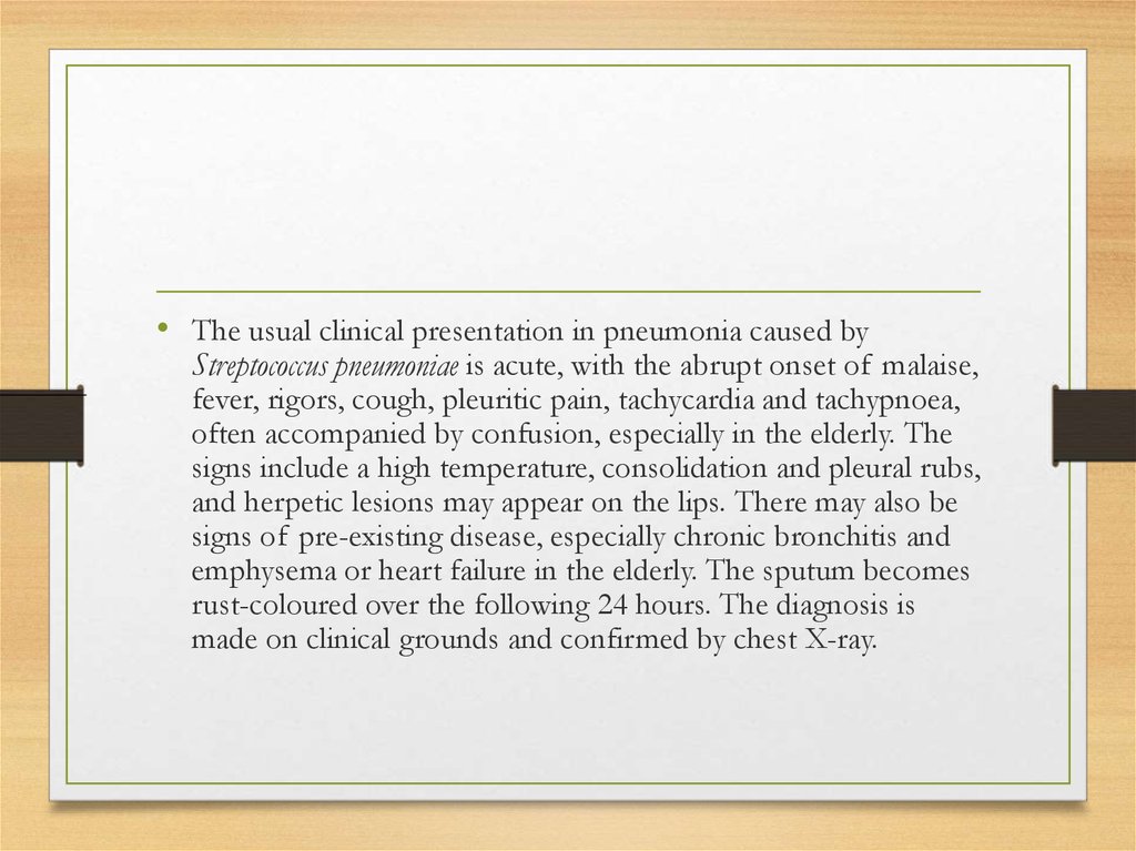
• The usual clinical presentation in pneumonia caused byStreptococcus pneumoniae is acute, with the abrupt onset of malaise,
fever, rigors, cough, pleuritic pain, tachycardia and tachypnoea,
often accompanied by confusion, especially in the elderly. The
signs include a high temperature, consolidation and pleural rubs,
and herpetic lesions may appear on the lips. There may also be
signs of pre-existing disease, especially chronic bronchitis and
emphysema or heart failure in the elderly. The sputum becomes
rust-coloured over the following 24 hours. The diagnosis is
made on clinical grounds and confirmed by chest X-ray.
8. Pneumococci are typically asociated with pneumonia
9. Lobar pneumonia: stage of onset
• Morphology. Congestion stage — extensive serous exudation,vascular engorgement, rapid bacterial proliferation.
Inspection. An increased respiratory rate is usually evident. Pain is a
frequent accompaniment, and with it the involved side shows a lag
of respiratory motion.
Palpation. Palpation confirms the findings on inspection. Tactile
fremitus is normal or even slightly decreased, and a pleural friction
rub may be present.
Percussion. Impaired resonance may be elicited with light
percussion. This finding is extremely important.
Auscultation. Although the breath sounds may be diminished,
expiration is prolonged and crepitation (crepitus indux) is heard.
With pleural involvement, a pleural friction sound is determined.
10. Lobar pneumonia: stage of consolidation
• Morphology. Red hepatization stage — airspaces are filled with PMN cells, vascular congestion,extravasation of RBC. Grey hepatization stage — accumulation of fibrin, inflammatory WBCs
and RBCs in various stages of disintegration, alveolar spaces filled with inflammatory exudate.
Complaints. Coughing may be associated with i sharp pain in the affected side. Mucoid sputum
be comes rusty brown (prune juice color).
General inspection. Cyanosis of the lips and fin gers. When the fever is high, the face may be
flushed The patient's nostrils dilate on inspiration, and expi ration is often grunting.
Inspection. Dyspnea is invariably present. Respi ratory movements are generally decreased on
the af fected side.
Palpation. Diminished respiratory excursions, i pleural friction rub may be felt. Tactile fremitus
is in creased.
Percussion. Dullness.
Auscultation. Bronchial breathing, bronchophony, pectoriloquy and whispered bronchophony
are evident with consolidation provided the bronchus to the in volved area is open. Rales are
less numerous and dis tinct than in the stages of engorgement or resolution,
11.
12. Lobar pneumonia: stage of resolution
• Morphology. Resolution stage — resorption of the exudate.• Inspection. The patient looks more comfortable and the cyanosis
disappears. The dyspnea disappears and the affected lung begins to
expand again.
• Palpation. The previously increased tactile fremitus becomes less
marked and gradually findings become normal.
• Percussion. The dullness gradually disappears and normal
resonance returns.
• Auscultation. The bronchial breathing is gradually replaced by
bronchovesicular breathing and later by normal vesicular breathing.
Crepitation reappears (crepitus redux). Small and large moist rales are
heard in increasing numbers.
13.
14. Complications
Lung abscess
Pleurisy
Toxic shock
Myocarditis
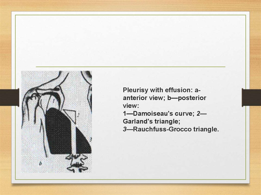
15. Pleurisy
16.
Pleurisy with effusion: aanterior view; b—posteriorview:
1—Damoiseau's curve; 2—
Garland's triangle;
3—Rauchfuss-Grocco triangle.
17.
• Mycoplasma pneumoniae is the most common cause ofthe «atypical» pneumonias. Infection usually occurs in
older children and young adults, who present with
pharyngitis and bronchitis; pneumonia occurs in minority and is rarely severe. Psittacosis is acquired
from birds and Q-fever from animals, commonly
farm livestock; they also cause «atypical pneumonia»,
although the Q-fever organism, Coxiella burnetti, may
also cause endocarditis. The diagnosis of the atypical
pneumonia is usually made by serology.
18.
• Staphylococcal pneumonia typically occurs as acomplication of influenza, especially in the elderly
and, although uncommon, is important because of
the attendant high mortality. It is a destructive
pneumonia, which frequently leads to the formation
of cavities within the lung. Such cavitating
pneumonia was most frequently caused by
tuberculosis in the past but now Staphylococci, Klebsiella
and anaerobic organisms are the most common
causes.
19.
• Legionnaires' disease is pneumonia caused by Le-gionella pneumophila. Infection is most common in
debilitated or immunocompromised patients. Most
cases are sporadic, but outbreaks occur from
contaminated water droplet sources. Patients may
present with a wide spectrum of additional
symptoms, such as headache, cerebellar ataxia, renal
failure or hepatic involvement. Special medium is
necessary for the culture of the organism and the
diagnosis is usually made by serology.
20.
• Aspiration pneumonia results from the aspiration of gastric contentsinto the lung and is associated with impaired consciousness {e.g.
anaesthesia; epilepsy, alcoholism) or dysphagia. Multiple organisms may
be isolated.
• Nosocomial pneumonia occurs when infection takes place in hospital;
patients may be debilitated, immunocompromised or have just
undergone a major operation. The causative organism(s) are often
Gram-negative or Gram-positive coccus, Staphylococcus au-reus. The high
mortality is usually related to the severity of the underlying disease.
Lung abscess or empye-ma (a collection of pus within the thoracic
cavity), or both, may be caused by specific organisms or may complicate any aspiration pneumonia. Septic pulmonary emboti can lead to
multiple lung abscesses, pulmonary infarcts may become infected
cavities and abscesses can develop distal to lesions obstructing a
bronchus.
21.
• Treatment of all pneumonias should be started immediately and the antibioticchosen should be the «best guess» (decided on by the origin of the pneumonia
and its clinical severity). If community-acquired, then a high-dose parenteral
penicillin (or erythromycin) will usually be effective. If legionnaires'disease is
suspected on epidemiological grounds, rifampicin should be given with
erythromycin. If staphylococcal pneumonia is suspected, because of preceding
influenza, flu-cioxacillin should be added to the regime. In hospital-acquired
pneumonia, combination therapy is required to cover the range of possible
pathogenic organisms (especially Gram-negative bacilli).
• Combinations such as gentamicin with piperacillin or a cephalosporin may be
used. In aspiration pneumonia, in which anaerobes may be present,
metronidasole should be added to these combinations. Supportive measures
should include oxygen, intravenous fluids, inotropic agents when necessary,
bronchial suction and assisted ventilation. Physiotherapy and bronchod-ilators are
of value in pneumonia complicating chronic bronchitis and emphysema.
22. Infection in the immunocompromised host
• There has been a steady increase in the number of patientswhose immune system has been damaged by malignancy, organ
failure, drugs or the HIV virus. In such immunocompromised
patients, infections of the lung are common and may be caused
by organisms that are not usually pathogenic in the normal
host. Invasive fungal infections tend to occur in neutropenic
patients, whereas T-cell defects often lead to infection with
viruses, mycobacteria and protozoa such as Pneumocystis carinii.
The tempo of infection in the immunocompromised patients
can be extremely rapid; it is important to take steps to identify
the pathogen and to start therapy as soon as possible.
23.
• Pneumocystis carinii is the most important cause offatal pneumonia in immunosuppressed patients. It is
believed that the infection is acquired in early childhood, and that reactivation occurs when immune system becomes damaged. The incubation period is approximately 1-2 months before the insidious appearance of a low-grade progressive pneumonia, which
manifest itself as severe dyspnoea with, at first, only
minimal chest signs and X-ray changes.
24.
• In Pneumocystis carinii pneumonia the changes on X-ray may bevery minor, but the patient is markedly hypoxic and a
transbronchial biopsy usually reveals P. carinii. Unless the
infection is treated promptly, most severe pneumonic changes
follow.
• The pneumonia progresses rapidly, and within a few days
obvious pneumonic changes may be seen on the chest X-ray.
Diagnosis depends on demonstrating the organism in sputum,
bronchial lavage or lung tissue, which may require a lung biopsy.
Treatment is with cotrimoxazole or pentamidine; both of these
may be used in prophylaxis. Mortality remains high despite
treatment.
25. Principles of treatment
Antibiotics
Expectorants
Desintoxication
Oxygen
Antigistamine agents
Symptomatic therapy