


")


 Медицина
Похожие презентации:


Lecture pulp and periapical disease
1. Diseases Of Pulp & Periapical Tissues
Diseases Of Pulp& Periapical
Tissues
2. PULPITIS
P U L P IT ISPulpitis is the most common cause of pain and
loss of teeth in younger persons.
The usual cause is caries penetrating the dentine
but there are other possibilities of pulpitis .
If untreated, is followed by death of the pulp and
spread of infection through the apical foramina
into the periapical tissue.
https://dentistrykey.com/library/examination-and-diagnosis-of-pulp-rootcanal-and-periapical-periradicular-conditions/
3. CAUSES OF PULP DISEASE
The causes of pulp disease arePhysical, Chemical and Bacterial.
Physical
a. Mechanical
- Trauma:
. Accidental
. Iatrogenic dental procedures
- Pathological wear
- Crack through body of tooth
a. Thermal
- Heat from cavity preparation
- Exothermic heat from setting of cements
b.Electrical ( galvanic current from dissimilar metallic
filling)
4.
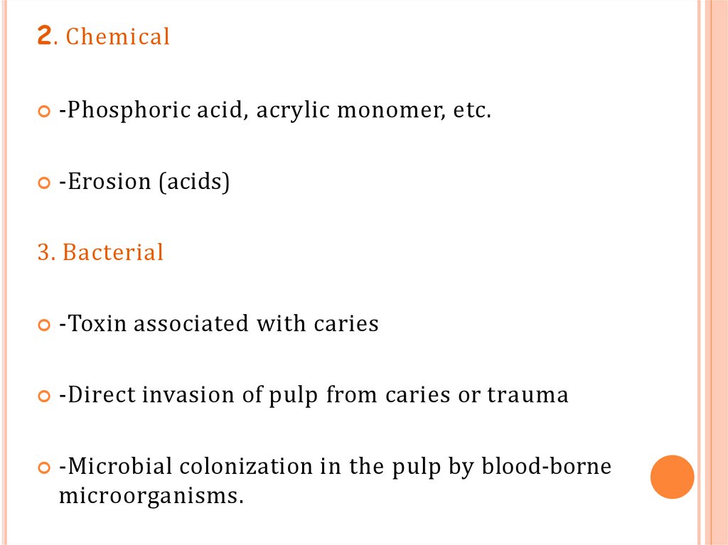
2 . Chemical-Phosphoric acid, acrylic monomer, etc.
-Erosion (acids)
3. Bacterial
-Toxin associated with caries
-Direct invasion of pulp from caries or trauma
-Microbial colonization in the pulp by blood-borne
microorganisms.
5. CLASSIFICATION
C L A S S IF IC ATIO NI. According to pathological condition: Focal or acute reversible
pulpitis (Pulp hyperaemia)
- Irreversible pulpitis
II. According to its duration: - Acute pulpitis
- Chronic pulpitis
III. According to presence of dentin covering the
pulp chamber: - Open pulpitis
- Closed pulpitis
6. CLASSIFICATION
C L A S S IF IC AT IO NIV. According to extension of inflammation in pulp
tissue: - Partial pulpitis
- Complete /total pulpitis
V. According to amount of pus formation: - Exudative pulpitis
- Suppurative pulpitis
7. Pulp state
8.
FOCAL REVERSIBLE PULPITIS (PULP HYPEREMIA)Mild, transient, localized inflammatory response.
It is a reversible condition .
CLINICAL FEATURES:
Caries - pain from cold test disappeares immediately.
Hyperemia -pain from cold test does not linger more than 30 s. No
percussion sensitivity, no spontaneous pain, no heat sensitivity.
Affected tooth responds to stimulation of electric pulp tester at
lower level of current indicating low pain threshold.
Teeth usually show deep caries, metallic restoration with
defective margins.
9.
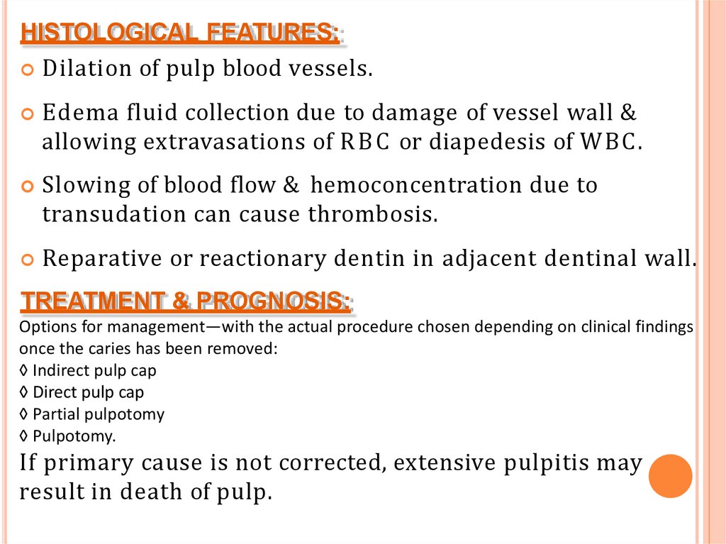
HISTOLOGICAL FEATURES:Dilation of pulp blood vessels.
Edema fluid collection due to damage of vessel wall &
allowing extravasations of R B C or diapedesis of WBC .
Slowing of blood flow & hemoconcentration due to
transudation can cause thrombosis.
Reparative or reactionary dentin in adjacent dentinal wall.
TREATMENT & PROGNOSIS:
Options for management—with the actual procedure chosen depending on clinical findings
once the caries has been removed:
◊ Indirect pulp cap
◊ Direct pulp cap
◊ Partial pulpotomy
◊ Pulpotomy.
If primary cause is not corrected, extensive pulpitis may
result in death of pulp.
10.
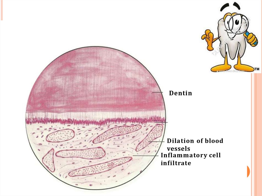
DentinDilation of blood
vessels
Inflammatory cell
infiltrate
11. PULP HYPEREMIA
12. ACUTE PULPITIS
ACUTE P ULPITISIrreversible condition characterized by acute, intense
inflammatory response in pulp .
It is a frequent immediate sequela of focal reversible pulpitis, it
may occur as an acute exacerpation of a chronic process.
Acute pulpitis may be either closed where the dentinal wall of
the pulp is intact or open where the dentinal wall is broken.
CLINICAL FEATURES:
o Pain from cold test lingers more than 30 s. May get pain from heat test
May have spontaneous pain. May be percussion sensitive.
Radiographically or clinically visible deep caries.
Pain - poorly localized since pulp of individual tooth is not
represented in sensory cortex.
Intrapulpal abscess formation cause severe pain lancinating or
throbbing type. (10 – 15mins).(acute total pulpitis)
Intensity of pain can increase when patient lies down.
13.
Acute pulpitis withIntrapulpal abscess
14.
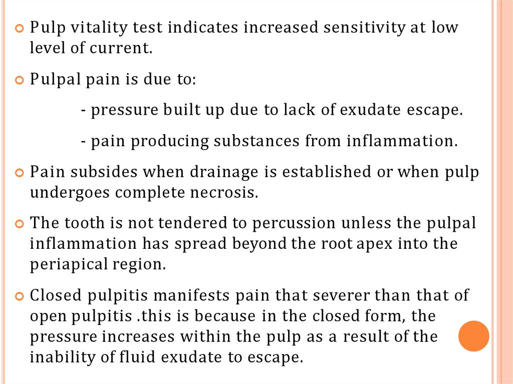
Pulp vitality test indicates increased sensitivity at lowlevel of current.
Pulpal pain is due to:
- pressure built up due to lack of exudate escape.
- pain producing substances from inflammation.
Pain subsides when drainage is established or when pulp
undergoes complete necrosis.
The tooth is not tendered to percussion unless the pulpal
inflammation has spread beyond the root apex into the
periapical region.
Closed pulpitis manifests pain that severer than that of
open pulpitis .this is because in the closed form, the
pressure increases within the pulp as a result of the
inability of fluid exudate to escape.
15. HISTOLOGIC FEATURES:
Edema in pulp with vasodilation.Infiltration of polymorphonuclear leukocytes along
vascular channels & migrate through endothelium lined
structures.
Destruction of odontoblasts at pulp dentin border.
Rise in pressure due to inflammatory exudate
local
collapse of venous part of circulation
Tissue hypoxia
&
Destruction of pulp & abscess formation.
Abscess consists of pus, leukocytes & bacteria.
Numerous abscess formation cause pulp liquefaction &
necrosis. (acute suppurative pulpitis)
16. Acute pulpitis
Acute pulpitis. Beneath the carious exposure (topright) a dense inflammatory inflammatory infiltrate is
accumulating. More deeply. The pulp is intensely
hyperaemic.
Acute pulpitis. Infection has penetrated
dentine
Causing inflammation to spread down the pulp and pus
to form in corner
17. PULPITIS
Acute pulpitis stage. The entire pulphas been destroyed and replaced by
inflammatory cells and dilated vessels
Acute caries and pulpitis. Infection
has penetrated to the pulp. Part of
the pulp has been destroyed, and
an abscess has formed containing a
bead of pus
Localized pulpitis with localized pulp abscess
18. Acute PULPITIS
19. Pulp abscess
20. Pulp abscess
21. TREATMENT & PROGNOSIS:
TREATMENT & PROGNOSIS:Options for management:
◊ Extraction
◊ Pulpectomy and root canal treatment—with the
following considerations/variations:
≫≫ Pulp tissue is present and needs to be removed
≫≫ Sodium hypochlorite is used to dissolve pulp tissue
remnants
≫≫ Anti-inflammatory medicament should be used to
reduce periapical nerve sprouting and neuropathic pain,
e.g., a corticosteroid/antibiotic compound.
If the tooth also has acute apical periodontitis—consider
postoperative systemic NSAID’s and analgesics.
22. Chronic Pulpitis
Chronic Pul pitisPersistent inflammatory reaction in pulp with little or non
symptoms.
It can arise from a previous acute
Pulpitis or occurs as the chronic type from the onset.
o It may be open or closed form .
CLINICAL FEATURES:
Pain is not prominent, mild, dull ache which is intermittent.
Reaction to thermal changes is reduced because of
degeneration of nerves.
Response to pulp vitality tester is reduced.
Wide open carious lesion & with exposure of pulp cause
relatively little pain.
Manipulation with small instruments often elicits bleeding
but with little pain.
23. HISTOLOGIC FEATURES:
Infiltration of mononuclear cells, lymphocytes & plasmacells, with vigorous connective tissue reaction.
Capillaries are prominent; fibroblastic activity & collagen
fibers in bundles.(chronic closed pulpitis)
When granulation tissue formation occurs in wide open
exposed pulp surface – ulcerative pulpitis. (with bacterial
stains & micro org. in carious lesion)
Chronic open ulcerative pulpitis , is characterized by the
presence of an ulcer on the exposed pulp surface, with a
large number of PMNIs below the surface, there is adense
chronic inflammatory cell infiltration with increased
fibroblastic activity.
24. Chronic Pulpitis
Chronic Pul pitis25. TREATMENT & PROGNOSIS:
TREATMENT & PROGNOSIS:Root canal therapy
Extraction of tooth.
26. Chronic Hyperplastic Pulpitis (pulp polyp)
It is a form of a chronic pulp disease.Overgrowth of pulp tissue outside the boundary of pulp
chamber as protruding mass.
CLINICAL FEATURES:
It occurs almost exclusively in children & young adults and
involves teeth with large open carious cavity.
Pulp - pinkish red globule of tissue protruding from
chamber & extend beyond caries.
Most commonly affected are deciduous molar & Ist
permanent molars.
Pulp is relatively insensitive because few nerves in
hyperplastic tissue.
27.
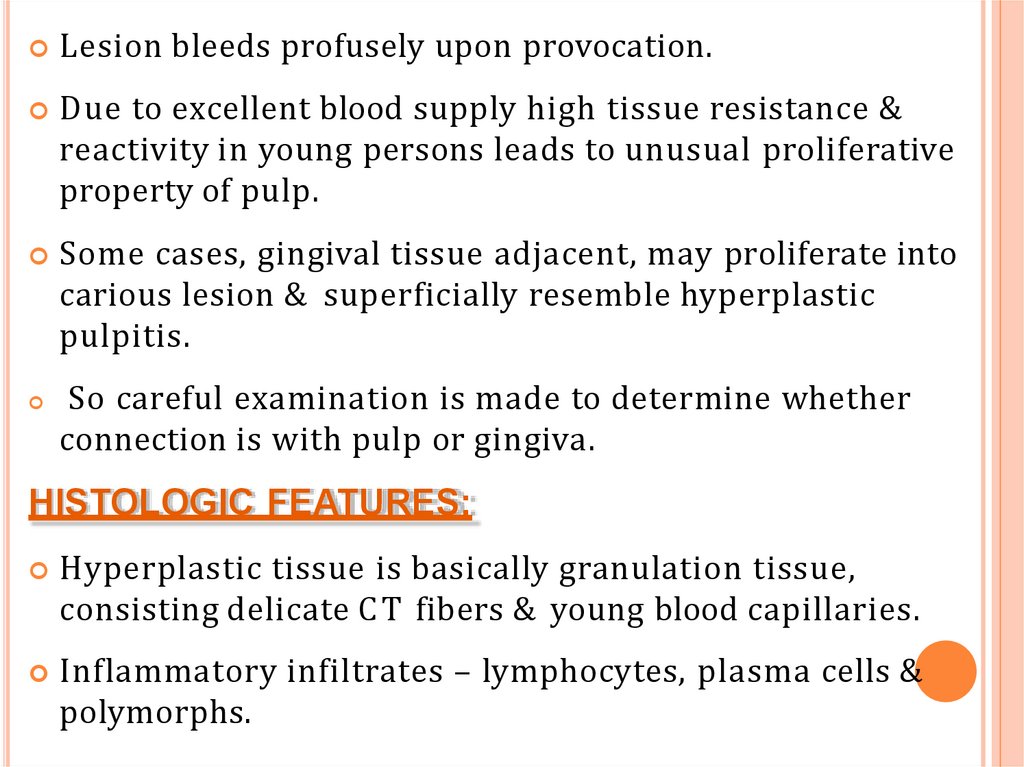
Lesion bleeds profusely upon provocation.Due to excellent blood supply high tissue resistance &
reactivity in young persons leads to unusual proliferative
property of pulp.
Some cases, gingival tissue adjacent, may proliferate into
carious lesion & superficially resemble hyperplastic
pulpitis.
So careful examination is made to determine whether
connection is with pulp or gingiva.
HISTOLOGIC FEATURES:
Hyperplastic tissue is basically granulation tissue,
consisting delicate C T fibers & young blood capillaries.
Inflammatory infiltrates – lymphocytes, plasma cells &
polymorphs.
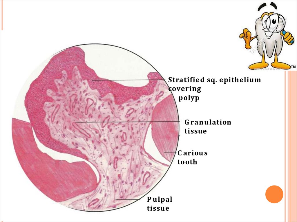
28.
Stratified sq. epitheliumcovering
polyp
G ranu lation
tissue
C ariou s
tooth
P u lpal
tissue
29.
Stratified squamous type epithelial lining resembles oralmucosa with well formed rete pegs.
Grafted epithelial cells are believed to be desquamated
epith. Cells, which carried by saliva.
TREATMENT & PROGNOSIS:
Extraction of tooth or pulp extripation .
30. Pulp Polyp
31. Pulp Polyp
32. Gangrenous Necrosis of Pulp
Untreated pulpitispulp.
results complete necrosis of
As this is associated with bacterial infection – pulp
gangrene.
It is associated with foul odor when pulp is opened for
endodontic treatment.
In sickle cell anemia, blockage of pulp vessels be defective
R B C results pulp necrosis.
Non vital pulp maintain general histology being non
purulent.
This may be due to trauma or infarct.
33.
Necrosis ofpulp
34. IRREVERSIBLE PULPITIS
IRREVERSIBLE PULPIT ISREV ERSIBLE PULPIT IS
Mild – moderate inflammatory
condition.
Nature of pain is mild &
diffuse.
Brief duration & can be
produce cold stimuli that
elicits the pain mostly,
although hot, sweet or sour
food may also initiate the pain.
Sharp, severe, radiating pain
of long duration & varying
intensity.
Pain continues even after the
stimulus is removed.
Pain may exacerbate with
bending over or lying down.
It may progress to more severe
pain )throbbing(.
Once stimulus is removed,
pain is usually subsides.
Tooth responds to electric pulp
tester at lower currents.
Increased by stimulus, like
heat or without stimulus.
Reversible pulpitis if allowed
to progress can led to
irreversible pulpitis.
When infection extends into
P D L - apical periodontitis.
35.
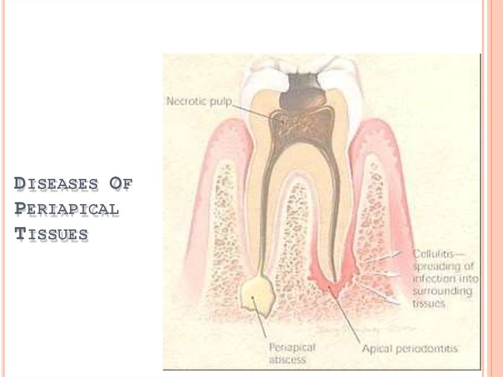
DISEASES OFPERIAPICAL
TISSUES
36. Diseases of the periapical tissues
Inflammation of P D L around apical portion of root.Cause:1. spread of infection following pulp necrosis,2. occlusal
trauma,3. inadvertent endodontic procedures ,4.infection
through the gingival crevice.
In P D L inflammation the patient can locate the symptoms to a
particular tooth due to stimulation of the proprioceptive nerve
ending in P D L .
Types: 1.Acute Apical Periodontitis
2.Chronic Apical Periodontitis
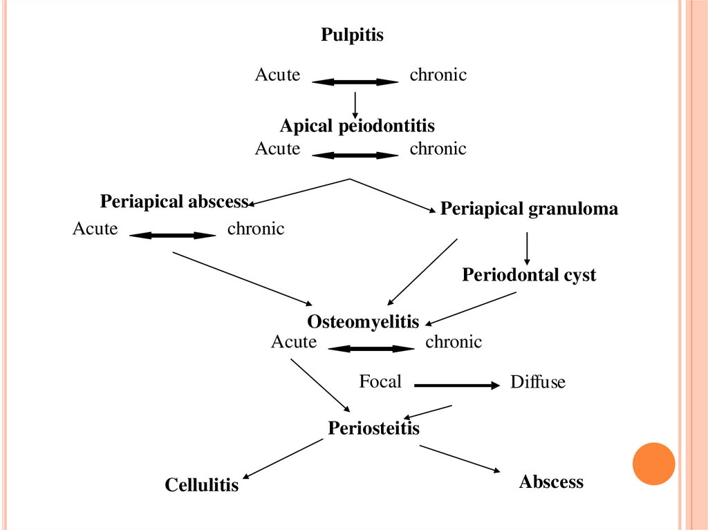
37.
PulpitisAcute
chronic
Apical peiodontitis
Acute
chronic
Periapical abscess
Acute
chronic
Periapical granuloma
Periodontal cyst
Osteomyelitis
Acute
chronic
Focal
Diffuse
Periosteitis
Cellulitis
Abscess
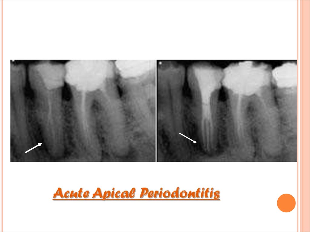
38. Acute Apical Periodontitis
A cuteApical PeriodontitisCLINICAL FEATURES:
Thermal changes does not induce pain.
Slight extrusion of tooth from socket.
Cause tenderness on mastication due to inflammatory edema
collected in P D L .
Due to external pressure, forcing of edema fluid against already
sensitized nerve endings results in severe pain.
RADIOGRAPHIC FEATURES:
• A ppear normal except for widening of P D L space.
39.
40. HISTOLOGIC FEATURES:
P D L shows signs of inflammation -vascular dilation-infiltration of P M N s
Inflammation is transient, if caused by acute trauma.
If irritant not removed, progress into surrounding bone
resorption.
Abscess formation may occur if it is associated with bacterial infection
periapical abscess /Alveolar abscess.
Acute
TREATMENT & PROGNOSIS:
• Selective grinding if inflammation due to occlusal trauma.
Options for management:
◊ Extraction
◊ Root canal treatment—with the following considerations/variations:
≫≫ There is no pulp tissue present
≫≫ Sodium hypochlorite is used as an antibacterial agent (i.e., tissue dissolution less important)
≫≫ Antibacterial medicament is required, e.g., calcium hydroxide
≫≫ If the tooth also has acute apical periodontitis—consider using a corticosteroid/antibiotic
compound in conjunction with the calcium hydroxide to reduce periapical inflammation and pain.
≫≫ If the tooth also has an acute apical abscess—consider drainage; consider systemic antibiotics.
41. Chronic Apical Periodontitis
ChronicApical Periodontitis(Periapical Granuloma)
Most common sequelae of pulpitis or apical periodontitis.
If acute (exudative) left untreated
chronic (proliferative).
Periapical granuloma is localized mass of chronic granulation
tissue formed in response to infection.
CLINICAL FEATURES:
Tooth involved is non vital /slightly tender on percussion.
Percussion may produce dull sound due to granulation tissue at
apex.
42. Mild pain on chewing on solid food.
Tooth may be slightly elongated in socket.Sensitivity is due to hyperemia, edema & inflammation of P D L .
In many cases, asymptomatic.
Fully developed granuloma seldom presents more severe clinical
symptoms.
RADIOGRAPHIC FEATURES:
L esion can be either well /ill defined
Thickening of P D L at root apex.
As concomoitant bone resorption & proliferation of granulation
tissue appears to be radiolucent area.
Loss of apical lamina dura.
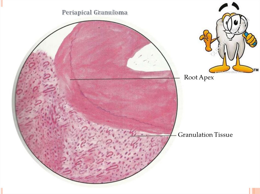
43. Periapical Granuloma
44. Thin radiopaque line or zone of sclerotic bone sometimes seen outlining lesion.
Long standing lesion may show varying degrees of rootresorption.
HISTOLOGIC FEATURES:
Granulation tissue mass consists proliferating fibroblasts,
endothelial cells & numerous immature blood capillaries with
bone resorption.
C apillaries lined with swollen endothelial cells.
Its is relatively homogenous lesion composed of macrophages,
lymphocytes & plasma cells.
45. Periapical Granuloma
46. Cholesterol clefts
47.
Collection of cholesterol clefts, with multinuclear gaint cells.Epithelial rests of Malassez may proliferate in response to
chronic inflammation & may undergo cystification.
The granulation tissue is outlined by a capsule of fibrous tissue
that is usually attached to the cementum.
TREATMENT & PROGNOSIS:
Extraction & RC T with /without apicoetomy.
If untreated
apical periodontal cyst formation .
48.
Periapical GranulomaRoot Apex
Granulation Tissue
49. Sequlae:-
Sequlae:1) The granuloma may continue to enlarge and be associated withresorption of the bone and root apex.
2 ) A cute exacerbation
3) Suppuration may occur
acute apical periodontitis .
acute or chronic periapical abscess.
4 ) P roliferation of epithelial cells rests of malassez
cyst .
5) Low grade irritation to the apical tissues
(osteosclerosis) .
radicular
bone apposition
6) Low grade irritation to the apical tissues
the apposition of
cementum on the root surface (hypercementosis).
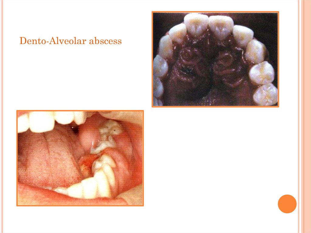
50. Periapical Abscess
Periapical A bscess(D ento-A lveolar abscess, A lveolar A bscess)
It is an acute or chronic localized suppurative process of the
dental periapical region.
Developed from acute periodontitis /periapical granuloma.
Acute exacerbation of chronic lesion
Phoenix Abscess
Cause due to – pulp infection, traumatic injury
irritation of periapical tissues ( endo procedures).
pulp necrosis,
CLINICAL FEATURES:
F eatures of acute inflammation.
Tenderness of tooth to percussion .
The tooth is slightly extruded from its socket.
Systemic manifestations like lymphadenitis & fever may
present.
51.
P eriapicalabscess
52.
53.
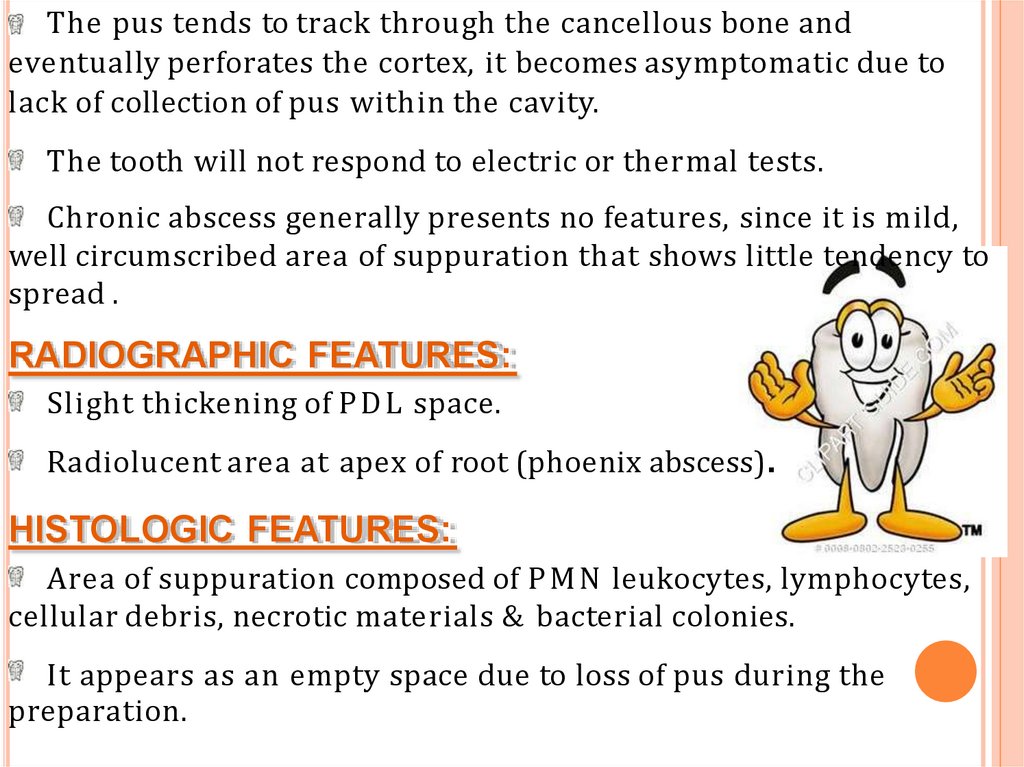
The pus tends to track through the cancellous bone andeventually perforates the cortex, it becomes asymptomatic due to
lack of collection of pus within the cavity.
The tooth will not respond to electric or thermal tests.
Chronic abscess generally presents no features, since it is mild,
well circumscribed area of suppuration that shows little tendency to
spread .
RADIOGRAPHIC FEATURES:
Slight thickening of P D L space.
Radiolucent area at apex of root (phoenix abscess).
HISTOLOGIC FEATURES:
Area of suppuration composed of P M N leukocytes, lymphocytes,
cellular debris, necrotic materials & bacterial colonies.
It appears as an empty space due to loss of pus during the
preparation.
54. Dento-Alveolar abscess
55. Dento-Alveolar abscess
ill defined radiolucency .56. Dento-Alveolar abscess
57. Inflammatory infiltrate, cellular debris, necrotic materials etc..
P eriapicalabscess
58. The abscess cavity is surrounded by acute inflammatory cell and few chronic inflammatory cells.
D ilation of blood vessels in P D LMarrow space show inflammatory infiltrates.
In chronic periapical abscess, the abscess cavity is
surrounded by dense layer of chronic inflammatory cell and few
acute inflammatory cells, and surrounded by dense bundle of
collagen fibers.
TREATMENT & PROGNOSIS:
Drainage of abscess by opening pulp chamber or extraction.
RC T.
If untreated, causes formation of fistulous tract opening to oral
mucosa (parulis) , osteomyelitis, cellulites & bacteremia .
C avernous sinus thrombosis has been reported.
59.
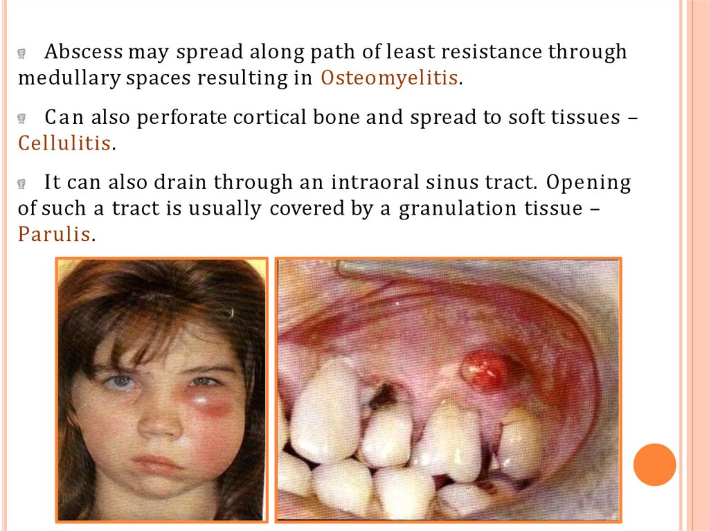
Abscess may spread along path of least resistance throughmedullary spaces resulting in Osteomyelitis.
C a n also perforate cortical bone and spread to soft tissues –
Cellulitis.
It can also drain through an intraoral sinus tract. Opening
of such a tract is usually covered by a granulation tissue –
Parulis.
60. COMPLICATIONS
Facial CellulitisL udwig's angina
Osteomyelitis
Septicaemia
Menengitis, brain abscess, cavernous sinus thrombosis
61. CELLULITIS
It is a rapidly spreading inflammation of the soft tissuescharacterized by diffuse pus formation, usually associated
with malaise and an elevated temperature.
This happens if an abscess is not able to establish drainage
through the skin surface or into oral cavity .
TYPES:
Cellulitis arising from dental infection and spreading
through soft tissues of head and neck can take various
forms.
Mostly, infection spreads through tissue spaces like canine
space, infratemporal space, pharyngeal space, buccal space,
submental and submandibular space etc.
62. CELLULITIS
Twoespecially dangerous forms of cellulitis are:-
cellulitis associated with mandibular teeth into
submandibular and cervical tissues may cause
( Ludwig’s angina).
cellulitis associated with maxillary teeth towards
the eye may cause( Cavernous sinus thrombosis)
63. LUDWIG’S ANGINA
Cellulitis of submandibular region involving sublingual,submandibular and submental spaces.
In 70% cases develops from spread of infection from
mandibular teeth.
Increased prevalence in immunocompromised patients
like A I D S , aplastic anemia, organ transplantation etc .
64. CLINICAL FEATURES
It produces a broad –like swelling of thefloor of the mouth .
Involvement of the sublingual space results
in elevation and posterior displacement of
the tongue, leading to difficulty in eating,
swelling (dysphagia) and
breathing(dyspnea) .
A fter reaching submandibular region,
infection extends to lateral pharyngeal and
retropharyngeal spaces.
65.
Lateral pharyngeal space involvement may causerespiratory obstruction due to laryngeal
edema(suffocation).
In sever cases – tachypnea, dyspnea, tachycardia, may
also be noted.
General signs – fever, malaise, leukocytosis, and raised
Erythrocyte Sedimation Rate E S R .
TREATMENT
1.
2.
3.
maintenance of the airway, tracheostomy may be
indicated.
Antibiotic therapy.
Surgical drainage.
66. CAVERNOUS SINUS THROMBOSIS
The infection from the posterior maxillary teeth reach theorbit via the maxillary sinus , while infection from the
anterior maxillary teeth reach the orbit via the ophthalmic
veins.
Infection from orbit reaches the cavernous sinus through the
communicating veins between them.
67. CLINICAL FEATURES
Periorbital edema including lateral border of nose,protrusion and fixation of eyeball.
Pupil dilatation, lacrimation, photophobia and loss of
vision may also occur.
Pain along distribution of ophthalmic and maxillary
branches of Vth cranial nerve.
Proptosis, chemosis seen in 90% cases.
Fever, chills, headache, sweating, tachycardia, nausea
and vomiting also occur.
68. Treatment
1.2.
3.
High dose of penicillin.
Extraction and drainage(if fluctuant).
Corticosteroid and anticoagulant to prevent
thrombosis and septic emboli formation.