


")
")


")


 Медицина
Похожие презентации:


")


Coma
1. Coma
ZSMUDepartment of general practice –
family medicine
2. Neural basis of consciousness
Consciousnesscannot be readily
defined in terms of anything else
A
state of awareness of self and
surrounding
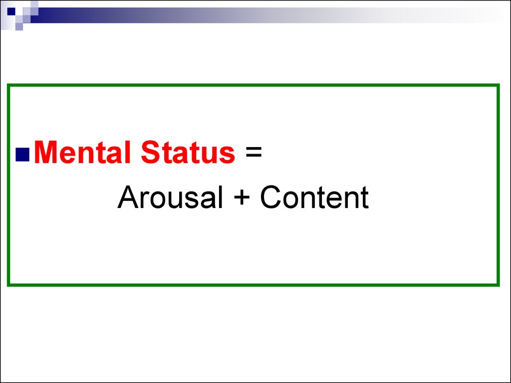
3.
MentalStatus =
Arousal + Content
4. Anatomy of Mental Status
Ascending reticular activating system (ARAS)Activating
systems of upper brainstem, hypothalamus,
thalamus
Determines
Cerebral hemispheres and interaction
between functional areas in cerebral
hemispheres
Determines
the level of arousal
the intellectual and emotional functioning
Interaction between cerebral hemispheres and
activating systems
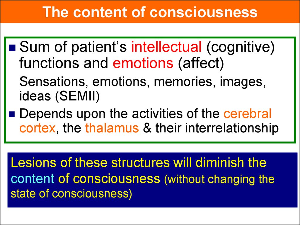
5.
The content of consciousnessSum
of patient’s intellectual (cognitive)
functions and emotions (affect)
Sensations, emotions, memories, images,
ideas (SEMII)
Depends upon the activities of the cerebral
cortex, the thalamus & their interrelationship
Lesions of these structures will diminish the
content of consciousness (without changing the
state of consciousness)
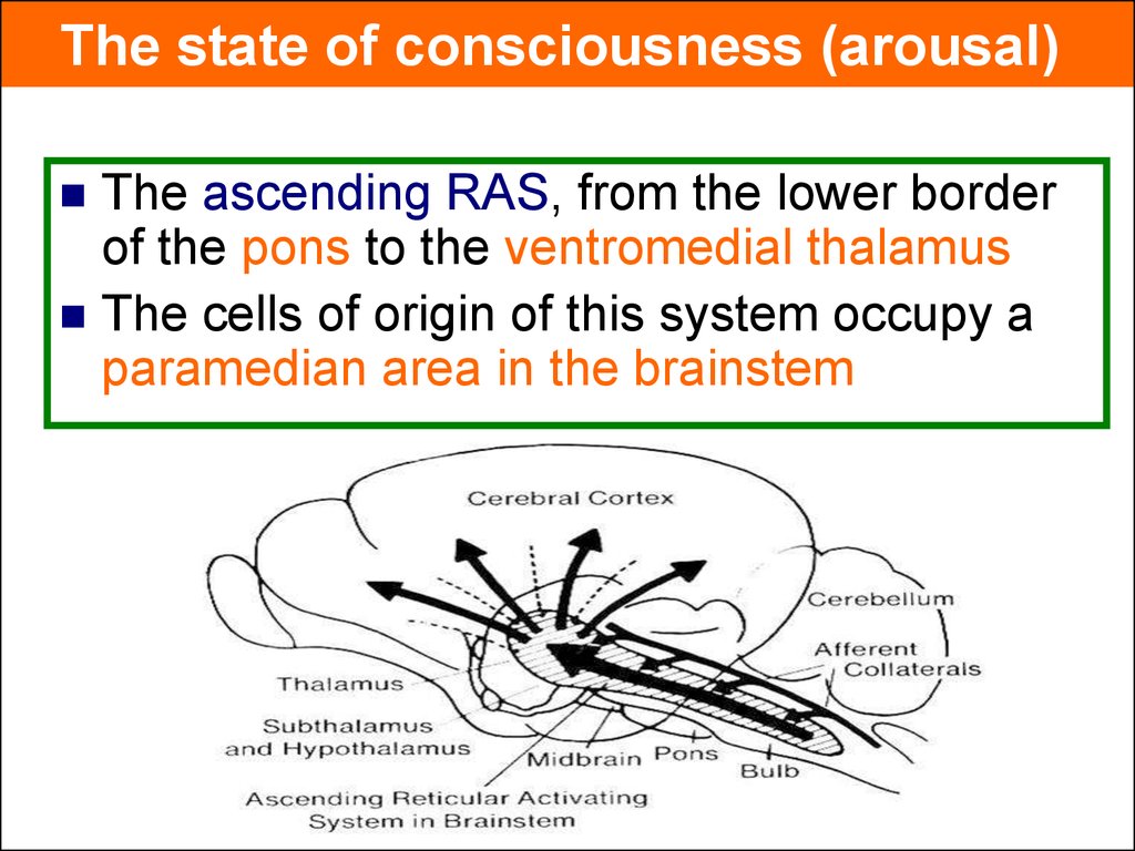
6.
The state of consciousness (arousal)The ascending RAS, from the lower border
of the pons to the ventromedial thalamus
The cells of origin of this system occupy a
paramedian area in the brainstem
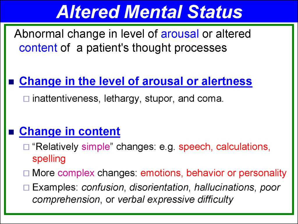
7.
Altered Mental StatusAbnormal change in level of arousal or altered
content of a patient's thought processes
Change in the level of arousal or alertness
inattentiveness,
lethargy, stupor, and coma.
Change in content
“Relatively
simple” changes: e.g. speech, calculations,
spelling
More complex changes: emotions, behavior or personality
Examples: confusion, disorientation, hallucinations, poor
comprehension, or verbal expressive difficulty
8. Definitions of levels of arousal (conciousness)
Alert (Conscious) - Appearance of wakefulness, awarenessof the self and environment
Lethargy - mild reduction in alertness
Obtundation - moderate reduction in alertness. Increased
response time to stimuli.
Stupor - Deep sleep, patient can be aroused only by
vigorous and repetitive stimulation. Returns to deep sleep
when not continually stimulated.
Coma (Unconscious) - Sleep like appearance and
behaviorally unresponsive to all external stimuli
(Unarousable unresponsiveness, eyes closed)
9. Semicoma was defined as complete loss of consciousness with a response only at the reflex level (now obsolete)
10. Psychogenic unresponsiveness
The patient, although apparentlyunconscious, usually shows some response
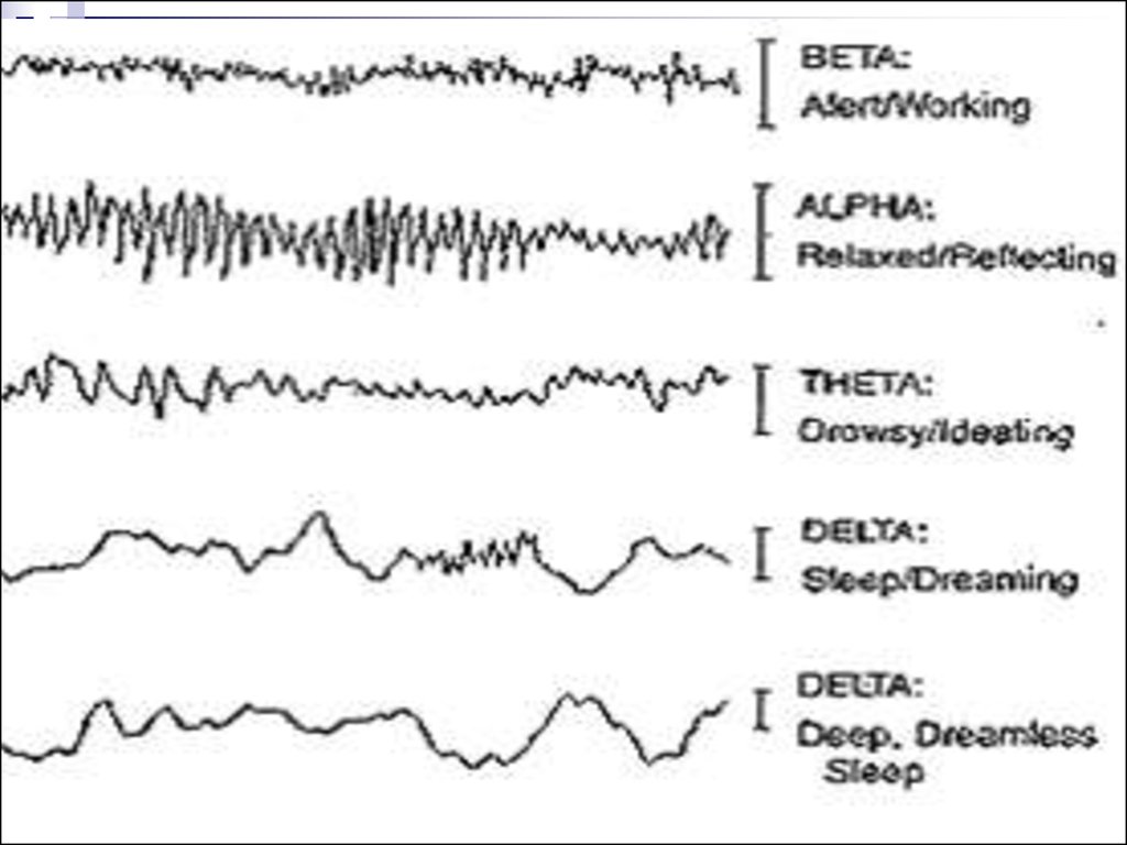
to external stimuli
An attempt to elicit the corneal reflex may
cause a vigorous contraction of the
orbicularis oculi
Marked resistance to passive movement of
the limbs may be present, and signs of
organic disease are absent
11.
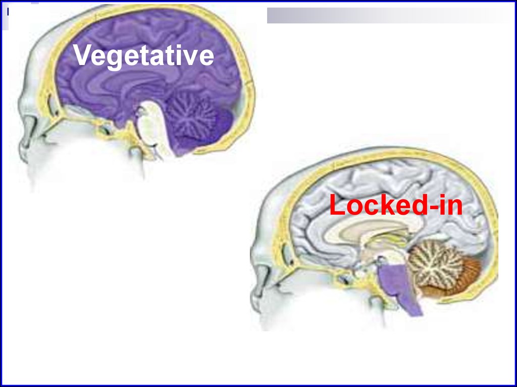
Vegetative state (coma vigil, apallic syndrome)Patients who survive coma do not remain in
this state for > 2–3 weeks, but develop a
persistent unresponsive state in which sleep–
wake cycles return.
After severe brain injury, the brainstem
function returns with sleep–wake cycles, eye
opening in response to verbal stimuli, and
normal respiratory control.
12. Locked in syndrome
Patient is awake and alert, but unable tomove or speak.
Pontine lesions affect lateral eye movement
and motor control
Lesions often spare vertical eye movements
and blinking.
13.
VegetativeLocked-in
14. Confusional state
Major defect: lack of attentionDisorientation
Patient
to time > place > person
thinks less clearly and more slowly
Memory
faulty (difficulty in repeating
numbers (digit span)
Misinterpretation of external stimuli
Drowsiness may alternate with hyper excitability and irritability
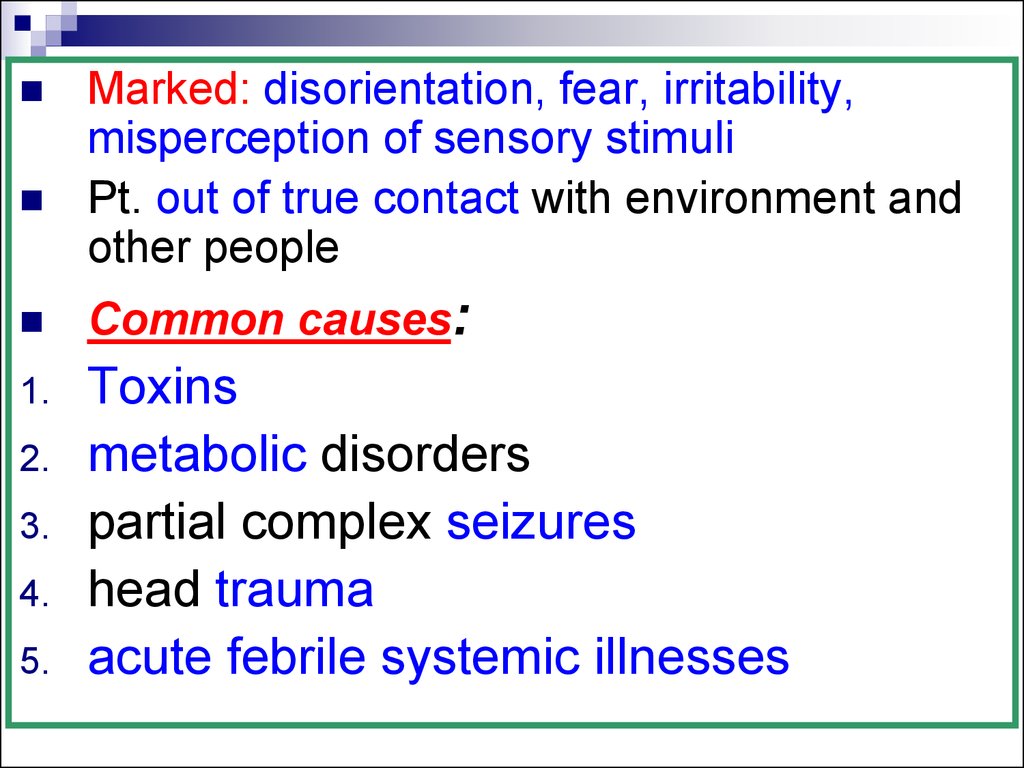
15. Delirium
Markedlyabnormal mental state
Severe
confusional state
PLUS Visual hallucinations &/or
delusions
(complex systematized dream like state)
16.
Marked: disorientation, fear, irritability,misperception of sensory stimuli
Pt. out of true contact with environment and
other people
Common causes:
1.
Toxins
metabolic disorders
partial complex seizures
head trauma
acute febrile systemic illnesses
2.
3.
4.
5.
17.
To cause coma, as defined as a state ofunconsciousness in which the eyes are
closed and sleep–wake cycles absent
Lesion of the cerebral hemispheres
extensive and bilateral
Lesions of the brainstem: above the
lower 1/3 of the pons and destroy both
sides of the paramedian reticulum
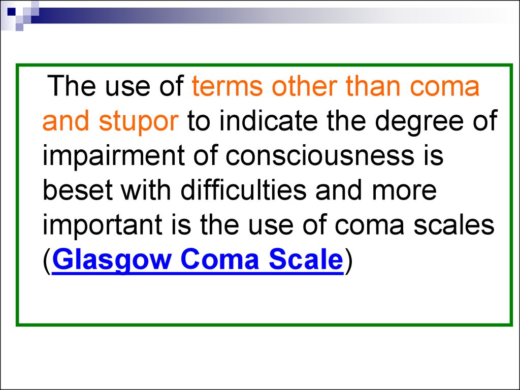
18.
The use of terms other than comaand stupor to indicate the degree of
impairment of consciousness is
beset with difficulties and more
important is the use of coma scales
(Glasgow Coma Scale)
19. Glasgow Coma Scale (GCS)
Best eyeresponse (E)
4 Eyes opening
Best verbal
response (V)
Best motor
response (M)
5 Oriented
6 Obeys commands
4 Confused
5 Localizes to pain
3 Inappropriate words
4 Withdraws from pain
2 Incomprehensible
3 Flexion in response to
sounds
pain
1 None
2 Extension to pain
spontaneously
3 Eye opening to
speech
2 Eye opening in
response to pain
1 No eye opening
1 No motor response
20.
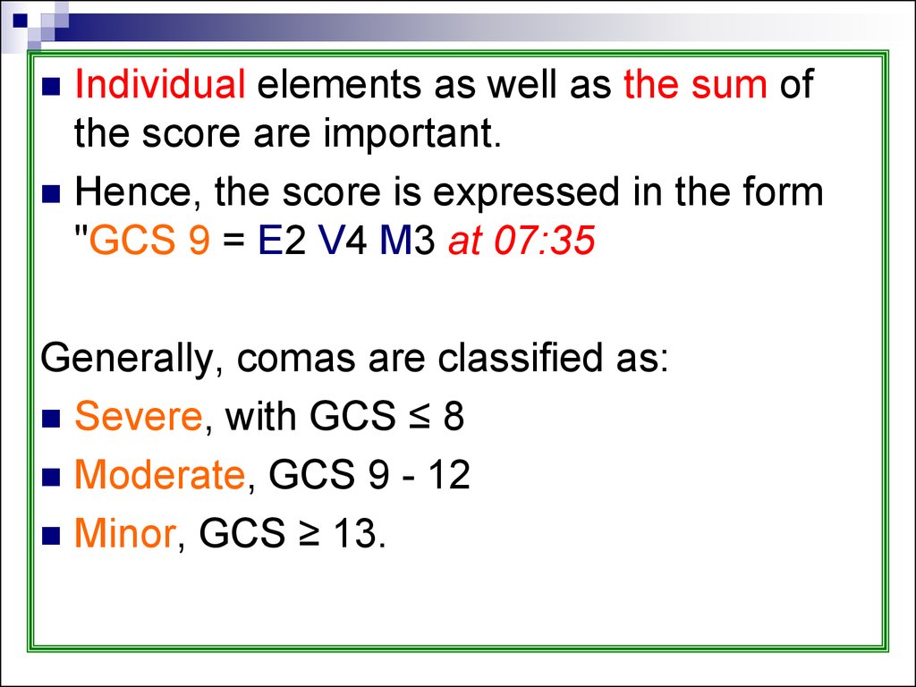
Individual elements as well as the sum ofthe score are important.
Hence, the score is expressed in the form
"GCS 9 = E2 V4 M3 at 07:35
Generally, comas are classified as:
Severe, with GCS ≤ 8
Moderate, GCS 9 - 12
Minor, GCS ≥ 13.
21. Approaches to DD
UnresponsiveABCs
Glucose, ABG, Lytes, Mg,
Ca, Tox, ammonia
IV D50, narcan,
flumazenil
Brainstem N
or other
Focal signs
Y
CT
Y
N
Unconscious
Diffuse brain dysfunction
metabolic/ infectious
Focal lesions
Tumor, ICH/SAH/ infarction
Pseudo-Coma
Psychogenic,
Looked-in,
NM paralysis
LP± CT
22. Approaches to DD
General examination:On arrival to ER immediate attention to:
1. Airway
2. Circulation
3. establishing IV access
4. Blood should be withdrawn: estimation of
glucose # other biochemical parameters #
drug screening
23.
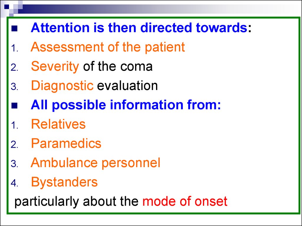
Attention is then directed towards:1. Assessment of the patient
2. Severity of the coma
3. Diagnostic evaluation
All possible information from:
1. Relatives
2. Paramedics
3. Ambulance personnel
4. Bystanders
particularly about the mode of onset
24.
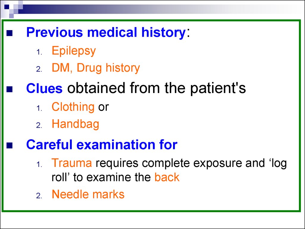
Previous medical history:1.
2.
Clues obtained from the patient's
1.
2.
Epilepsy
DM, Drug history
Clothing or
Handbag
Careful examination for
1.
2.
Trauma requires complete exposure and ‘log
roll’ to examine the back
Needle marks
25.
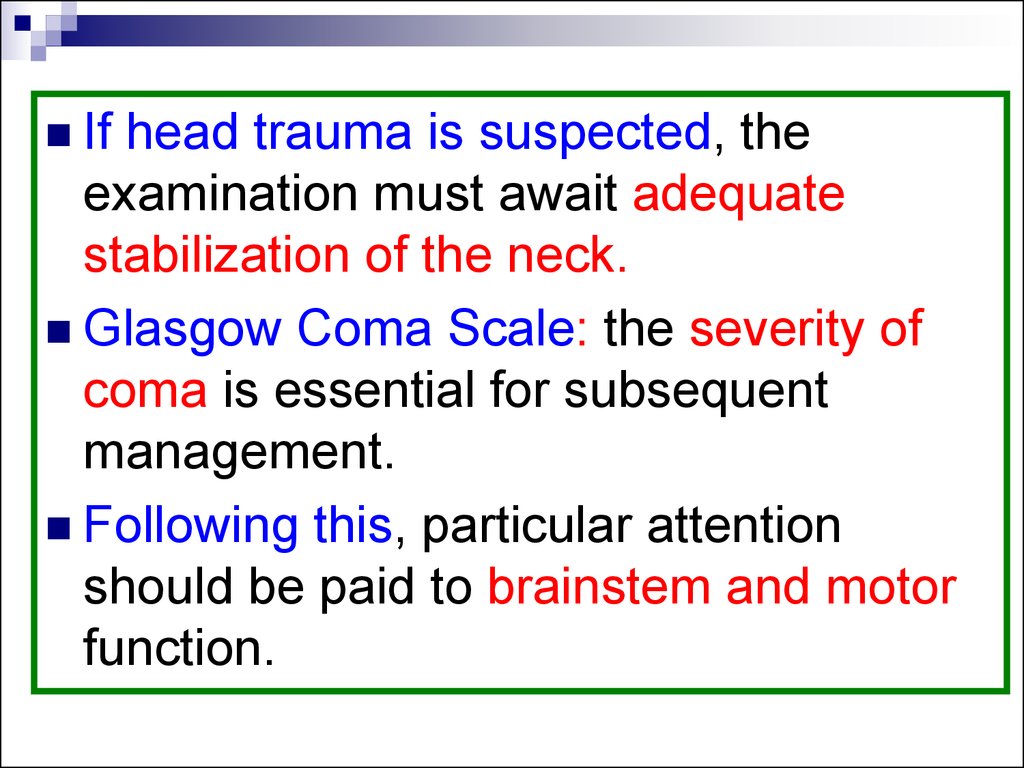
Ifhead trauma is suspected, the
examination must await adequate
stabilization of the neck.
Glasgow Coma Scale: the severity of
coma is essential for subsequent
management.
Following this, particular attention
should be paid to brainstem and motor
function.
26.
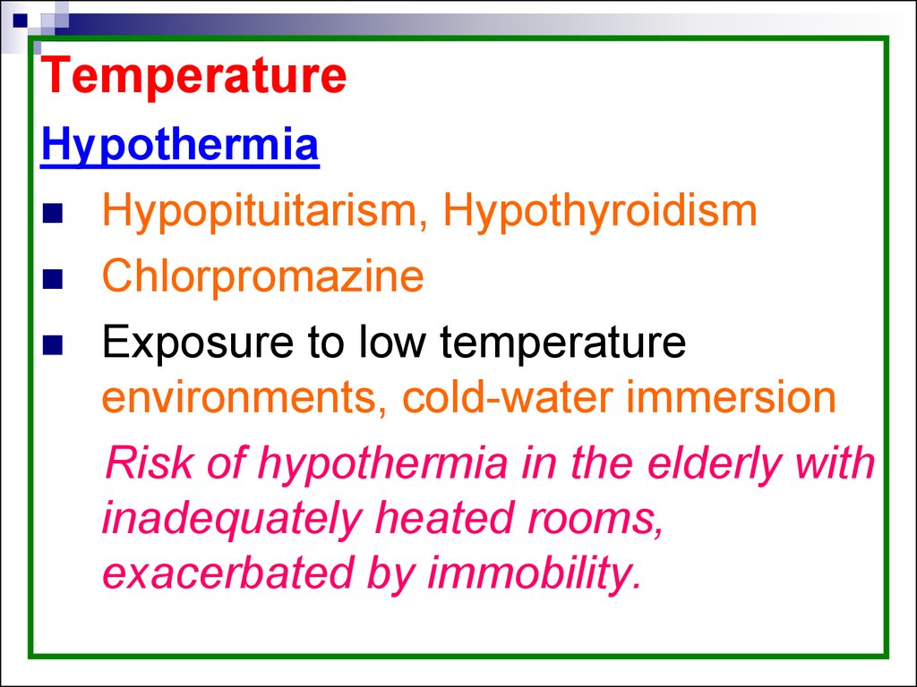
TemperatureHypothermia
Hypopituitarism, Hypothyroidism
Chlorpromazine
Exposure to low temperature
environments, cold-water immersion
Risk of hypothermia in the elderly with
inadequately heated rooms,
exacerbated by immobility.
27.
1.2.
C/P: generalized rigidity and muscle
fasciculation but true shivering may be
absent. (a low-reading rectal
thermometer is required).
Hypoxia and hypercarbia are common.
Treatment:
Gradual warming is necessary
May require peritoneal dialysis with warm
fluids.
28.
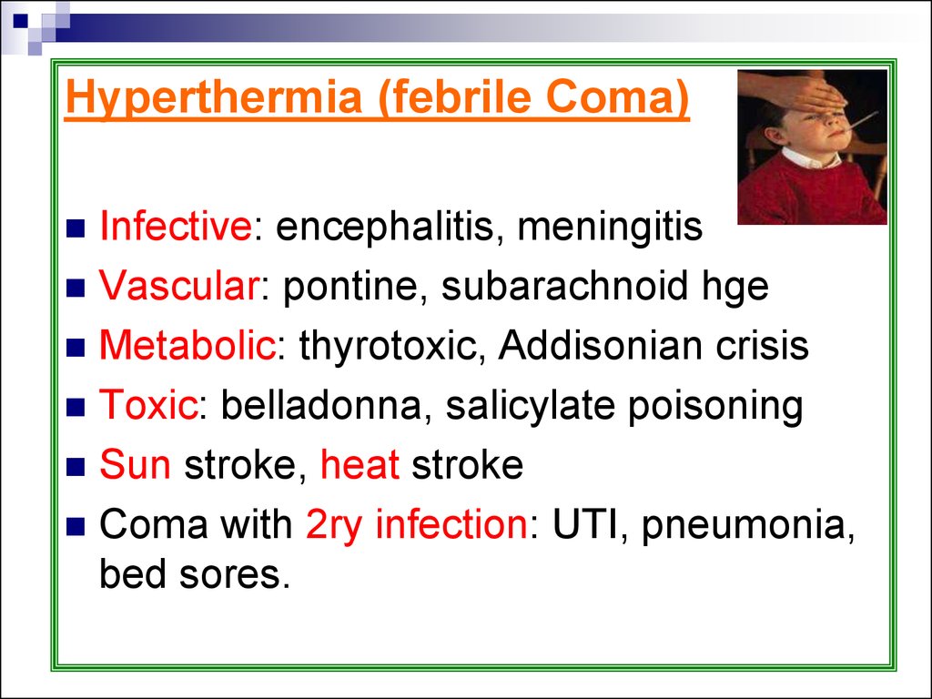
Hyperthermia (febrile Coma)Infective: encephalitis, meningitis
Vascular: pontine, subarachnoid hge
Metabolic: thyrotoxic, Addisonian crisis
Toxic: belladonna, salicylate poisoning
Sun stroke, heat stroke
Coma with 2ry infection: UTI, pneumonia,
bed sores.
29.
Hyperthermia or heat strokeLoss of thermoregulation dt. prolonged
exertion in a hot environment
Initial ↑ in body temperature with profuse
sweating followed by
hyperpyrexia, an abrupt cessation of
sweating, and then
rapid onset of coma, convulsions, and death
30.
This may be exacerbated by certain drugs,‘Ecstasy’ abuse—involving a loss of the thirst
reaction in individuals engaged in prolonged
dancing.
Other causes
Tetanus
Pontine hge
Lesions in the floor of the third ventricle
Neuroleptic malignant syndrome
Malignant hyperpyrexia with anaesthetics.
31.
Heat stroke neurological sequelaeParaparesis.
Cerebellar
ataxia.
Dementia (rare)
32.
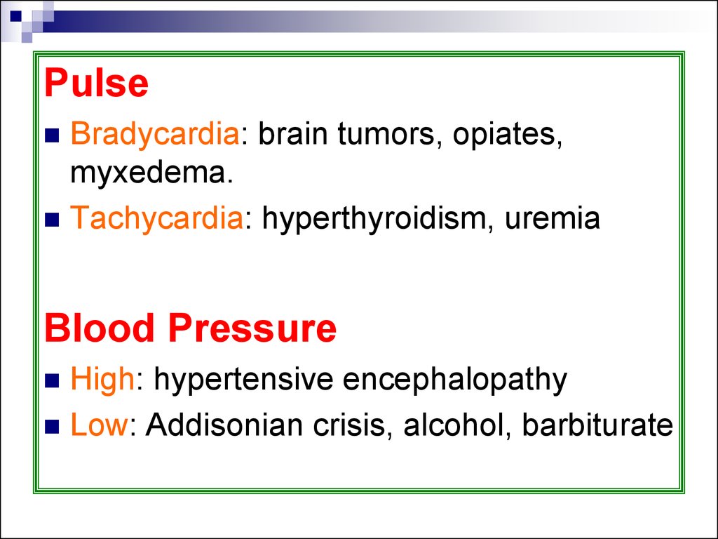
PulseBradycardia: brain tumors, opiates,
myxedema.
Tachycardia: hyperthyroidism, uremia
Blood Pressure
High: hypertensive encephalopathy
Low: Addisonian crisis, alcohol, barbiturate
33.
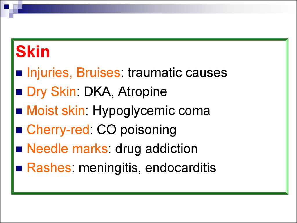
SkinInjuries, Bruises: traumatic causes
Dry Skin: DKA, Atropine
Moist skin: Hypoglycemic coma
Cherry-red: CO poisoning
Needle marks: drug addiction
Rashes: meningitis, endocarditis
34.
PupilsSize, inequality, reaction to a bright light.
An important general rule: most metabolic
encephalopathies give small pupils with
preserved light reflex.
Atropine, and cerebral anoxia tend to
dilate the pupils, and opiates will constrict
them.
35.
Structural lesions are more commonlyassociated with pupillary asymmetry and with
loss of light reflex.
Midbrain tectal lesions : round, regular,
medium-sized pupils, do not react to light
Midbrain nuclear lesions: medium-sized
pupils, fixed to all stimuli, often irregular and
unequal.
Cranial n III distal to the nucleus: Ipsilateral
fixed, dilated pupil.
36.
Pons (Tegmental lesions) : bilaterally smallpupils, {in pontine hge, may be pinpoint,
although reactive} assess the light response
using a magnifying glass
Lateral medullary lesion: ipsilateral Horner's
syndrome.
Occluded carotid artery causing cerebral
infarction: Pupil on that side is often small
37.
DiencephalonsSmall, reactive
Midbrain
Medium-sized, fixed
Dilated, Fixed
Pons
Ipsilateral dilated, Fixed
small, pinpoint
In hge reactive
.
38. Ocular movements
The position of the eyes at restPresence of spontaneous eye movement
The reflex responses to oculocephalic and
oculovestibular maneuvers
In diffuse cerebral disturbance but intact
brainstem function, slow roving eye
movements can be observed
Frontal lobe lesion may cause deviation of
the eyes towards the side of the lesion
39.
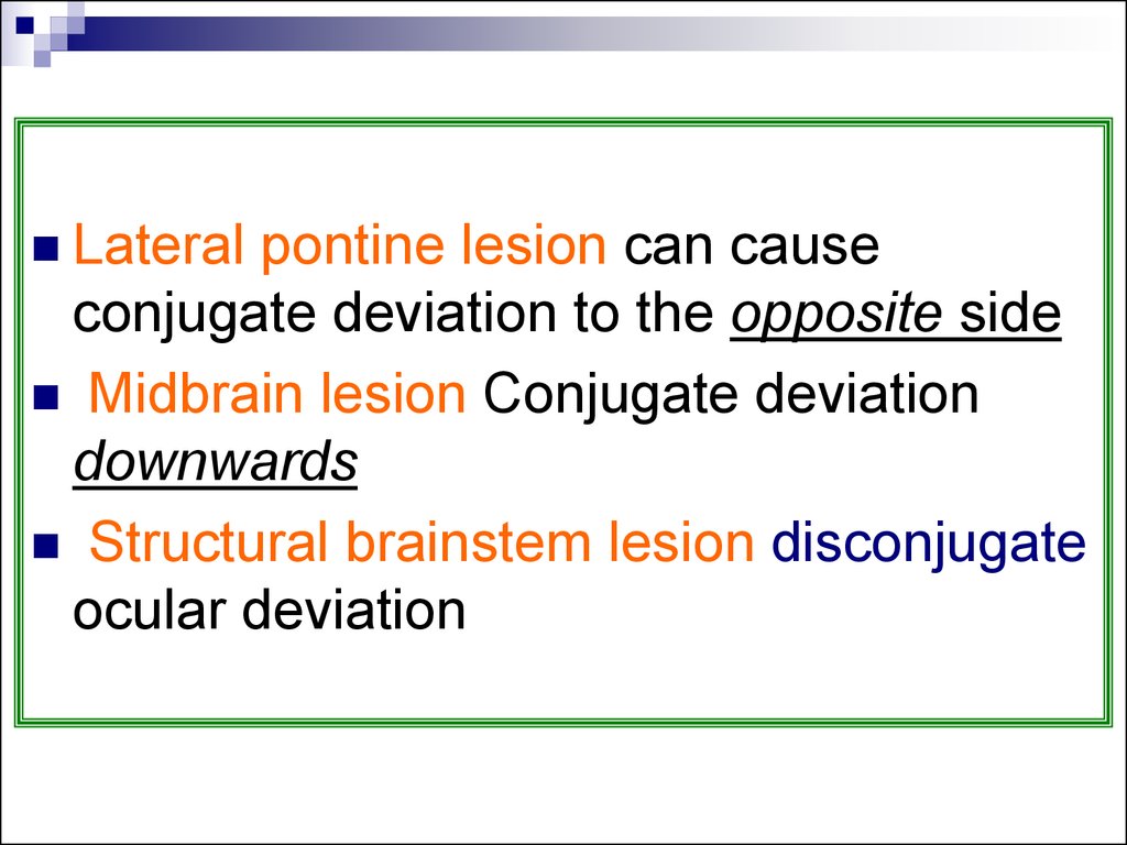
Lateralpontine lesion can cause
conjugate deviation to the opposite side
Midbrain lesion Conjugate deviation
downwards
Structural brainstem lesion disconjugate
ocular deviation
40.
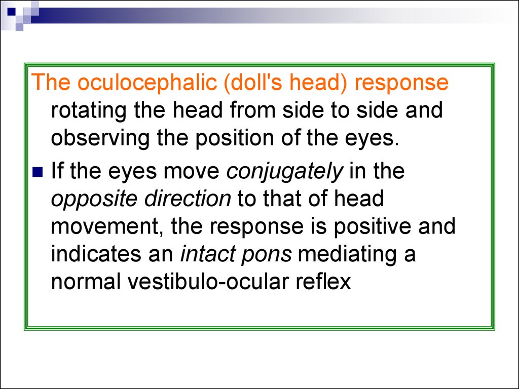
The oculocephalic (doll's head) responserotating the head from side to side and
observing the position of the eyes.
If the eyes move conjugately in the
opposite direction to that of head
movement, the response is positive and
indicates an intact pons mediating a
normal vestibulo-ocular reflex
41.
Caloric oculovestibular responses These aretested by the installation of ice-cold water
into the external auditory meatus, having
confirmed that there is no tympanic
rupture.
A normal response in a conscious patient
is the development of nystagmus with the
quick phase away from the stimulated side
This requires intact cerebropontine
connections
42.
Odour of breathAcetone: DKA
Fetor Hepaticus: in hepatic coma
Urineferous odour: in uremic coma
Alcohol odour: in alcohol intoxication
43.
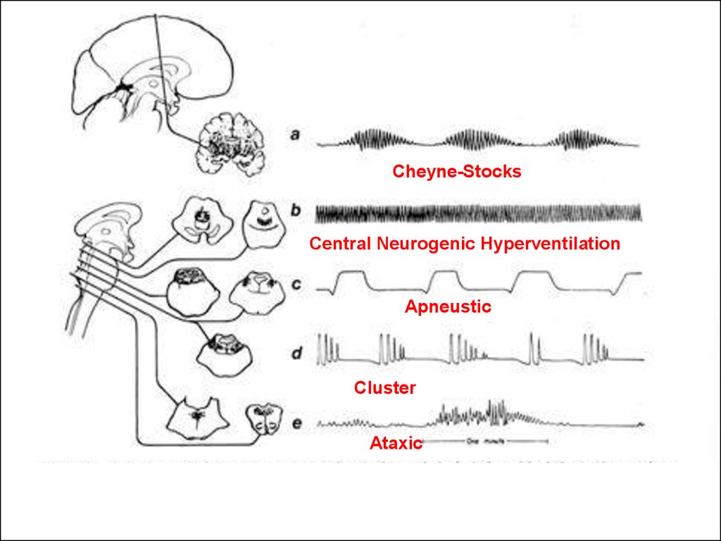
RespirationCheyne–Stokes respiration:
(hyperpnoea alternates with apneas) is
commonly found in comatose patients,
often with cerebral disease, but is
relatively non-specific.
Rapid, regular respiration is also common
in comatose patients and is often found
with pneumonia or acidosis.
44.
Central neurogenic hyperventilationBrainstem tegmentum (mostly tumors):
↑ PO2, ↓ PCO2, and
Respiratory alkalosis in the absence of any
evidence of pulmonary disease
Sometimes complicates hepatic encephalopathy
45.
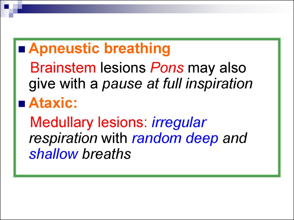
Apneusticbreathing
Brainstem lesions Pons may also
give with a pause at full inspiration
Ataxic:
Medullary lesions: irregular
respiration with random deep and
shallow breaths
46.
Cheyne-StocksCentral Neurogenic Hyperventilation
Apneustic
Cluster
Ataxic
47. Abnormal breathing patterns in coma
Cheynes - StokesCentral Neurogenic
Midbrain
Apneustic
Pons
Ataxic
Medulla
ARAS
48.
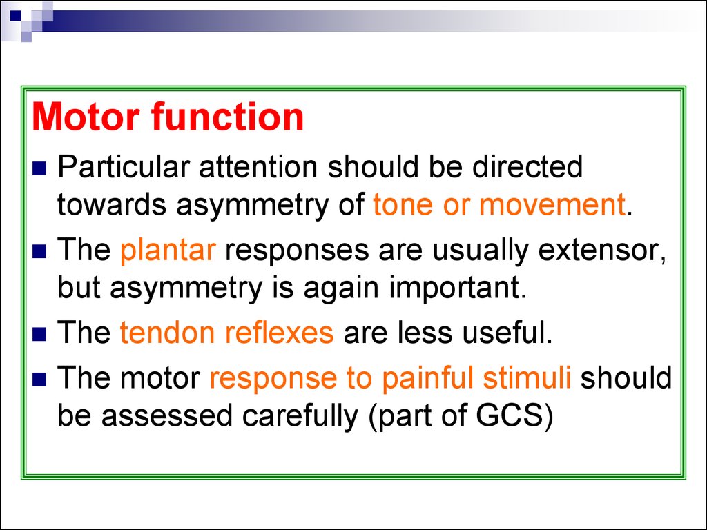
Motor functionParticular attention should be directed
towards asymmetry of tone or movement.
The plantar responses are usually extensor,
but asymmetry is again important.
The tendon reflexes are less useful.
The motor response to painful stimuli should
be assessed carefully (part of GCS)
49.
Painful stimuli: supraorbital nerve pressureand nail-bed pressure. Rubbing of the
sternum should be avoided (bruising and
distress to the relatives)
Patients may localize or exhibit a variety of
responses, asymmetry is important
50.
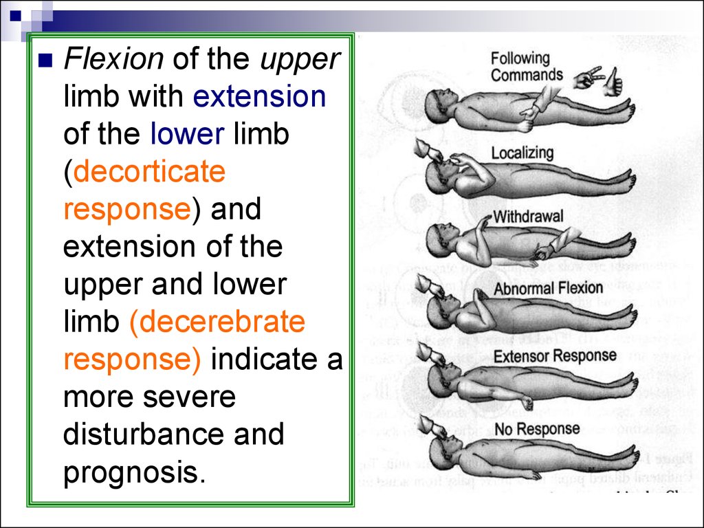
Flexion of the upperlimb with extension
of the lower limb
(decorticate
response) and
extension of the
upper and lower
limb (decerebrate
response) indicate a
more severe
disturbance and
prognosis.
51. Signs of lateralization
Unequal pupilsDeviation of the eyes to one side
Facial asymmetry
Turning of the head to one side
Unilateral hypo-hypertonia
Asymmetric deep reflexes
Unilateral extensor plantar response (Babinski)
Unilateral focal or Jacksonian fits
52.
Head and neck1.
2.
The head
Evidence of injury
Skull should be palpated for depressed
fractures.
The ears and nose: haemorrhage and
leakage of CSF
The fundi: papilloedema or subhyaloid or
retinal haemorrhages
53.
1.2.
Neck: In the presence of trauma to the
head, associated trauma to the neck
should be assumed until proven
otherwise.
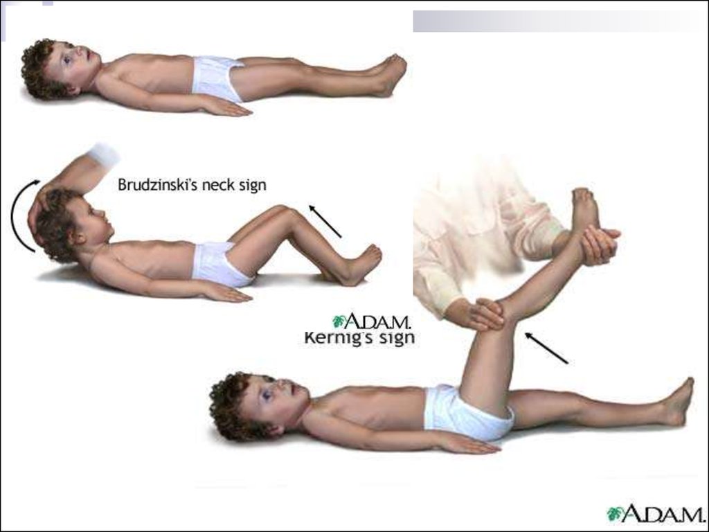
Positive Kernig's sign : a meningitis or
SAH. If established as safe to do so, the
cervical spine should be gently flexed
Neck stiffness may occur:
↑ ICP
incipient tonsillar herniation
54.
55. Causes of COMA
56.
CNS causes of comaCerebrovascular disease is a frequent cause
of coma.
Mechanism:
Impairment of perfusion of the RAS
With hypotension
Brainstem herniation ( parenchymal hge,
swelling from infarct, or more rarely,
extensive brainstem infarction)
57.
Subarachnoid haemorrhageLoss of consciousness is common with
SAH
only about 1/2 of patients recover
from the initial effects of the
haemorrhage.
Causes of coma:
1. Acute ↑ICP and
2. Later, vasospasms, hyponatraemia
58.
Parenchymal haemorrhage1.
2.
1.
2.
May cause a rapid decline in consciousness,
from
Rupture into the ventricles
or subsequent herniation and brainstem
compression.
Cerebellar haemorrhage or infarct with
Subsequent oedema
Direct brainstem compression, early
decompression can be lifesaving.
59.
Hypotension1.
2.
The critical blood flow in humans
required to maintain effective cerebral
activity is about 20 ml/100 g/min and
any fall below this leads rapidly to
cerebral insufficiency.
The causes:
syncope in younger patients
cardiac disease in older patients.
60.
Hypertensive encephalopathyNow
rare with better control of blood
pressure.
C/P: impaired consciousness, grossly
raised blood pressure, papilloedema.
Neuropathologically: fibrinoid necrosis,
arteriolar thrombosis, microinfarction,
and cerebral oedema (failure of
autoregulation)
61.
Raised intracranial pressureMass
effects: tumours, abscesses,
haemorrhage, subdural, extradural
haematoma, brainstem herniation→
distortion of the RAS.
C/P: depends on normal variation in
the tentorial aperture, site of lesion,
and the speed of development.
62.
Herniation and loss of consciousnessLesions located deeply, laterally, or in the
temporal lobes > located at a distance, such
as the frontal and occipital lobes.
Rate of growth: slowly growing tumours may
achieve a substantial size and distortion of
cerebral structure without impairment of
consciousness, in contrast to small rapidly
expanding lesions
63.
Centralherniation involves
downward displacement of the upper
brainstem
Uncal herniation in which the medial
temporal lobe herniates through the
tentorium
64.
Central herniation: small pupils are followedby midpoint pupils, and irregular respiration
gives way to hyperventilation as coma
deepens.
Uncal herniation: a unilateral dilated pupil,
due to compression of the III nerve, and
asymmetric motor signs. As coma deepens,
the opposite pupil loses the light reflex and
may constrict briefly before enlarging.
Rarely, Upward herniation can occur with
posterior fossa masses
65.
Head injuryThe leading cause of death below the age
of 45, head injury accounts for 1/2 of all
trauma deaths
A major cause of patients presenting with
coma.
A history is usually available and, if not,
signs of injury such as bruising of the scalp
or skull fracture lead one to the diagnosis
66.
Alcoholon the breath provides a direct
clue to a cause of coma, evidence of
head injury need not necessarily imply
that this is the cause.
Epileptic seizure, may have resulted in
a subsequent head injury
67.
Damagecan be diffuse or focal.
Rotational forces of the brain cause
surface cortical contusions and even
lacerations, most obvious
frontotemporally because of the
irregular sphenoidal wing and orbital
roof.
Subdural bleeding due to tearing of
veins
68.
Diffuse axonal injury is now seen as themajor consequence of head injury and
associated coma.
Mild degrees of axonal injury also occur
with concussion and brief loss of
consciousness
69.
Secondary damage can occur fromparenchymal haemorrhage, brain oedema,
and vascular dilatation, all of which will lead
to ↑ICP→ ↓perfusion pressure, which can be
accentuated by systemic hypoxia and blood
loss.
Subdural and extradural haematomata may
cause impairment of consciousness
following apparent recovery are important to
diagnose, as they are readily treatable
surgically.
70.
InfectionsSystemic infections may result in coma as
an event secondary to metabolic and
vascular disturbance or seizure activity.
Direct infections of the CNS, as with
meningitis and encephalitis, can all be
associated with coma.
Meningitis: the onset is usually subacute,
intense headache, associated with fever and
neck stiffness. meningococcal meningitis
may be rapid in onset
71.
Diagnosis is confirmed by identifying thechanges in the CSF, from which it may be
possible to isolate the causative organism.
Prompt treatment of acute meningitis is,
however, imperative and may precede
diagnostic confirmation.
Encephalitis: usually subacute, and often
associated with fever and/or seizures,
herpes simplex encephalitis may be
explosive at onset, leading to coma within
a matter of hours Treatment with aciclovir,
precedes definitive diagnosis.
72.
Parasitic infectionsCerebral malaria
25 % mortality rate.
Associated with 2–10 % of cases of
infection with Plasmodium falciparum.
C/P: acute profound mental obtundation or
psychosis, leading to coma with extensor
plantar responses
CSF: may show increased protein,
characteristically there is no pleocytosis
73.
Hypoglycaemia and lactic acidosis, whichmay contribute to the coma.
Treatment: intravenous quinine.
Steroids, which were at one time prescribed
widely for oedema, are now contraindicated
as they prolong the coma.
74.
Septic patientsCommonly develop an encephalopathy.
In some patients this can be severe, with a
prolonged coma.
Lumbar puncture in such patients is usually
normal or only associated with a mildly
elevated protein level.
EEG is valuable and is abnormal, ranging
from diffuse theta through to triphasic waves
and suppression or burst-suppression
75.
Althoughthere is a high mortality,
there is the potential for complete
reversibility
Presence of coma should not prevent
an aggressive approach to
management of such patients
including, for example, haemodialysis
to deal with acute renal failure
76. Metabolic causes of coma
Hepatic comaThe patient is known to be suffering from liver
failure
May occur in patients with chronic liver failure
and portosystemic shunting (In these cases
jaundice may be absent)
77.
Precipitation: GIT hge, infection, certaindiuretics, sedatives, analgesics, general
anaesthesia, high-protein food or ammonium
compounds
Subacute onset, although it can be sudden,
with an initial confusional state often bilateral
asterixis or flapping tremor.
Asterixis, a -ve myoclonus jerk, results in
sudden loss of a maintained posture. elicited
by asking the subject to maintain extension at
the wrist
78.
79.
As coma supervenes, there is oftendecerebrate and/or decorticate posturing with
extensor plantar responses
Diagnosis: signs of liver disease hepatic fetor,
and biochemical evidence of disturbed liver
function. EEG with paroxysms of bilaterally
synchronous slow waves in the delta range or
with occasional triphasic waves
80.
The disturbance of consciousness due toraised ammonia, and indeed treatments to
reduce ammonia
endogenous benzodiazepine ligands may
contribute to the hepatic coma,
benzodiazepine antagonist, flumazenil, in
hepatic coma would support this view
81.
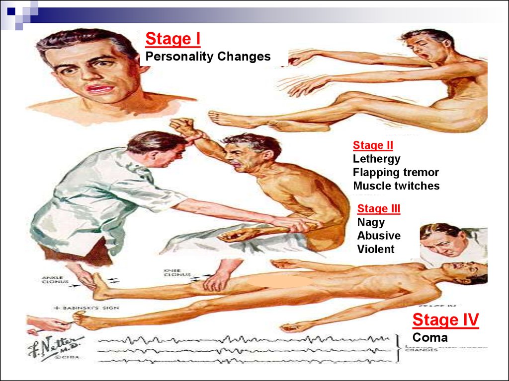
Stage IPersonality Changes
Stage II
Lethergy
Flapping tremor
Muscle twitches
Stage III
Nagy
Abusive
Violent
Stage IV
Coma
82.
Renal comaMay occur in acute or chronic renal failure
Raised blood urea alone cannot be
responsible for the loss of consciousness
but the
Metabolic acidosis, electrolyte disturbances
and Water intoxication due to fluid retention
may be responsible
83.
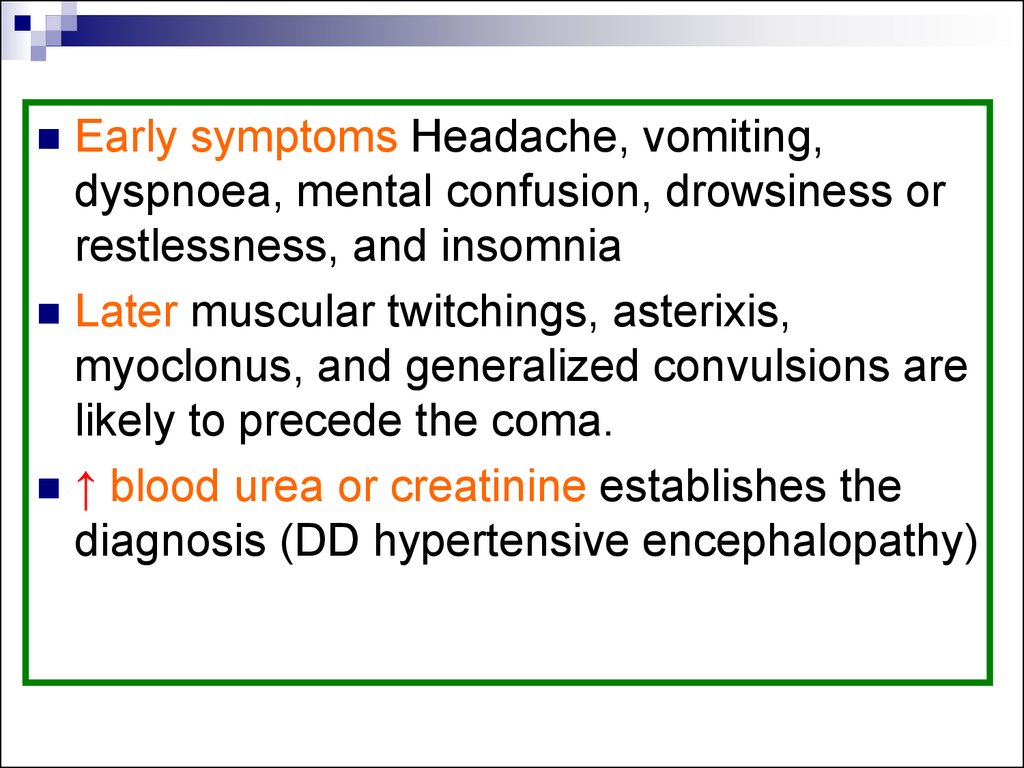
Early symptoms Headache, vomiting,dyspnoea, mental confusion, drowsiness or
restlessness, and insomnia
Later muscular twitchings, asterixis,
myoclonus, and generalized convulsions are
likely to precede the coma.
↑ blood urea or creatinine establishes the
diagnosis (DD hypertensive encephalopathy)
84.
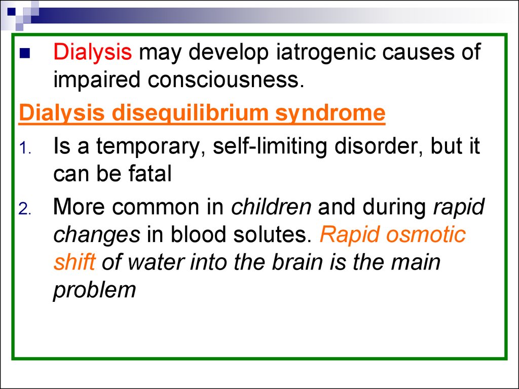
Dialysis may develop iatrogenic causes ofimpaired consciousness.
Dialysis disequilibrium syndrome
1. Is a temporary, self-limiting disorder, but it
can be fatal
2. More common in children and during rapid
changes in blood solutes. Rapid osmotic
shift of water into the brain is the main
problem
85.
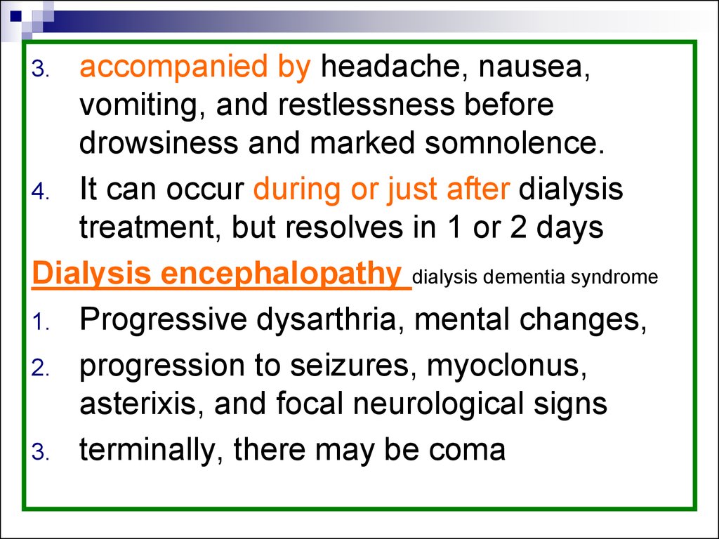
accompanied by headache, nausea,vomiting, and restlessness before
drowsiness and marked somnolence.
4. It can occur during or just after dialysis
treatment, but resolves in 1 or 2 days
Dialysis encephalopathy dialysis dementia syndrome
1. Progressive dysarthria, mental changes,
2. progression to seizures, myoclonus,
asterixis, and focal neurological signs
3. terminally, there may be coma
3.
86.
4.5.
6.
EEG: paroxysmal bursts of irregular,
generalized spike and wave activity.
has been attributed to the neurotoxic
effects of aluminium: aluminium-containing
antacids and a high aluminium content in
the water
Reached its peak prevalence in the mid
1970s, before preventive action was taken.
87.
Disturbance of glucose metabolismDiabetic Ketoacidosis
Subacute onset with late development of
coma.
Marked ketoacidosis, usually above 40
mmol/l, together with ketonuria.
Secondary lactic acidosis (DD severe anoxia
or methyl alcohol or paraldehyde poisoning)
Patients are dehydrated, rapid, shallow
breathing, occasionally acetone on the breath.
The plantar responses are usually flexor until
coma supervenes.
88.
Hyperglycaemic non-ketotic diabetic comaMore commonly seen in the elderly.
Coma is more common than with
ketoacidosis.
Profound cellular dehydration, risk of
developing cerebral venous thrombosis,
which may contribute to the disturbance of
consciousness.
It may be induced by drugs, acute
pancreatitis, burns, and heat stroke
89.
Hypoglycaemic comaMuch more rapid onset.
Symptoms appear with blood sugars of less
than 2.5 mmol/l
Initially autonomic: sweating and pallor, and
then inattention and irritability progressing to
stupor, coma, and frequent seizures.
May present with a focal onset (hemiparesis)
Plantar responses are frequently extensor.
Patients may be hypothermic.
90.
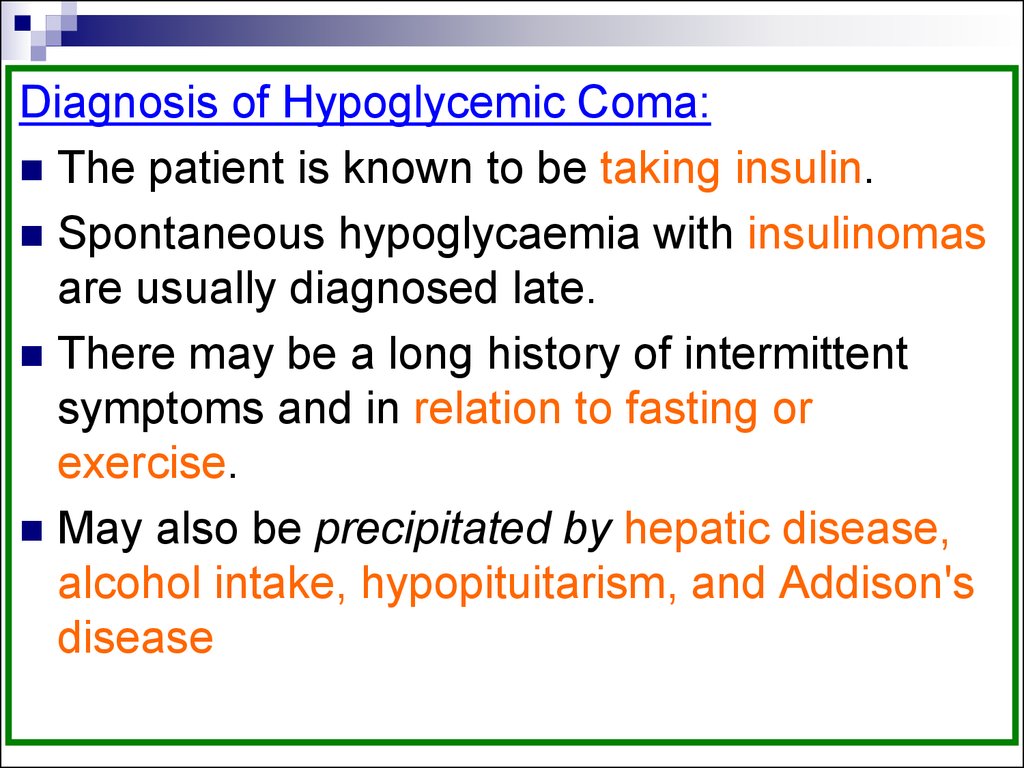
Diagnosis of Hypoglycemic Coma:The patient is known to be taking insulin.
Spontaneous hypoglycaemia with insulinomas
are usually diagnosed late.
There may be a long history of intermittent
symptoms and in relation to fasting or
exercise.
May also be precipitated by hepatic disease,
alcohol intake, hypopituitarism, and Addison's
disease
91.
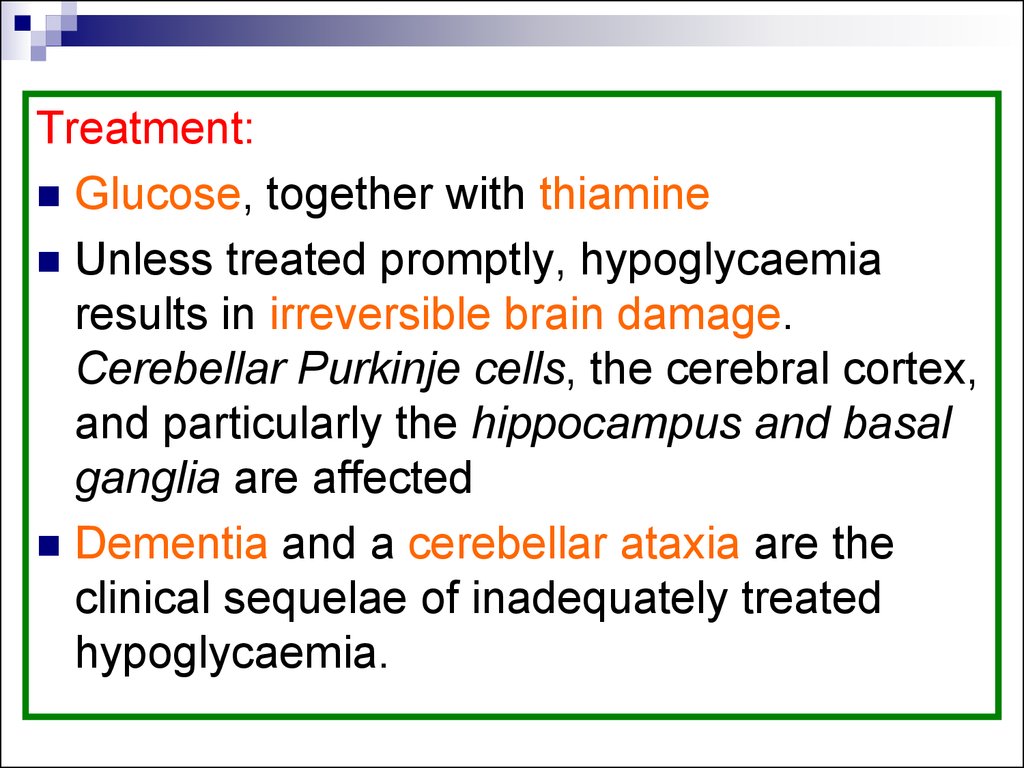
Treatment:Glucose, together with thiamine
Unless treated promptly, hypoglycaemia
results in irreversible brain damage.
Cerebellar Purkinje cells, the cerebral cortex,
and particularly the hippocampus and basal
ganglia are affected
Dementia and a cerebellar ataxia are the
clinical sequelae of inadequately treated
hypoglycaemia.
92.
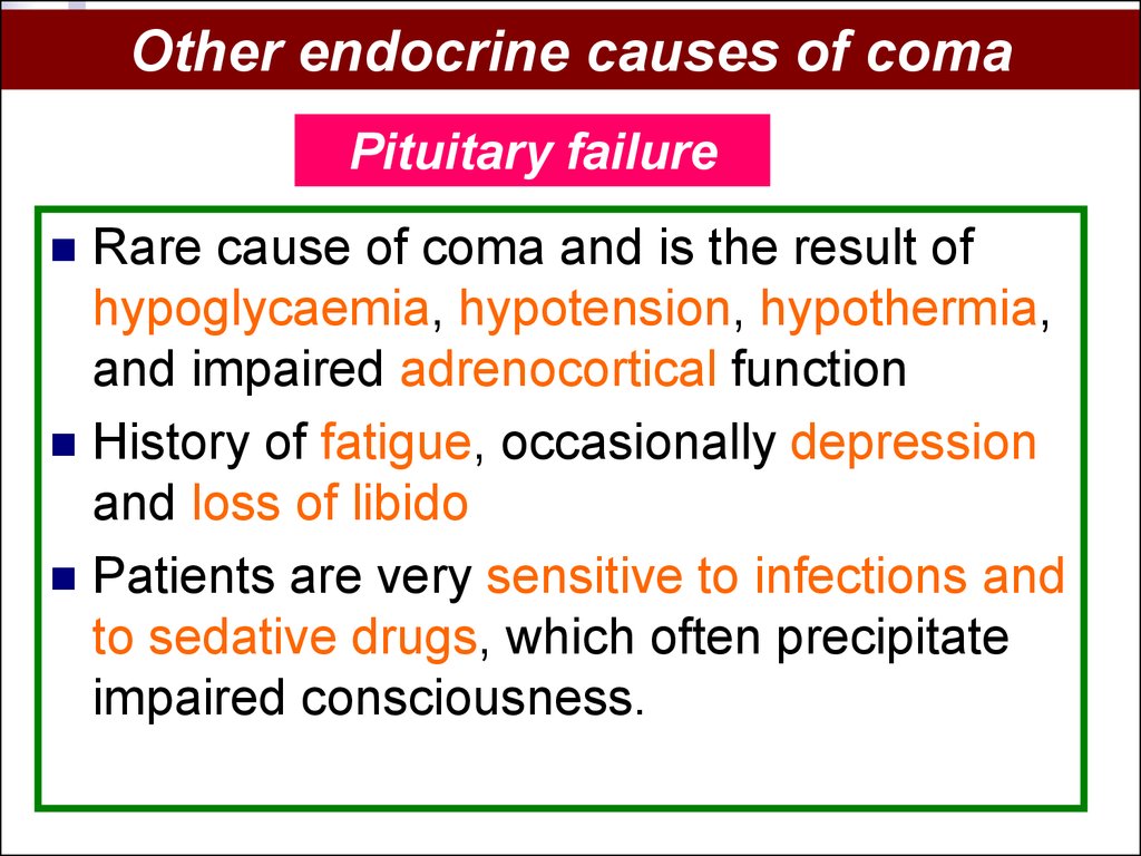
Other endocrine causes of comaPituitary failure
Rare cause of coma and is the result of
hypoglycaemia, hypotension, hypothermia,
and impaired adrenocortical function
History of fatigue, occasionally depression
and loss of libido
Patients are very sensitive to infections and
to sedative drugs, which often precipitate
impaired consciousness.
93.
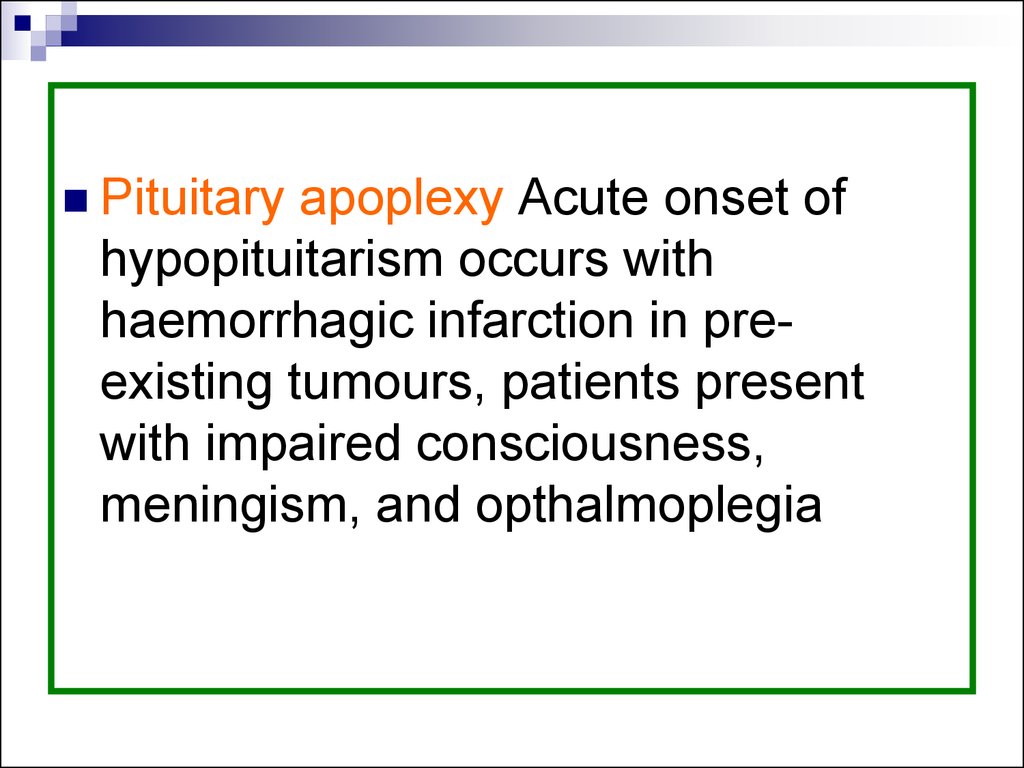
Pituitaryapoplexy Acute onset of
hypopituitarism occurs with
haemorrhagic infarction in preexisting tumours, patients present
with impaired consciousness,
meningism, and opthalmoplegia
94.
HypothyroidismMental symptoms are common, with
headaches, poor concentration, and apathy;
this is frequently diagnosed as depression.
With progression there is increasing
somnolence and, patients become sensitive
to drugs and infections.
These and cold weather, particularly in the
elderly, may precipitate myxoedemic coma.
95.
Myxoedemic coma has a high mortality andis associated with hypoglycaemia and
hyponatraemia.
low-reading thermometer to detect
hypothermia
Treatment: support of ventilation and blood
pressure and cautious correction of the
thyroid deficiency with tri-iodothyronine
96.
HyperthyroidismMild mental symptoms: anxiety,
restlessness,reduced attention.
‘Thyroid storm’ with agitated delirium, which
can progress to coma, may have bulbar
paralysis
Apathetic form of thyrotoxicosis: particularly
the elderly, with depression leading to
apathy, confusion, and coma without any
signs of hypermetabolism
97.
Adrenocortical failureMental changes are common in Addison's
disease and secondary hypoadrenalism.
Undiagnosed Addison's disease is frequently
associated with behavioural changes and
fatigue.
Infection or trauma may precipitate coma and
associated hypotension, hypoglycaemia, and
dehydration
98.
Tendon reflexes are often absent↑ ICP, papilloedema
Friedrichsen–Waterhouse syndrome acute
adrenal failure due to meningococcal
septicaemia a cause of sudden coma in
infants.
Acute adrenal failure due to HIV infection can
occur
99.
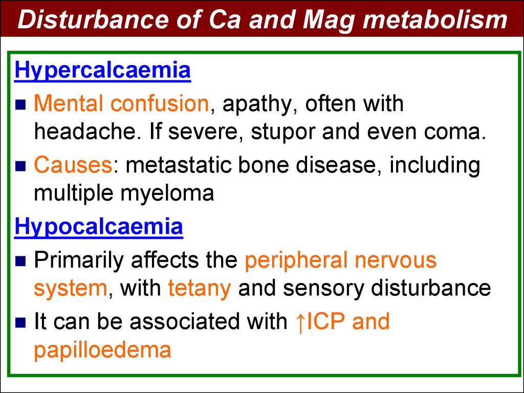
Disturbance of Ca and Mag metabolismHypercalcaemia
Mental confusion, apathy, often with
headache. If severe, stupor and even coma.
Causes: metastatic bone disease, including
multiple myeloma
Hypocalcaemia
Primarily affects the peripheral nervous
system, with tetany and sensory disturbance
It can be associated with ↑ICP and
papilloedema
100.
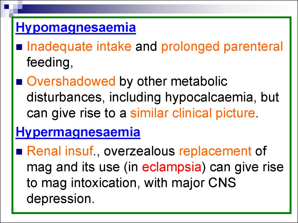
HypomagnesaemiaInadequate intake and prolonged parenteral
feeding,
Overshadowed by other metabolic
disturbances, including hypocalcaemia, but
can give rise to a similar clinical picture.
Hypermagnesaemia
Renal insuf., overzealous replacement of
mag and its use (in eclampsia) can give rise
to mag intoxication, with major CNS
depression.
101.
DrugsPoisoning, drug abuse, and alcohol
intoxication accounting for up to 30 % of
those presenting through accident and
emergency departments.
80 % require only simple observation in
their management.
102.
1.2.
3.
1.
2.
3.
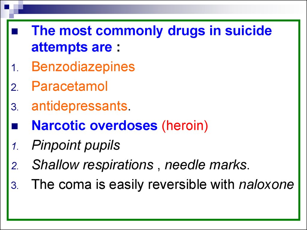
The most commonly drugs in suicide
attempts are :
Benzodiazepines
Paracetamol
antidepressants.
Narcotic overdoses (heroin)
Pinpoint pupils
Shallow respirations , needle marks.
The coma is easily reversible with naloxone
103.
1.2.
Solvent abuse and glue sniffing should
be considered in the undiagnosed patient
with coma.
Drugs may also result in disturbed
consciousness due to
secondary metabolic derangement
the acidosis associated with ethylene
glycol and carbon monoxide poisoning
104.
Alcohol intoxicationApparent from the history, flushed face,
rapid pulse, and low blood pressure. The
smell of alcohol on the breath.
Intoxicated are at increased risk of
hypothermia and of head injury can be the
cause of coma.
At low plasma concentrations of alcohol,
mental changes, at higher levels, coma
ensues, >350 mg/dl may prove fatal.
105.
106. Miscellaneous causes of coma
107.
SeizuresCommon cause of coma, with a period of
unconsciousness following a single
generalized seizure commonly lasting
between 30 and 60 minutes.
Following status epilepticus, there may be a
prolonged period of coma. History, trauma to
the tongue or inside of the mouth.
Seizures secondary to metabolic
disturbances may have a longer period of
coma.
108.
Extensive neurological diseasePMLE
severe
end-stage multiple sclerosis.
Prion disease may lead to coma over a
short period of 6–8 weeks, but this is
following a progressive course of
widespread neurological disturbance.
109.
EclampsiaIn
the second half of pregnancy and
represents a failure of autoregulation,
with raised blood pressure.
Neuropathologically: there are ring
haemorrhages around occluded small
vessels with fibrinoid deposits.
110.
CP: seizures, cortical blindness, and coma.Management: control of convulsions and
raised blood pressure. Parental magnesium
is commonly employed, may give rise to
hypermagnesaemia.
Postpartum complications of pregnancy
cerebral angiitis and venous sinus
thrombosis, may also lead to coma
111. Investigation of coma
At presentation blood will be taken fordetermination of glucose, electrolytes, liver
function, calcium, osmolality, and blood
gases.
Blood should also be stored for a
subsequent drug screen if needed
112.
Following the clinical examination, a broaddistinction between a metabolic cause,
with preserved pupillary responses, or a
structural cause of coma is likely to have
been established
Although most patients with coma will
require CT scanning, or indeed all with
persisting coma, clearly this is of greater
urgency when a structural lesion is
suspected
113.
In the absence of focal signs, but withevidence of meningitis, a lumbar puncture
may need to be performed before
scanning, as a matter of clinical urgency.
In other situations, lumbar puncture should
be delayed until after the brain scan
because of the risk of precipitating a
pressure cone secondary to a cerebral
mass lesion
114.
All patients will require chest radiographyand ECG, detailed investigations of
systemic disease will be directed by the
clinical examination.
The EEG is of value in identifying the
occasional patient with subclinical status
epilepticus, and is clearly of value in
assessing the patient who has been
admitted following an unsuspected seizure
115.
Fast activity is commonly found with drugoverdose and slow wave abnormalities
with metabolic and anoxic coma.
An isoelectric EEG may occur with druginduced comas, but otherwise indicates
severe cerebral damage.
116.
117. Management of the unconscious patient
Treatment of the underlying causeMaintenance of normal physiology: respiration,
circulation, and nutrition
Patient should be nursed on his or her side
without a pillow
Attention will clearly need to be paid to the
airway, requiring an oral airway as a minimum
118.
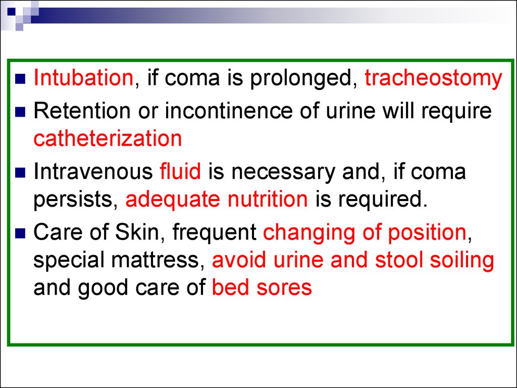
Intubation, if coma is prolonged, tracheostomyRetention or incontinence of urine will require
catheterization
Intravenous fluid is necessary and, if coma
persists, adequate nutrition is required.
Care of Skin, frequent changing of position,
special mattress, avoid urine and stool soiling
and good care of bed sores
119. Prognosis in coma
In general, coma carries a serious prognosis.This is dependent to a large extent on the
underlying cause.
Coma due to depressant drugs carries an
excellent prognosis provided that resuscitative
and supportive measures are available and no
anoxia has been sustained
Metabolic causes, apart from anoxia, carry a
better prognosis than structural lesions and
head injury
120.
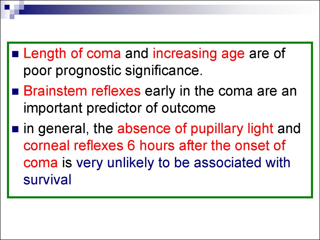
Length of coma and increasing age are ofpoor prognostic significance.
Brainstem reflexes early in the coma are an
important predictor of outcome
in general, the absence of pupillary light and
corneal reflexes 6 hours after the onset of
coma is very unlikely to be associated with
survival
121.
The chronic vegetative state usually carries auniformly poor prognosis, although a partial
return of cognition, or even restoration to
partial independence, has been reported
very rarely.
Although unassociated with coma, the
‘locked-in’ syndrome also carries a poor
prognosis, with only rare recoveries reported.