


 Медицина
Похожие презентации:


ECG complication of treatment
1. ZSMU Department of general practice – family medicine
ECG complicationof treatment
ZSMU
Department of general practice –
family medicine
2. Case 1
3.
A 65-year-old woman presents to theEmergency Department (ED) with
generalized fatigue and palpitations.
She was started on an angiotensinconverting enzyme (ACE) inhibitor 2
months ago but has missed her followup appointments.
What life-threatening metabolic
abnormality could be responsible for
the findings shown in her ECG tracing?
4.
5.
6.
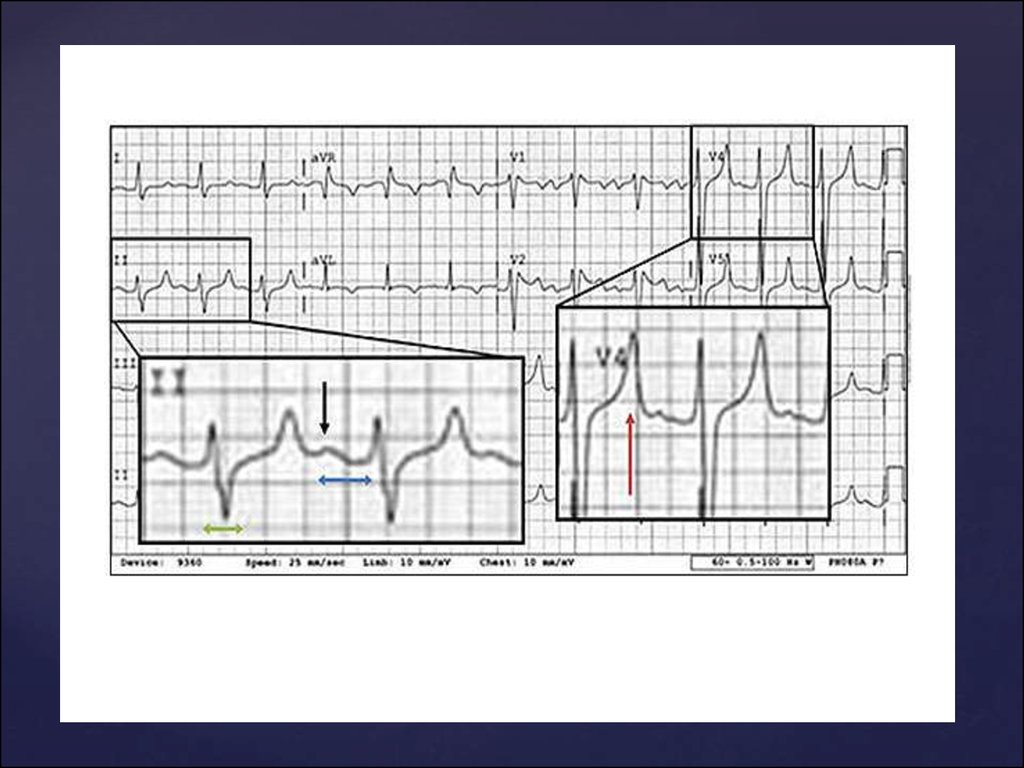
Life-Threatening Condition (I): Hyperkalemia.The tracing shows a regular rhythm at 75
beats/min.
A P wave is present in front of each QRS complex,
indicating that the rhythm is sinus.
A flattened P wave (black arrow), a prolonged PR
interval (blue bar), borderline widened QRS
complexes (green bar), and—more
pathognomonic—pointed, narrow, and tented tall T
waves (red arrow) are all features of hyperkalemia.
The patient's serum potassium concentration when
the tracing was recorded was 7.2 mEq/L.
7. Case 2
8.
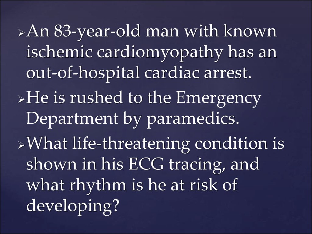
An 83-year-old man with knownischemic cardiomyopathy has an
out-of-hospital cardiac arrest.
He is rushed to the Emergency
Department by paramedics.
What life-threatening condition is
shown in his ECG tracing, and
what rhythm is he at risk of
developing?
9.
10.
11.
Life-Threatening Condition (II): Long QTInterval and T-Wave Alternans.
The tracing shows a sinus rhythm at 60
beats/min.
The QT interval (black bar) is prolonged to
680 msec (normal, 300-440 msec), with a
QTc also of 680 msec (normal, <460 msec).
The T-wave heights alternate (blue arrows),
and such alternation is often a precursor to
the more severe rhythm of torsades de
pointes.
12. Case 3
13.
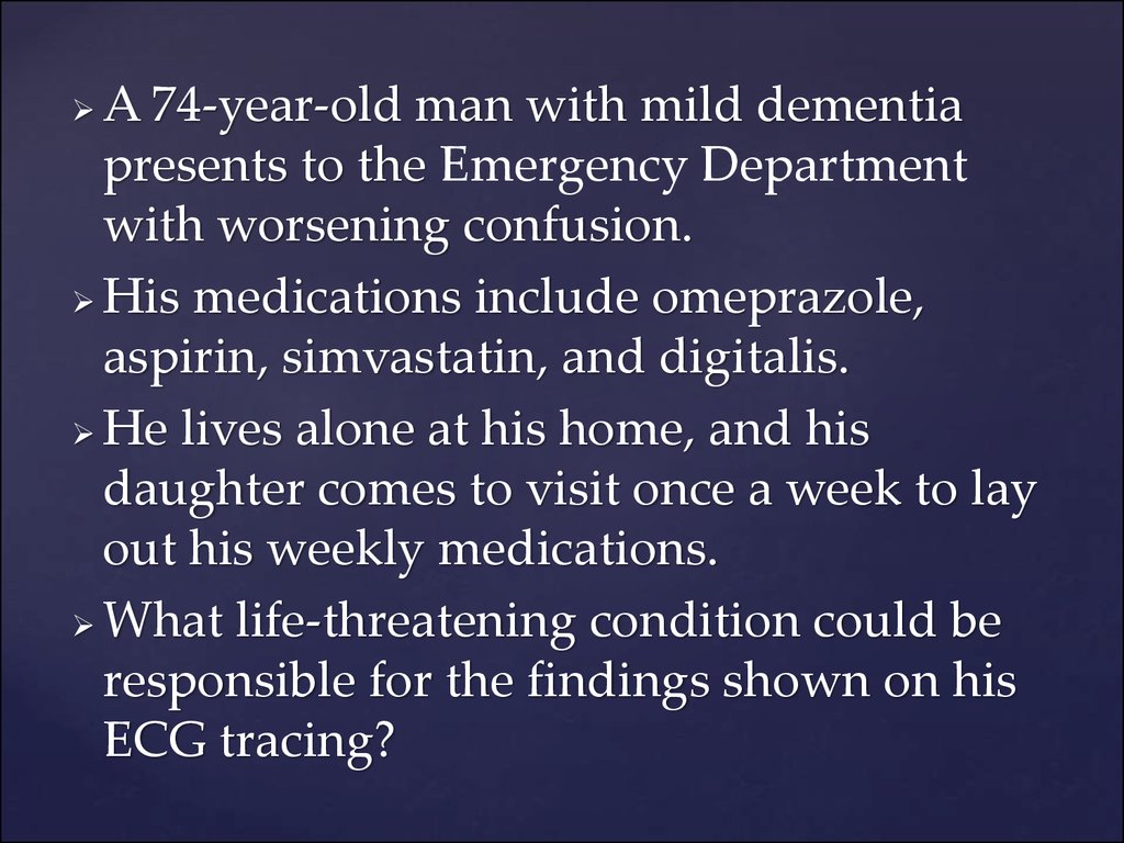
A 74-year-old man with mild dementiapresents to the Emergency Department
with worsening confusion.
His medications include omeprazole,
aspirin, simvastatin, and digitalis.
He lives alone at his home, and his
daughter comes to visit once a week to lay
out his weekly medications.
What life-threatening condition could be
responsible for the findings shown on his
ECG tracing?
14.
15.
16.
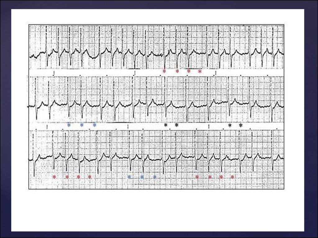
Life-Threatening Condition (III):
Digitalis Toxicity. The tracing shows no P waves,
with a baseline of irregular, fine undulations,
reflecting atrial fibrillation.
The QRS complex is narrow and occurs regularly
sometimes (in the latter part of the middle strip) and
in groups at other times.
This tracing is an example of junctional tachycardia
with variable conduction to the ventricle.
Conducted and skipped QRS complexes are present
in patterns of 2:1 (black asterisk), 3:2 (blue asterisk), or
4:3 (red asterisk).
The tracing is highly suggestive of digitalis toxicity,
especially in this clinical context.
17. Case 4
18.
• A 25-year-old man arrives at theEmergency Department with a heavy
cough after getting caught outside in a
snowstorm while hiking.
• A routine ECG is performed.
• The concerned intern takes one look at it
and rushes over to show you what he
believes to be a serious problem.
• Do you agree with the intern's
assessment of a life-threatening
condition seen on the tracing?
19.
20.
21.
• Life-Threatening Condition (IV):• Artifact Simulating a Run of Ventricular
Tachycardia.
• At first glance, this ECG suggests a run of
ventricular tachycardia.
• However, sharp deflections occur regularly
at the same rate as the sinus rhythm seen at
the beginning and at the end of the tracing
(black bar).
• These deflections are undoubtedly QRS
complexes of the sinus rhythm and provide
an example of an artifact simulating
ventricular tachycardia
22. Case 5
23.
• A 32-year-old woman comes to theEmergency Department complaining
of light-headedness and sweating.
• She is 5'6" tall and weighs less than
100 lb.
• An ECG is immediately obtained,
which evolves while you are
watching.
• What life-threatening condition is
seen on the ECG tracing?
24.
25.
26.
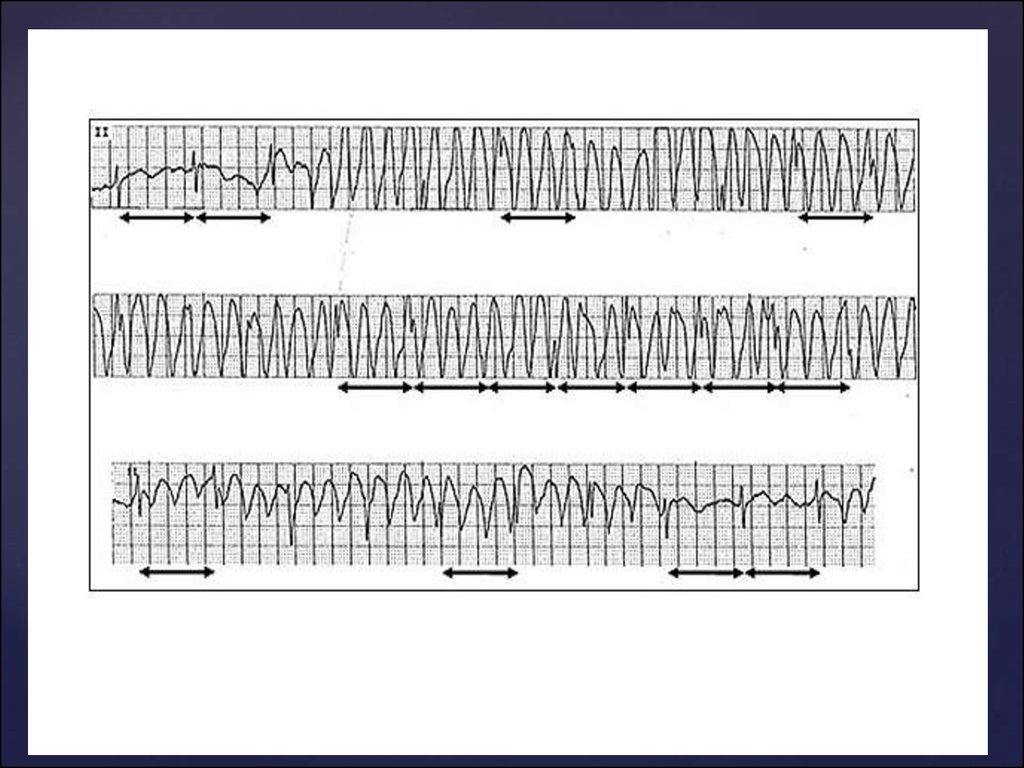
• Life-Threatening Condition (V):• Torsades de Pointes.
• Sinus rhythm is present at the beginning
(blue box), but the QT interval of the sinus
beats is long (black bar).
• This is followed by a wide QRS
tachycardia at a rate of approximately 200
beats/min (red box).
• The QRS morphology and axis
continuously change, indicating torsades
de pointes, which is life-threatening.