


 Медицина
Похожие презентации:


 Causes, symptoms, & treatment")


Ophthalmology
1.
Ophthalmology2.
3.
Orbital cellulitisa systemically unwell patient
proptosis
peri-ocular swelling and erythema
tenderness over the sinuses
ocular nerve compromise (reduced vision,
impaired colour vision or abnormal pupils)
restricted and painful eye movements
In peri-orbital cellulitis, which usually follows an
abrasion, there is no pain or restriction of eye
movement
Treatment is with IV cefotaxime until afebrile, then
amoxycillin/clavulanate for 7–10 days for peri-orbital cellulitis
and for orbital cellulitis, IV cefotaxime + di(flu) cloxacillin
together followed by amoxycillin/clavulanate (o) 10 days
4.
Conjunctivitis “Pink eye”Risk factors: exposure to someone infected, rubbing
eyes, contact lenses.
Symptoms:
Marked, diffuse redness
Watery, stringy, purulent discharge
Treatment
Viral
Artificial tears, cool compresses,
antihistamines
Bacterial
Erythromycin ophthalmic ointment
Or Polytrim, Azithromycin,
Ciprofloxacin
Allergic
Self-limiting
Zyrtec, Claritin
5.
Scleritis and episcleritisManagement
Corticosteroids or
NSAIDs
Episcleritis:
itching
a red and sore eye
no discharge
no watering
Episcleritis
Salmon-pink or red
discoloration
vision normal (usually)
often sectorial
usually self-limiting
Scleritis:
painful
loss of vision
urgent referral
Scleritis
Violaceous or purplish
discoloration
6.
Corneal abrasionCauses:
Symptoms:
Trauma
Ocular pain
Contact lens wear/injury
Foreign body sensation
Infection—microbial keratitis:
Watering of the eye (epiphora)
bacterial (e.g. Pseudomonas [contact lens])
Neurotrophic (e.g. trigeminal nerve defect)
Blepharospasm
Blurred vision
Immune-related (e.g. rheumatoid arthritis)
Spontaneous corneal erosion
Management
Chronic blepharitis
Check for a foreign body
Overexposure (e.g. eyelid defects)
Treat with chloramphenicol 1% ointment ± homatropine 2%
(if pain due to ciliary spasm)
Diagnosis is best performed with a slit
lamp using a cobalt blue filter and
flourescein staining
Double eye pad (if not infected)
A 6 mm defect heals in 48 hours
7.
Uveitis (iritis)Clinical feature
Eye redness, esp. around the edge of the iris
Eye discomfort or pain
Increased tearing
Blurred vision
Sensitivity to light
Floaters in the field of vision
Small pupil
Treatment
pupil dilatation with atropine drops
Causes include autoimmune-related diseases such as the
seronegative arthropathies (e.g. ankylosing spondylitis), SLE,
IBD, sarcoidosis and some infections (e.g. toxoplasmosis and
syphilis)
Diagnosis: Slit-lamp examination an increase in the protein
content of the aqueous (flare) in the anterior chamber
Keratic precipitates it’s when WBC display on the back surface
of the conea.
topical steroids to suppress inflammation
systemic corticosteroids
8.
CataractCauses: advancing age, diabetes mellitus,
smoking cigarettes, steroids (topical or oral),
radiation: long exposure to UV light, TORCH
organisms → congenital cataracts, trauma,
uveitis, dystrophia myotonica, galactosaemia
Symptoms:
Blurred vision:
Diagnosis
Reduced visual acuity (sometimes
improved with pinhole)
Diminished red reflex on
ophthalmoscopy
A change in the appearance of the
lens
reading difficulty
difficulty in recognising faces
Management
problems with driving, especially at night
The removal of the cataractous lens
and optical correction to restore
vision with an intraocular lens implant
difficulty with television viewing
reduced ability to see in bright light
may see haloes around lights
9.
Cataract10.
Hypertensive retinopathyRisk factors – increasing age, obesity,
family history, alcohol, smoking
Systemic hypertension directly affects
the retinal, choroidal and optic nerve
vasculature
Diagnosis: fundoscopic exam or digital
retinal photography, findings usually
bilateral
Treatment: blood pressure control
11.
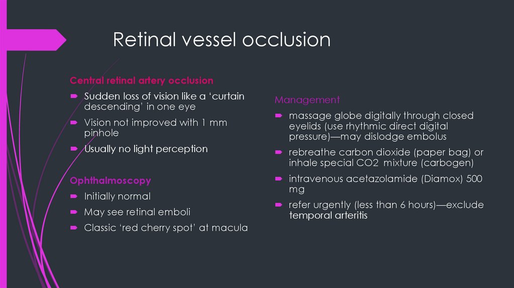
Retinal vessel occlusionCentral retinal artery occlusion
Sudden loss of vision like a ‘curtain
descending’ in one eye
Vision not improved with 1 mm
pinhole
Management
massage globe digitally through closed
eyelids (use rhythmic direct digital
pressure)—may dislodge embolus
Usually no light perception
rebreathe carbon dioxide (paper bag) or
inhale special CO2 mixture (carbogen)
Ophthalmoscopy
intravenous acetazolamide (Diamox) 500
mg
Initially normal
May see retinal emboli
Classic ‘red cherry spot’ at macula
refer urgently (less than 6 hours)—exclude
temporal arteritis
12.
CRAO and BRAO13.
Retinal vessel occlusionCentral retinal vein thrombosis
Sudden loss of central vision in one eye
(if macula involved): can be gradual
over days
Vision not improved with 1 mm pinhole
Ophthalmoscopy shows swollen disc and
multiple retinal haemorrhages, ‘stormy
sunset’ appearance.
Management
No immediate treatment is effective.
fibrinolysin treatment
Laser photocoagulation may be
necessary in later stages
14.
CRVO and BRVO15.
GlaucomaClosed-angle glaucoma
Normal IOP 10-21mmHG
Open-angle glaucoma
Gradual increases resistance through
the trabecular meshwork
Risk factors: advancing age, family
history, black ethnic origin, myopia
Symptoms: asymptomatic, loss of
peripheral vision, fluctuating pain,
blurred vision, halos surrounding lights
(worse at night)
The iris bulges forward and seals off
the trabecular meshwork from the
anterior chamber
Risk factors: increasing age, female,
family history, Chinese/east Asian
ethnic origin, shallow anterior
chamber, medications (Noradrenalin,
oxybutynin, amitriptyline)
Symptoms: severe painful red eye,
blurred vision, patient >50 years, hazy
cornea, fixed semidilated pupil, eye
feels hard, halos around lights,
associated headache, nausea and
vomiting
16.
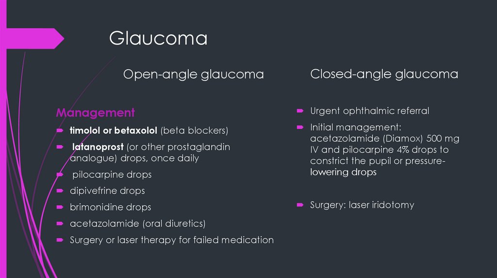
GlaucomaOpen-angle glaucoma
Closed-angle glaucoma
Management
Urgent ophthalmic referral
timolol or betaxolol (beta blockers)
Initial management:
acetazolamide (Diamox) 500 mg
IV and pilocarpine 4% drops to
constrict the pupil or pressurelowering drops
latanoprost (or other prostaglandin
analogue) drops, once daily
pilocarpine drops
dipivefrine drops
brimonidine drops
acetazolamide (oral diuretics)
Surgery or laser therapy for failed medication
Surgery: laser iridotomy
17.
Glaucoma18.
GlaucomaInvestigations
Tonometry (Goldmann applanation
tonometry)
Upper limit of normal is 22 mmHg
Ophthalmoscopy
Optic disc cupping >30% of total disc area
Visual fields
peripheral visual loss
19.
KeratitisKeratitis is inflammation of the cornea
pain, impaired eyesight, photophobia (light
sensitivity), red eye and a 'gritty' sensation
Causes: viral (HSV, Herpes zoster keratitis),
bacterial (staph), fungal, amoebic
(Acanthamoebic keratitis), parasitic
(Onchocercal keratitis,)
Treatment
depends on the cause of the
keratitis
antibacterial, antifungal, or
antiviral therapyantibacterial,
antifungal, or antiviral therapy
20.
BlepharitisAssociated with secondary ocular effects such as
styes, chalazia and conjunctival or corneal
ulceration
The two types are:
Anterior - around the skin, eyelashes, and lash
follicles
Posterior blepharitis involves the meibomian
gland orifices, meibomian glands, tarsal plate,
and blepharo-conjunctival junction
anterior blepharitis—staphylococcal
posterior blepharitis—seborrhoeic and rosacea
Clinical features
Persistent sore eyes or eyelids
Irritation, grittiness, burning, dryness and
‘something in the eye’ sensation
Lid or conjunctival swelling and redness
Crusts or scales around the base of the
eyelids
Discharge or stickiness, especially in
morning
Inflammation and crusting of the lid
margins
21.
BlepharitisManagement
Anterior blepharitis
A systematic and long-term commitment to a program of eyelid margin hygiene
Or apply chloromycetin 1% ointment once or twice daily for 4 weeks and review
Posterior blepharitis
Eyelid hygiene
Ocular lubricants
short-term use of a mild topical corticosteroid ointment
antibiotic ointment tetracycline hydrochloride 1% or framycetin 0.5% or
chloramphenicol 1% ointment to lid margins 3–6-hourly
systemic antibiotics: doxycycline 50 mg daily for at least 8 weeks (erythromycin for
children
<8 years), or flucloxacillin may be required for lid abscess.
22.
Subconjunctival hemorrhageA beefy red localised haemorrhage with a definite
posterior margin, it is pain free.
Usually causes by sudden increase in intrathoracic
pressure such as coughing and sneezing
No local therapy is necessary. The haemorrhage
absorbs over 2 weeks.
23.
Hypopyon and hyphemainflammatory cells in the anterior
chamber of the eye.
The most common cause of
hypopyon is endophthalmitis.
Blood within the aqueous fluid of the
anterior chamber.
The most common cause of hyphema is
trauma