


 Медицина
Похожие презентации:


")


Interesting case
1.
INTERESTINGCASE
Sirinthip Kittivisuit, MD.
Fellowship in pediatric rheumatology, Ramathibodi hospital
2.
A 12-year-old girl• CC: Blurred vision 1 day
History taking
3.
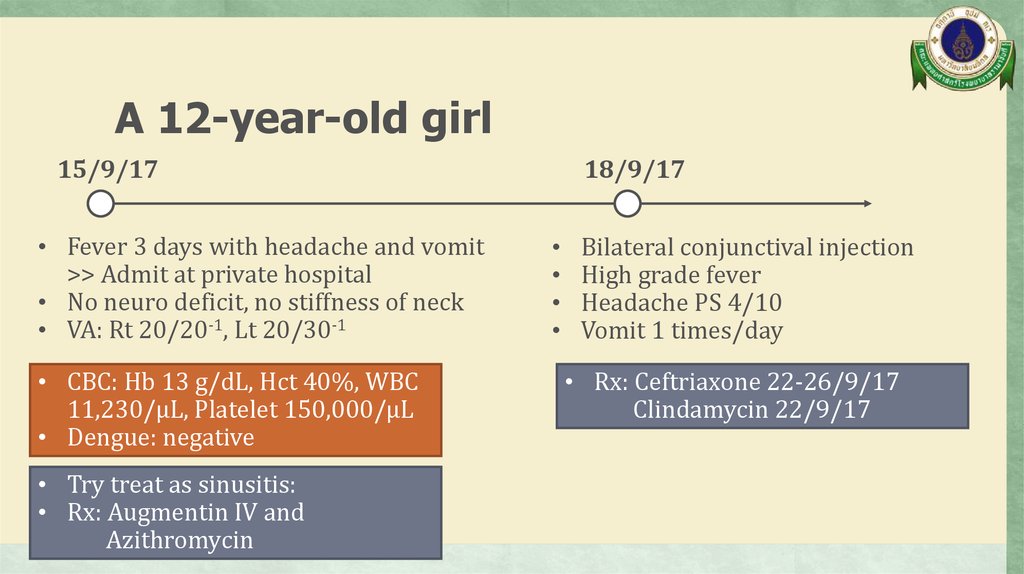
A 12-year-old girl15/9/17
• Fever 3 days with headache and vomit
>> Admit at private hospital
• No neuro deficit, no stiffness of neck
• VA: Rt 20/20-1, Lt 20/30-1
• CBC: Hb 13 g/dL, Hct 40%, WBC
11,230/µL, Platelet 150,000/µL
• Dengue: negative
• Try treat as sinusitis:
• Rx: Augmentin IV and
Azithromycin
18/9/17
Bilateral conjunctival injection
High grade fever
Headache PS 4/10
Vomit 1 times/day
• Rx: Ceftriaxone 22-26/9/17
Clindamycin 22/9/17
4.
A 12-year-old girl23/9/17
• Severe headache, night awakening pain
• CT brain: normal
• Lumbar puncture:
OP/CP 17/- cmH2O,
WBC 8, RBC 250/HPF,
protein 37 g/dL, sugar 89 mg%
• CSF culture: no growth
• Add Doxycycline 23-26/9/60
• Dexamethaxone 4 mg IV x 1 dose
25/9/17
• Fever subsite but still had headache
• Discharge 26/9/17
5.
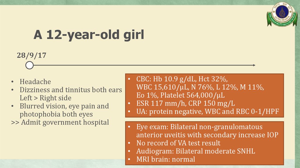
A 12-year-old girl28/9/17
• CBC: Hb 10.9 g/dL, Hct 32%,
• Headache
WBC 15,610/µL, N 76%, L 12%, M 11%,
• Dizziness and tinnitus both ears
Eo 1%, Platelet 564,000/µL
Left > Right side
• ESR 117 mm/h, CRP 150 mg/L
• Blurred vision, eye pain and
• UA: protein negative, WBC and RBC 0-1/HPF
photophobia both eyes
>> Admit government hospital
• Eye exam: Bilateral non-granulomatous
anterior uveitis with secondary increase IOP
• No record of VA test result
• Audiogram: Bilateral moderate SNHL
• MRI brain: normal
6.
A 12-year-old girl28/9/17
• Dx: Incomplete Vogt-KoyanagiHarada syndrome
• Rx: Pulse methylprednisolone
1 gm IV x 3 days (6-8/10/17)
then prednisolone (5) 3x2
>> Discharge 10/10/17
>> Refer to Ramathibodi hospital
14/12/17
Legs pain
• Left knee, bilateral ankles pain
• Bilateral thighs and legs pain
• Progression over 2 months with
severe pain and night awakening
pain within 2 weeks
• Precipitous by more walking
distance
• No morning stiffness, no joint
swollen/warmth/erythema,
no limit ROM
7.
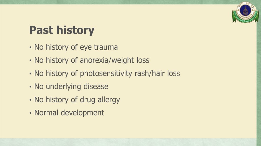
Past history• No history of eye trauma
• No history of anorexia/weight loss
• No history of photosensitivity rash/hair loss
• No underlying disease
• No history of drug allergy
• Normal development
8.
A 12-year-old girlPhysical
examination
9.
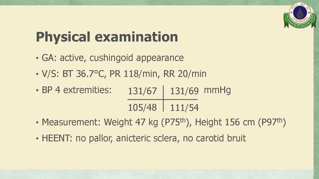
Physical examination• GA: active, cushingoid appearance
• V/S: BT 36.7°C, PR 118/min, RR 20/min
• BP 4 extremities:
131/67
131/69 mmHg
105/48
111/54
• Measurement: Weight 47 kg (P75th), Height 156 cm (P97th)
• HEENT: no pallor, anicteric sclera, no carotid bruit
10.
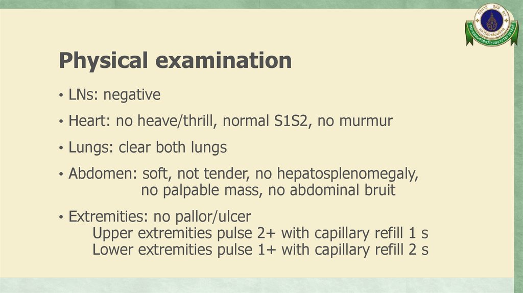
Physical examination• LNs: negative
• Heart: no heave/thrill, normal S1S2, no murmur
• Lungs: clear both lungs
• Abdomen: soft, not tender, no hepatosplenomegaly,
no palpable mass, no abdominal bruit
• Extremities: no pallor/ulcer
Upper extremities pulse 2+ with capillary refill 1 s
Lower extremities pulse 1+ with capillary refill 2 s
11.
Physical examination• Neuro signs:
Good consciousness
Intact CN functions
Normal tone, Motor power gr.V all extremities
Sensory intact
DTRs 2+ all extremities
BBK absent
Clonus negative
12.
A 12-year-old girlProblem list
13.
Problem list• Hypertension with arterial insufficiency: Lower limb
claudication with different BP between upper and lower
extremities
• Bilateral anterior uveitis
• Bilateral SNHL
14.
A 12-year-old girlDifferential
diagnosis
15.
Differential diagnosis• Takayasu arteritis
• Cogan’s syndrome
• Vogt-Koyanagi-Harada syndrome
16.
A 12-year-old girlInvestigation
17.
Investigation18.
Complete blood count• Hb 13.6 g/dL, Hct 43.3%
• WBC 13,300/µL, N 75%, L 15%, M 9%, Eo 1%
• Platelet 260,000/µL
19.
Blood chemistry• BUN 19, Cr 0.35 mg/dL
• Na 142, K 3.93, Cl 107, HCO3 22.2 mmol/L
• TB 0.3, DB 0.1 mg/dL
• AST 15, ALT 45, GGT 22 U/L
• Albumin 32.9 g/L
20.
Inflammatory markers• ESR 8 mm/hr
• CRP < 1 mg/L
21.
Urinalysis• Sp. gr. 1.019, pH 5, protein negative, blood negative
• WBC 0-1, RBC 0-1 /HPF
22.
Immunology• ANA negative
23.
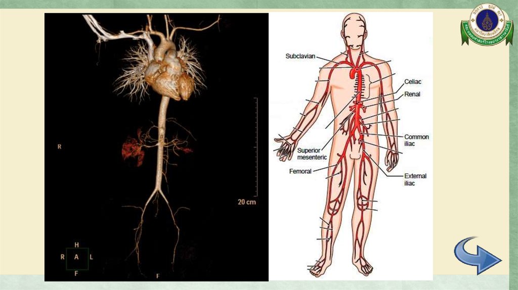
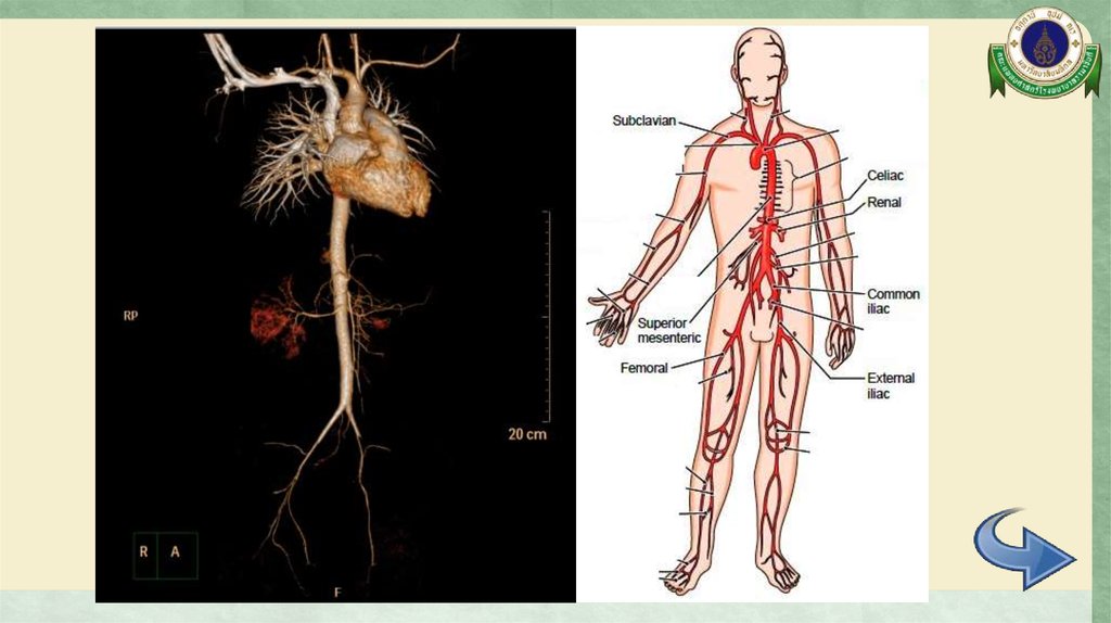
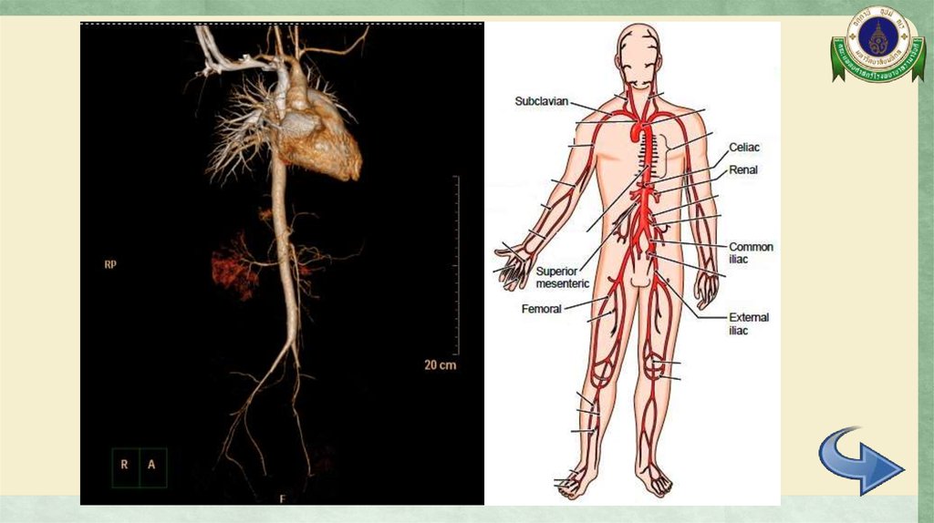
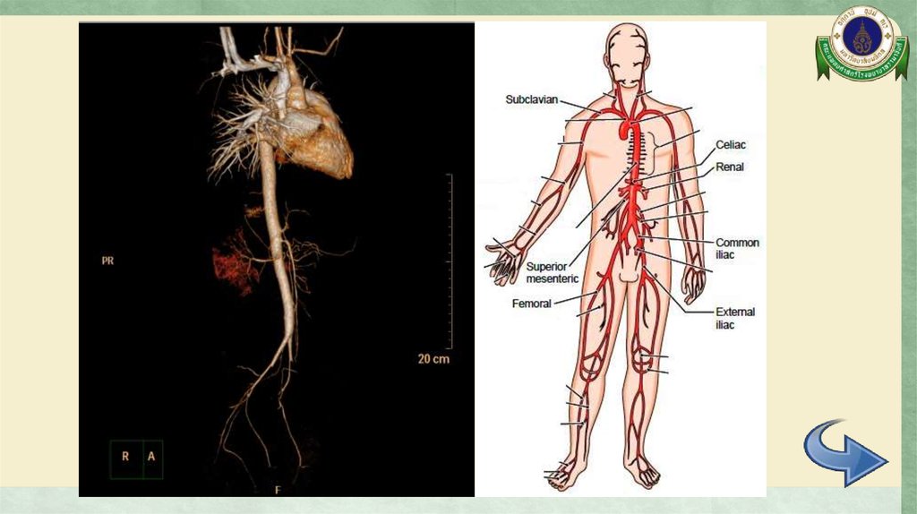
CTA whole aorta24.
CTA whole aorta25.
CTA whole aorta26.
CTA whole aorta27.
CTA whole aorta28.
CTA whole aorta29.
CTA whole aorta30.
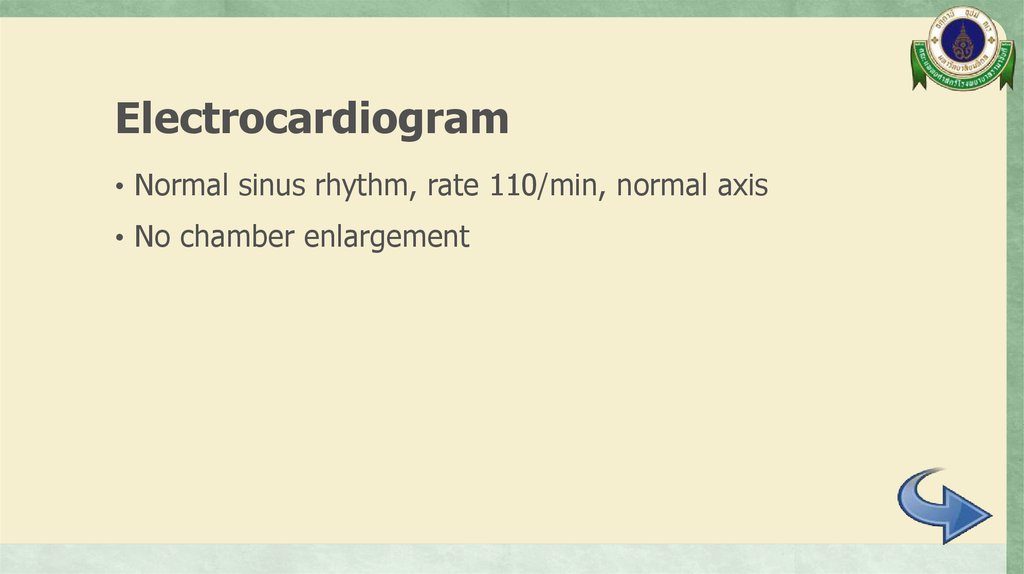
Electrocardiogram• Normal sinus rhythm, rate 110/min, normal axis
• No chamber enlargement
31.
Echocardiogram• Normal cardiac function
• Trivial to mild MR and AR
• No coarctation of aorta or aortic root dilatation
32.
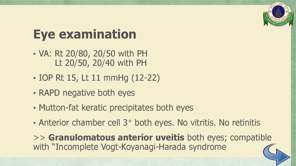
Eye examination• VA: Rt 20/80, 20/50 with PH
Lt 20/50, 20/40 with PH
• IOP Rt 15, Lt 11 mmHg (12-22)
• RAPD negative both eyes
• Mutton-fat keratic precipitates both eyes
• Anterior chamber cell 3+ both eyes. No vitritis. No retinitis
>> Granulomatous anterior uveitis both eyes; compatible
with “Incomplete Vogt-Koyanagi-Harada syndrome
33.
Audiogram• Moderately severe sensorineural hearing loss both ears
34.
A 12-year-old girlDifferential
diagnosis
35.
Differential diagnosis• Takayasu arteritis
• Cogan’s syndrome
• Vogt-Koyanagi-Harada syndrome
36.
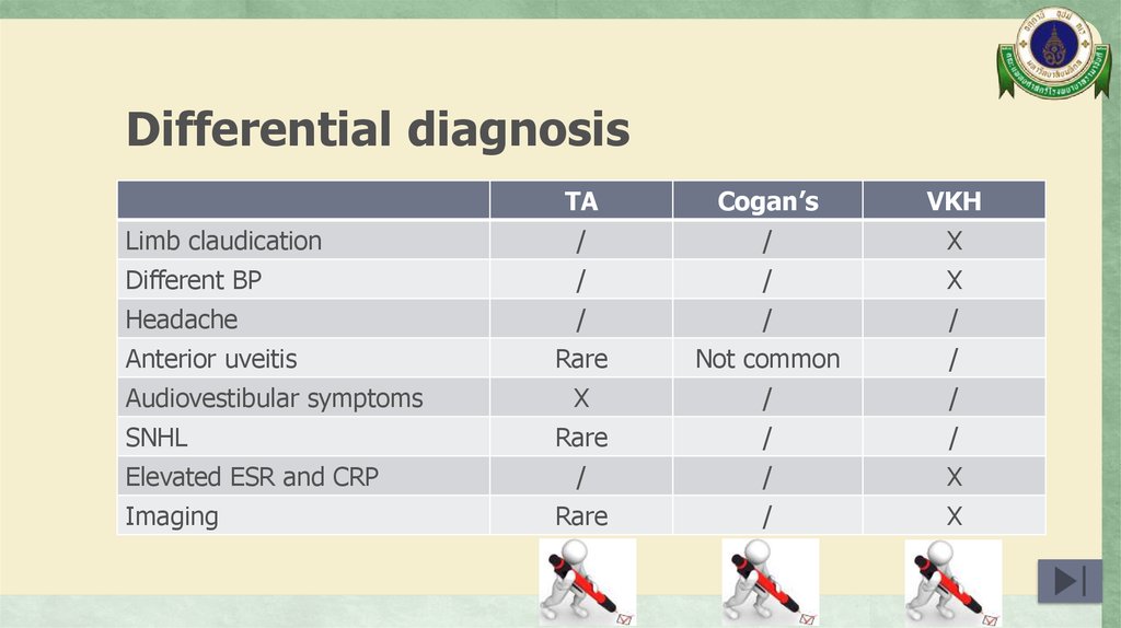
Differential diagnosisTA
Cogan’s
VKH
Limb claudication
/
/
X
Different BP
/
/
X
Headache
/
/
/
Rare
Not common
/
X
/
/
Rare
/
/
/
/
X
Rare
/
X
Anterior uveitis
Audiovestibular symptoms
SNHL
Elevated ESR and CRP
Imaging
37.
Takayasu arteritis (TA)• Arteritis, often granulomatous
• Predominantly affecting the aorta and/or its major
branches
Interesting points
• Diagnosis
• Ocular manifestations in TA
• SNHL in TA
• TA without involvement of aorta
Arthritis Rheum 2013;65:1-11.
38.
EULAR/PRINTO/PRES classificationcriteria of childhood TA
• Angiographic abnormalities
• Angiography (conventional, CT,
plus 1 of 5 following criteria
or MRI) of the aorta or its main
(sens 100%, spec 99.9%)
branches and pulmonary
arteries showing aneurysm/
1. Pulse deficit or claudication
dilatation, narrowing, occlusion
2. Four limbs blood pressure
or thickened arterial wall not due
discrepancy > 10 mmHg
to fibromuscular dysplasia, or
similar causes; changes usually
3. Bruit
focal or segmental
th
4. Hypertension >P95
5. Acute phase reactant
Ann Rheum Dis 2010;69:798-806.
39.
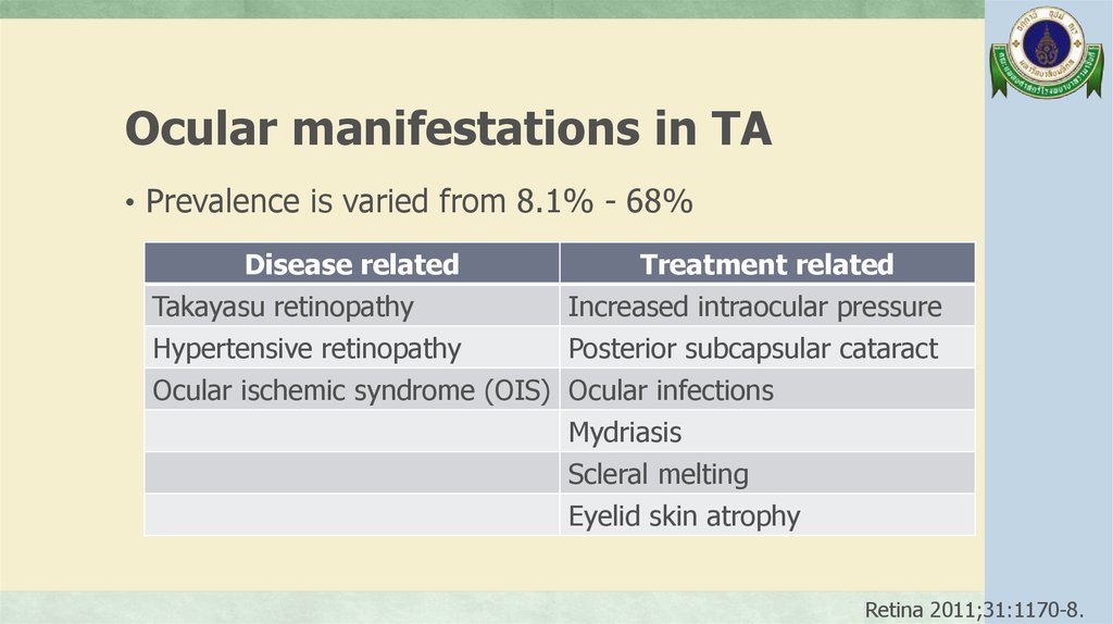
Ocular manifestations in TA• Prevalence is varied from 8.1% - 68%
Disease related
Treatment related
Takayasu retinopathy
Increased intraocular pressure
Hypertensive retinopathy
Posterior subcapsular cataract
Ocular ischemic syndrome (OIS) Ocular infections
Mydriasis
Scleral melting
Eyelid skin atrophy
Retina 2011;31:1170-8.
40.
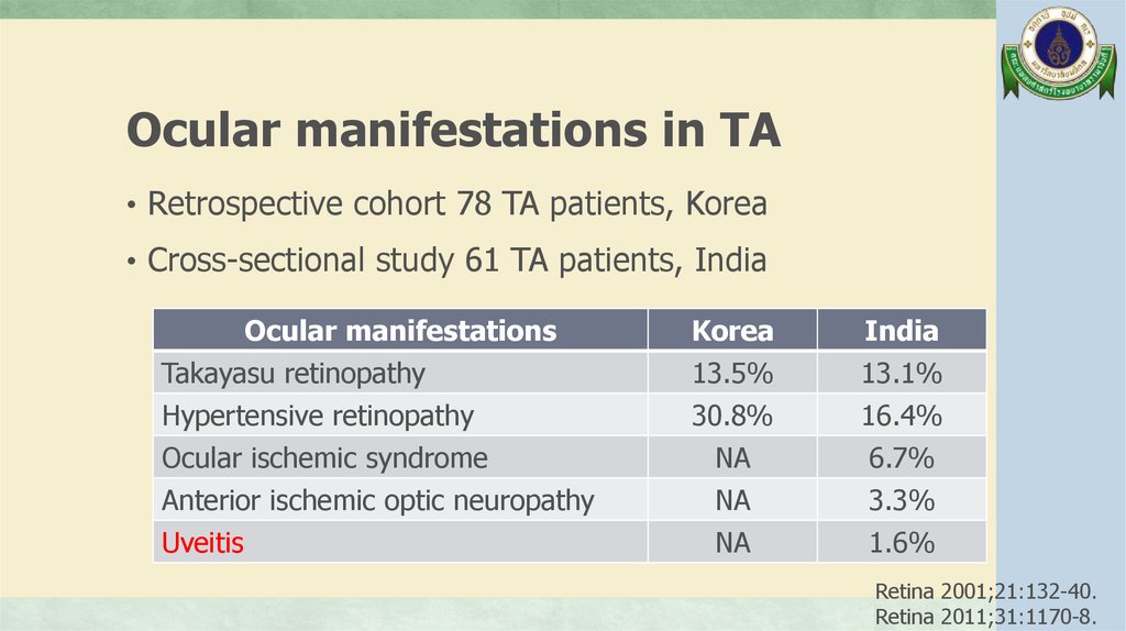
Ocular manifestations in TA• Retrospective cohort 78 TA patients, Korea
• Cross-sectional study 61 TA patients, India
Ocular manifestations
Takayasu retinopathy
Hypertensive retinopathy
Ocular ischemic syndrome
Anterior ischemic optic neuropathy
Uveitis
Korea
13.5%
30.8%
India
13.1%
16.4%
NA
NA
NA
6.7%
3.3%
1.6%
Retina 2001;21:132-40.
Retina 2011;31:1170-8.
41.
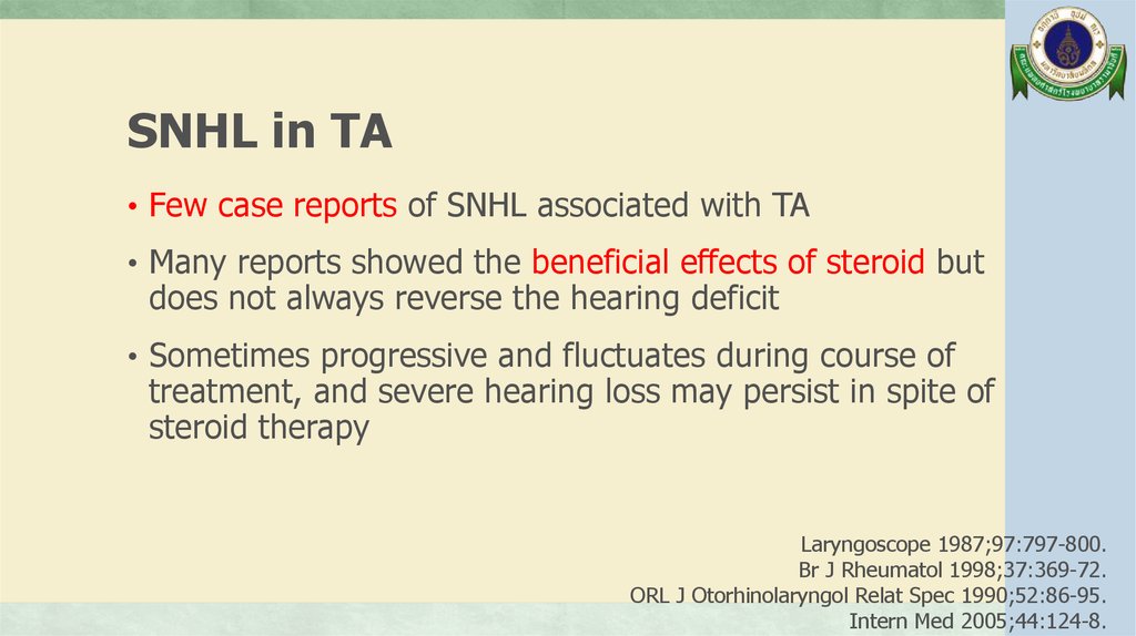
SNHL in TA• Few case reports of SNHL associated with TA
• Many reports showed the beneficial effects of steroid but
does not always reverse the hearing deficit
• Sometimes progressive and fluctuates during course of
treatment, and severe hearing loss may persist in spite of
steroid therapy
Laryngoscope 1987;97:797-800.
Br J Rheumatol 1998;37:369-72.
ORL J Otorhinolaryngol Relat Spec 1990;52:86-95.
Intern Med 2005;44:124-8.
42.
SNHL in TA• The mechanisms of the hearing loss in TA are reversible
circulatory disturbances due to vasculitis and/or some
autoimmune pathogenesis in the inner ear, especially in
hair cells
• Fujino et al. proposed the possibility of inner ear
dysfunction because of the vasculitis caused by the
adhesion of immune complex to the vessel wall
Practica Otologica 1985;78:2313-22.
Intern Med 2005;44:124-8.
43.
TA without involvement of aorta• Retrospective review 85 CT angiography in TA patient,
1994-2003, Korea
• 95% (81/85) Aortic involvement with or without major
aortic branch vessel involvement
• 5% (4/85) Only aortic branch involvement
- 3/85 Only left subclavian
- 1/85 Innominate artery, both common carotid artery
and superior mesenteric artery
J Vasc Surg 2007;45:906-14.
44.
Cogan’s syndrome (CS)• Characterized by ocular inflammatory lesions,
including interstitial keratitis, uveitis, and episcleritis,
and inner ear disease, including sensorineural hearing
loss and vestibular dysfunction
Interesting points
• Diagnosis
• Ocular manifestations in CS
• Audiovestibular manifestations in CS
• Vasculitic manifestations in CS
Arthritis Rheum 2013;65:1-11.
45.
Typical CSDefined by following 3 conditions:
(1) Ocular symptoms typically an isolated non-syphilitic
interstitial keratitis that could be associated with
conjunctivitis, conjunctival or subconjunctival bleeding
or iritis
(2) Audiovestibular symptoms usually progressing to
deafness in 1-3 months
(3) Interval between the onset of ocular and audiovestibular
manifestations of less than 2 year
Arch Ophthalmol 1945;33:144-9.
46.
Atypical CSAny of following conditions:
(1) Inflammatory ocular manifestations including episcleritis,
scleritis, retinal artery occlusion, choroiditis, retinal
hemorrhage, papilloedema, exophthalmos with or without
interstitial keratitis; patients with isolated conjunctivitis,
subconjunctival hemorrhage or iritis with Ménière’s episodes
within interval of 2 year
(2) Typical ocular manifestations associated within 2 year with
audiovestibular symptoms different from of Ménière-like episodes
(3) Delay of more than 2 year between onset of typical ocular and
audiovestibular manifestation
Medicine 1980;59:426-41.
47.
Ocular manifestations in CS• 80% Interstitial keratitis, mostly bilateral involvement;
inflamed small blood vessels invade the adjacent normally
avascular corneal stroma
• Target for inflammation is the small vessels in the
vascularized layers of the anterior globe: conjunctivitis,
episcleritis, scleritis, uveitis
• Retinitis, optic neuritis, glaucoma, papillary edema,
cataracts, ocular motor palsy, exophthalmia, central
retinal artery occlusion, xerophthalmia, ptosis
Rheumatology 2004;43:1007-15.
Arthritis Rheum 2013;65:1-11.
Autoimmune Rev 2014;13:351-4.
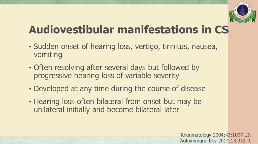
48.
Audiovestibular manifestations in CS• Sudden onset of hearing loss, vertigo, tinnitus, nausea,
vomiting
• Often resolving after several days but followed by
progressive hearing loss of variable severity
• Developed at any time during the course of disease
• Hearing loss often bilateral from onset but may be
unilateral initially and become bilateral later
Rheumatology 2004;43:1007-15.
Autoimmune Rev 2014;13:351-4.
49.
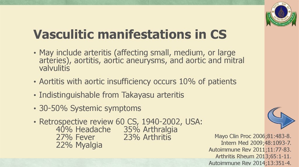
Vasculitic manifestations in CS• May include arteritis (affecting small, medium, or large
arteries), aortitis, aortic aneurysms, and aortic and mitral
valvulitis
• Aortitis with aortic insufficiency occurs 10% of patients
• Indistinguishable from Takayasu arteritis
• 30-50% Systemic symptoms
• Retrospective review 60 CS, 1940-2002, USA:
40% Headache
27% Fever
22% Myalgia
35% Arthralgia
23% Arthritis
Mayo Clin Proc 2006;81:483-8.
Intern Med 2009;48:1093-7.
Autoimmune Rev 2011;11:77-83.
Arthritis Rheum 2013;65:1-11.
Autoimmune Rev 2014;13:351-4.
50.
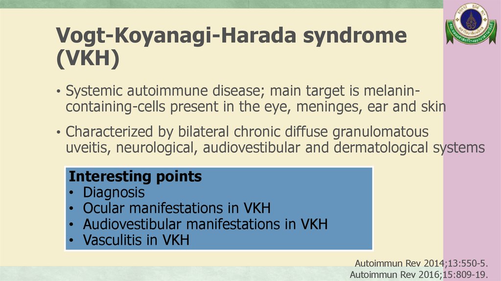
Vogt-Koyanagi-Harada syndrome(VKH)
• Systemic autoimmune disease; main target is melanin-
containing-cells present in the eye, meninges, ear and skin
• Characterized by bilateral chronic diffuse granulomatous
uveitis, neurological, audiovestibular and dermatological systems
Interesting points
• Diagnosis
• Ocular manifestations in VKH
• Audiovestibular manifestations in VKH
• Vasculitis in VKH
Autoimmun Rev 2014;13:550-5.
Autoimmun Rev 2016;15:809-19.
51.
Clinical course of VKH• Prodromal phase: Few days prior to ocular symptoms,
predominately neurological and auditory manifestations (severe
headache, nausea, meningismus, dysacusia, tinnitus, fever, orbital
pain, photophobia, pleocytosis of CSF)
• Acute uveitic phase: Bilateral vision blurring with choroiditis,
vitritis and papillitis which inflammatory cell infiltration into choroid
is hallmark. Mutton-fat keratic precipitates may be found
• Convalescent phase: Gradual abatement of uveitis;
depigmentation of skin, hair, choroid
• Chronic recurrent phase: predominately anterior uveitis
Semin Ophthalmol 2005;20:183-90.
52.
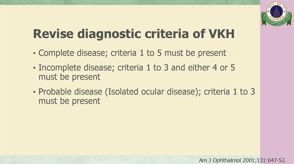
Revise diagnostic criteria of VKH• Complete disease; criteria 1 to 5 must be present
• Incomplete disease; criteria 1 to 3 and either 4 or 5
must be present
• Probable disease (Isolated ocular disease); criteria 1 to 3
must be present
Am J Ophthalmol 2001;131:647-52.
53.
Early manifestations:• Diffuse choroiditis (focal areas of
subretinal fluid, bullous serous
retinal detachment)
OR, characteristics
fluorescein
(1) No history of penetrating •ocular
trauma or surgery
preceding
angiography
findings
AND
onset of uveitis
echography evidence of diffuse
choroidal
thickening
(2) No clinical or laboratory evidence
suggestive
of other ocular disease
Revise diagnostic criteria of VKH
(3) Bilateral ocular involvement
(a manifestations:
or b must be met, depending on
Late
the stage of disease)
• History of suggestive of prior uveitis
a. Early manifestations with the above described
b. Late manifestations characteristics
• AND ocular depigmentation
(4) Neurological/audiotory findings:
or tinnitus
or CSF pleocytosis
• AND Meningismus
other ocular signs
(nummular
depigmented
(5) Integumentary finding (not chorioretinal
preceding onset
of CNS or scars
ocularordisease):
alopecia or poliosis or vitiligorecurrent or chronic anterior uveitis)
Am J Ophthalmol 2001;131:647-52.
54.
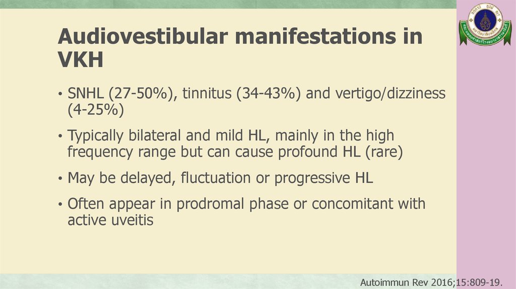
Audiovestibular manifestations inVKH
• SNHL (27-50%), tinnitus (34-43%) and vertigo/dizziness
(4-25%)
• Typically bilateral and mild HL, mainly in the high
frequency range but can cause profound HL (rare)
• May be delayed, fluctuation or progressive HL
• Often appear in prodromal phase or concomitant with
active uveitis
Autoimmun Rev 2016;15:809-19.
55.
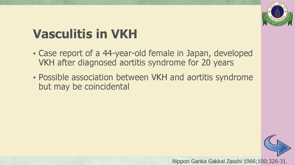
Vasculitis in VKH• Case report of a 44-year-old female in Japan, developed
VKH after diagnosed aortitis syndrome for 20 years
• Possible association between VKH and aortitis syndrome
but may be coincidental
Nippon Ganka Gakkai Zasshi 1966;100:326-31.
56.
A 12-year-old girlManagement
57.
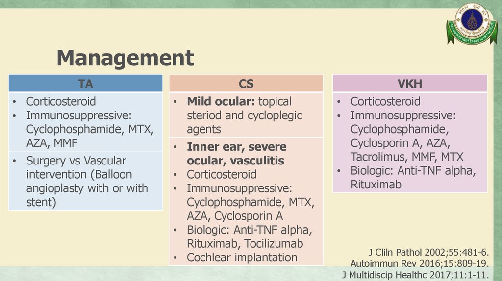
ManagementTA
• Corticosteroid
• Immunosuppressive:
Cyclophosphamide, MTX,
AZA, MMF
• Surgery vs Vascular
intervention (Balloon
angioplasty with or with
stent)
CS
• Mild ocular: topical
steriod and cycloplegic
agents
• Inner ear, severe
ocular, vasculitis
• Corticosteroid
• Immunosuppressive:
Cyclophosphamide, MTX,
AZA, Cyclosporin A
• Biologic: Anti-TNF alpha,
Rituximab, Tocilizumab
• Cochlear implantation
VKH
• Corticosteroid
• Immunosuppressive:
Cyclophosphamide,
Cyclosporin A, AZA,
Tacrolimus, MMF, MTX
• Biologic: Anti-TNF alpha,
Rituximab
J Cliln Pathol 2002;55:481-6.
Autoimmun Rev 2016;15:809-19.
J Multidiscip Healthc 2017;11:1-11.
58.
Management in this patient1/11/17
14/12/17
• VA: Rt 20/80, 20/50 with PH
Lt 20/50, 20/40 with PH
• AC cell 2+, mutton-fat BE
• VA: Rt 20/40-1, Lt 20/25-2 with PH
• AC trace, positive KP
• Audiogram: Rt mild, Lt mod SNHL
• Prednisolone (5) 4x2 [1 MKD]
• MTX (2.5) 5 tab PO weekly
titrate to 4 tab PO twice a week
[15 mg/m2/wk]
• Folic acid (5) 0.5x1
• 1% Pred forte 1 drop BE q 1 h
• 1% Atropine 1 drop BE bid
• 0.5% Glauco-oph 1 drop BE bid
Pulse methylprednisolone 1 gm
Prednisolone (5) 3x2 [0.6 MKD]
IV Cyclophosphamide [1st dose]
ASA (81) 1x1, Amlodipine (10) 1x1
CaCO3, Vitamin D
1% Pred forte 1 drop BE qid
1% Atropine 1 drop BE OD
0.5% Glauco-oph 1 drop BE bid
59.
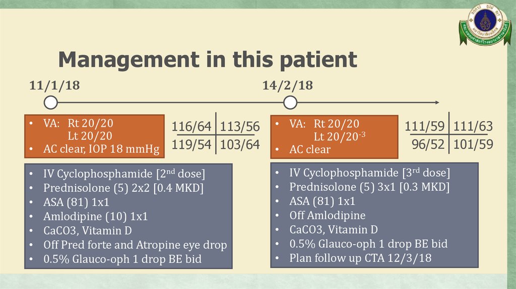
Management in this patient11/1/18
• VA: Rt 20/20
Lt 20/20
• AC clear, IOP 18 mmHg
14/2/18
116/64 113/56
119/54 103/64
IV Cyclophosphamide [2nd dose]
Prednisolone (5) 2x2 [0.4 MKD]
ASA (81) 1x1
Amlodipine (10) 1x1
CaCO3, Vitamin D
Off Pred forte and Atropine eye drop
0.5% Glauco-oph 1 drop BE bid
• VA: Rt 20/20
Lt 20/20-3
• AC clear
111/59 111/63
96/52 101/59
IV Cyclophosphamide [3rd dose]
Prednisolone (5) 3x1 [0.3 MKD]
ASA (81) 1x1
Off Amlodipine
CaCO3, Vitamin D
0.5% Glauco-oph 1 drop BE bid
Plan follow up CTA 12/3/18
60.
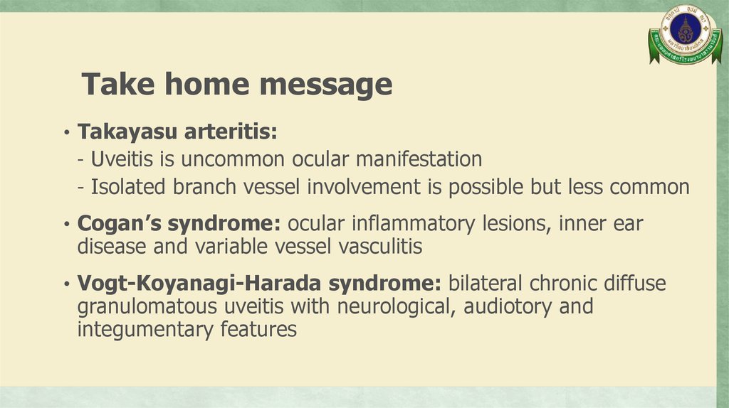
Take home message• Takayasu arteritis:
- Uveitis is uncommon ocular manifestation
- Isolated branch vessel involvement is possible but less common
• Cogan’s syndrome: ocular inflammatory lesions, inner ear
disease and variable vessel vasculitis
• Vogt-Koyanagi-Harada syndrome: bilateral chronic diffuse
granulomatous uveitis with neurological, audiotory and
integumentary features
61.
Thank you62.
Rheumatic diseases with reportedaortic involvement
Clin Exp Rheumatol 2006;24:S41-7.
63.
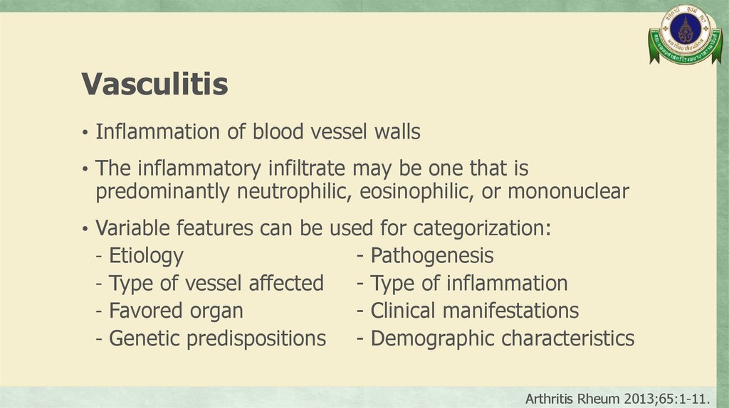
Vasculitis• Inflammation of blood vessel walls
• The inflammatory infiltrate may be one that is
predominantly neutrophilic, eosinophilic, or mononuclear
• Variable features can be used for categorization:
- Etiology
- Pathogenesis
- Type of vessel affected
- Type of inflammation
- Favored organ
- Clinical manifestations
- Genetic predispositions
- Demographic characteristics
Arthritis Rheum 2013;65:1-11.
64.
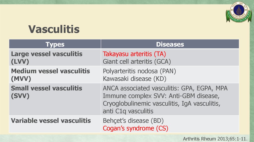
VasculitisArthritis Rheum 2013;65:1-11.
65.
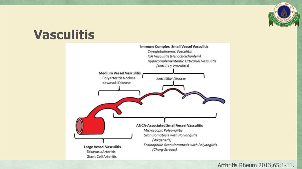
VasculitisTypes
Large vessel vasculitis
(LVV)
Diseases
Takayasu arteritis (TA)
Giant cell arteritis (GCA)
Medium vessel vasculitis
(MVV)
Small vessel vasculitis
(SVV)
Polyarteritis nodosa (PAN)
Kawasaki disease (KD)
ANCA associated vasculitis: GPA, EGPA, MPA
Immune complex SVV: Anti-GBM disease,
Cryoglobulinemic vasculitis, IgA vasculitis,
anti C1q vasculitis
Behçet’s disease (BD)
Cogan’s syndrome (CS)
Variable vessel vasculitis
Arthritis Rheum 2013;65:1-11.
66.
VasculitisTypes
Single organ vasculitis
(SOV)
Diseases
Cutaneous leukocytoclastic angiitis
Cutaneous arteritis
Primary central nervous system vasculitis
Isolated aortitis
Others
Arthritis Rheum 2013;65:1-11.
67.
VasculitisTypes
Vasculitis associated with
systemic disease
Diseases
Lupus vasculitis
Rheumatoid vasculitis
Sarcoid vasculitis
Others
Arthritis Rheum 2013;65:1-11.
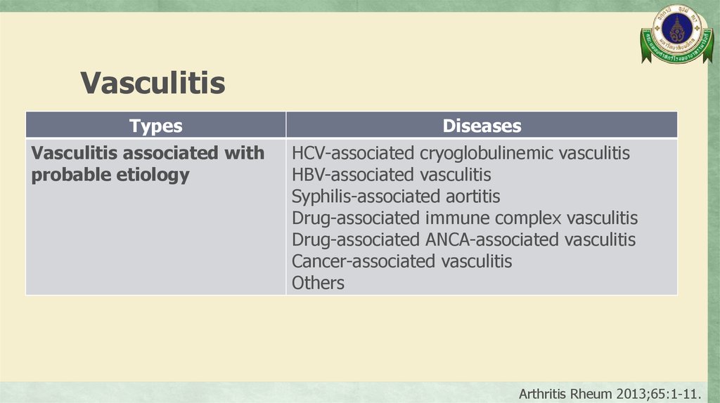
68.
VasculitisTypes
Vasculitis associated with
probable etiology
Diseases
HCV-associated cryoglobulinemic vasculitis
HBV-associated vasculitis
Syphilis-associated aortitis
Drug-associated immune complex vasculitis
Drug-associated ANCA-associated vasculitis
Cancer-associated vasculitis
Others
Arthritis Rheum 2013;65:1-11.
69.
Aortitis• Pathological term for inflammation of the aortic wall
• The classification of aortitis broadly includes underlying
rheumatologic and infectious diseases, along with isolated
aortitis
Circulation 2008;117:3039-51.
70.
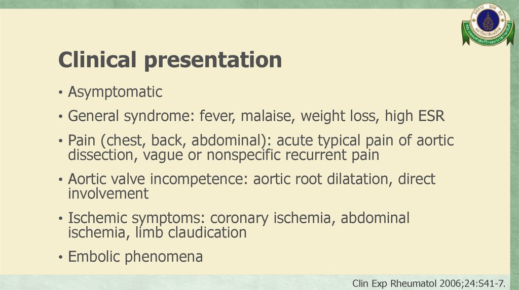
Clinical presentation• Asymptomatic
• General syndrome: fever, malaise, weight loss, high ESR
• Pain (chest, back, abdominal): acute typical pain of aortic
dissection, vague or nonspecific recurrent pain
• Aortic valve incompetence: aortic root dilatation, direct
involvement
• Ischemic symptoms: coronary ischemia, abdominal
ischemia, limb claudication
• Embolic phenomena
Clin Exp Rheumatol 2006;24:S41-7.
71.
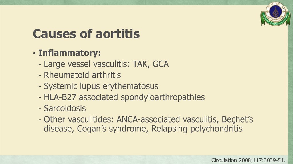
Causes of aortitis• Inflammatory:
- Large vessel vasculitis: TAK, GCA
- Rheumatoid arthritis
- Systemic lupus erythematosus
- HLA-B27 associated spondyloarthropathies
- Sarcoidosis
- Other vasculitides: ANCA-associated vasculitis, Beçhet’s
disease, Cogan’s syndrome, Relapsing polychondritis
Circulation 2008;117:3039-51.
72.
Causes of aortitis• Isolated aortitis:
- Isolated idiopathic (thoracic aortitis)
- Chronic periaortitis: Idiopathic retroperitoneal fibrosis,
Inflammatory abdominal aortic aneurysm,
Perianeurysmal aortitis, Idiopathic abdominal periaortitis
Circulation 2008;117:3039-51.
73.
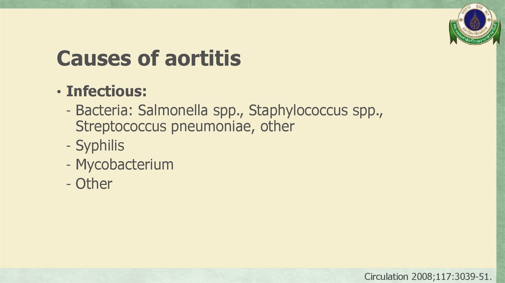
Causes of aortitis• Infectious:
- Bacteria: Salmonella spp., Staphylococcus spp.,
Streptococcus pneumoniae, other
- Syphilis
- Mycobacterium
- Other
Circulation 2008;117:3039-51.
74.
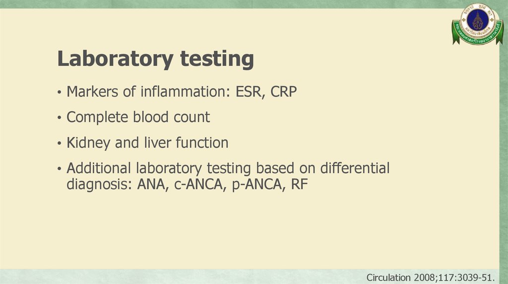
Laboratory testing• Markers of inflammation: ESR, CRP
• Complete blood count
• Kidney and liver function
• Additional laboratory testing based on differential
diagnosis: ANA, c-ANCA, p-ANCA, RF
Circulation 2008;117:3039-51.
75.
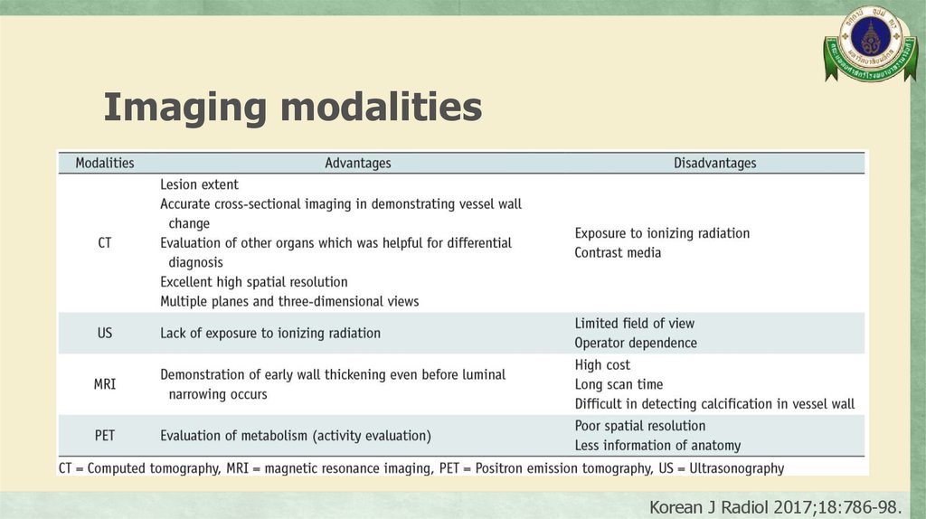
Imaging modalitiesKorean J Radiol 2017;18:786-98.
76.
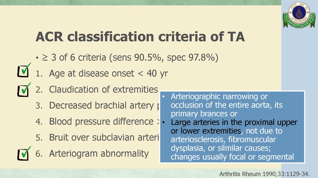
ACR classification criteria of TA• ≥ 3 of 6 criteria (sens 90.5%, spec 97.8%)
1. Age at disease onset < 40 yr
2. Claudication of extremities
3.
4.
5.
6.
• Arteriographic narrowing or
occlusion of the entire aorta, its
Decreased brachial artery pulse
primary brances or
Blood pressure difference >• 10
mmHg
Large
arteries in the proximal upper
or lower extremities, not due to
Bruit over subclavian arteries arteriosclerosis,
or aorta
fibromuscular
dysplasia, or silmilar causes;
Arteriogram abnormality
changes usually focal or segmental
Arthritis Rheum 1990;33:1129-34.
77.
Takayasu arteritis (TA)• Incidence 0.4-1 case/1,000,000/year
• Onset usually occurs before the age of 50 years, which is
a major distinction from giant cell arteritis, whose onset
usually occurs after age 50
• Average age of diagnosis 25-30 years
• 75-97% of patients are female
Curr Rheumatol Rep 2005;7:270-5.
Circulation 2008;117:3039-51.
Arthritis Rheum 2013;65:1-11.
78.
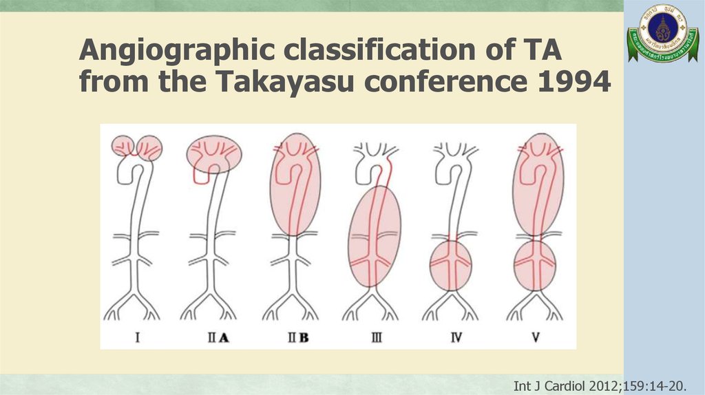
Angiographic classification of TAfrom the Takayasu conference 1994
Int J Cardiol 2012;159:14-20.