


 Медицина
Похожие презентации:


Omphalocele and Gastroschisis
1.
Omphalocele andGastroschisis
1
2.
Background:• Gastroschisis and omphalocele are among
the most frequently encountered
congenital anomalies in pediatric surgery.
• Combined incidence of these anomalies is
1 in 2000 births, which means, for
example, that a pediatric surgeon will see
2 such babies for every 1 born with
esophageal atresia or tracheoesophageal
fistula.
2
3.
Background:• Many babies have correctable lesions and simply
require routine pediatric care.
• For others, the abdominal wall defect is part of a larger
constellation of unresolved problems, and further care
by specialists is necessary.
• All of these children, however, require general
management by pediatricians who have knowledge of
their particular anomalies and their past surgical
histories.
• For example, physicians should know if an associated
malrotation was corrected (to prevent midgut volvulus)
and whether an abnormally located appendix was
removed (to prevent occurrence of atypical
appendicitis).
3
4.
Pathophysiology: Embryology• the human embryo initially is disc-shaped and
composed of 2 cell layers.
• It acquires a third cell layer as it grows above the
umbilical ring and becomes cylindrical by elongation
and inward folding.
• The body folds (cephalic, caudal, lateral) meet in the
center of the embryo where the amnion invests the
yolk sac.
• Defective development at this critical location results
in a spectrum of abdominal wall defects.
4
5.
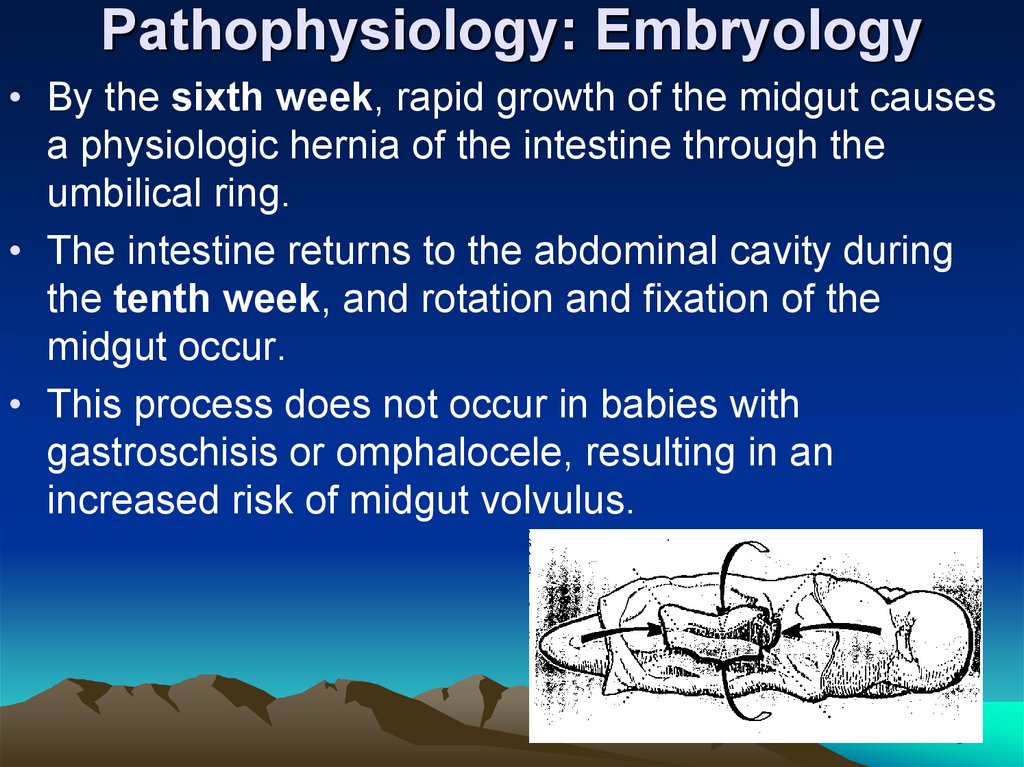
Pathophysiology: Embryology• By the sixth week, rapid growth of the midgut causes
a physiologic hernia of the intestine through the
umbilical ring.
• The intestine returns to the abdominal cavity during
the tenth week, and rotation and fixation of the
midgut occur.
• This process does not occur in babies with
gastroschisis or omphalocele, resulting in an
increased risk of midgut volvulus.
5
6.
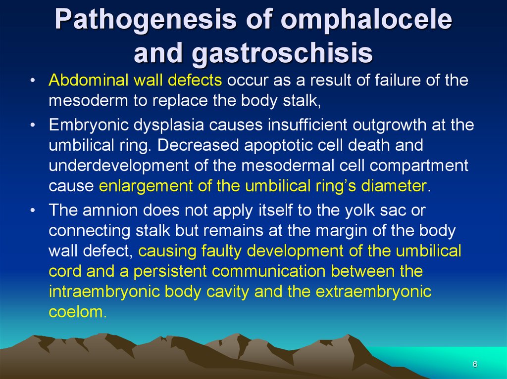
Pathogenesis of omphaloceleand gastroschisis
• Abdominal wall defects occur as a result of failure of the
mesoderm to replace the body stalk,
• Embryonic dysplasia causes insufficient outgrowth at the
umbilical ring. Decreased apoptotic cell death and
underdevelopment of the mesodermal cell compartment
cause enlargement of the umbilical ring’s diameter.
• The amnion does not apply itself to the yolk sac or
connecting stalk but remains at the margin of the body
wall defect, causing faulty development of the umbilical
cord and a persistent communication between the
intraembryonic body cavity and the extraembryonic
coelom.
6
7.
Pathogenesis of Omphalocele• In babies with omphalocele failure of central
fusion at the umbilical ring by growth of the
mesoderm causes defective abdominal wall
closure and persistent herniation of the midgut.
• The abdominal viscera are contained within a
translucent sac, which is composed of amnion,
Wharton jelly, and peritoneum.
• The umbilical vessels radiate onto the wall of the
sac. In 50% of cases, the liver, spleen, and
ovaries or testes accompany the extruded
midgut.
7
8.
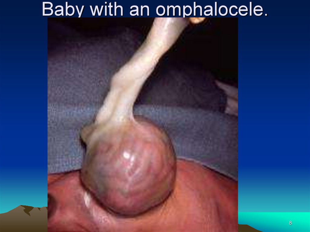
Baby with an omphalocele.8
9.
Baby with a ruptured omphalocele.9
10.
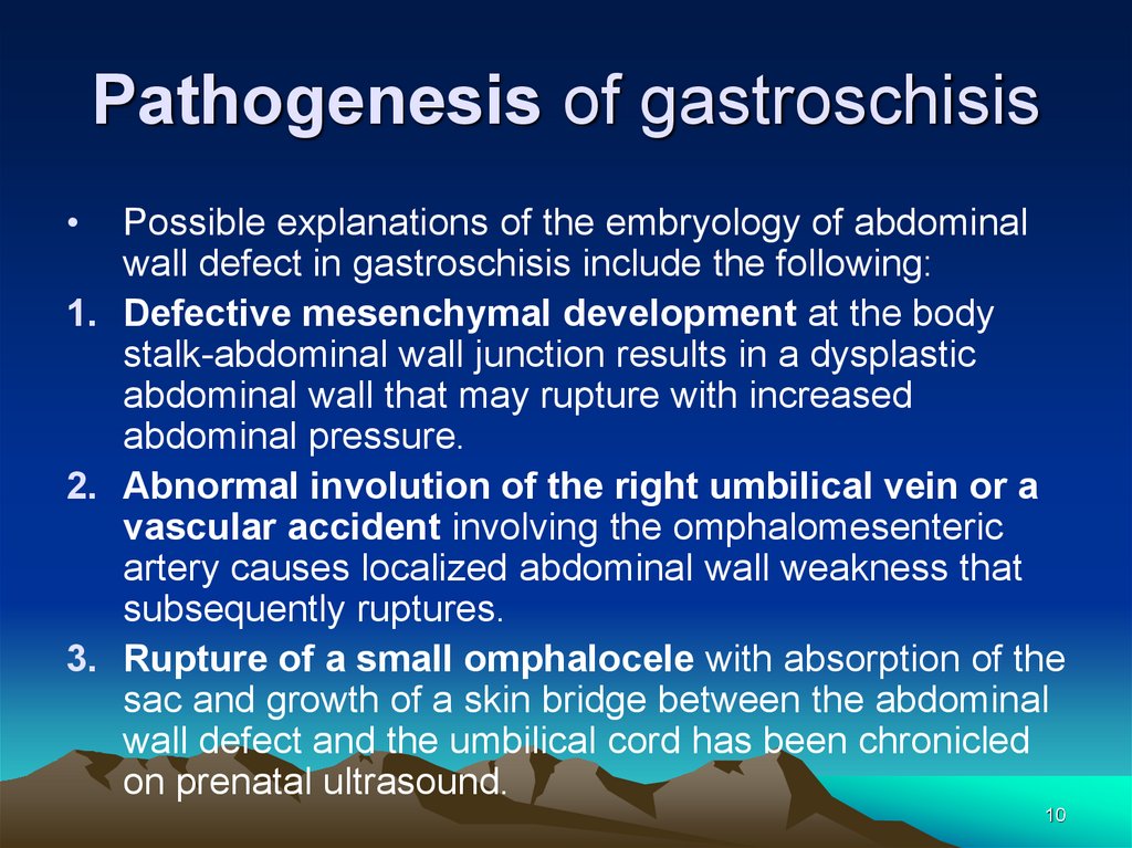
Pathogenesis of gastroschisisPossible explanations of the embryology of abdominal
wall defect in gastroschisis include the following:
1. Defective mesenchymal development at the body
stalk-abdominal wall junction results in a dysplastic
abdominal wall that may rupture with increased
abdominal pressure.
2. Abnormal involution of the right umbilical vein or a
vascular accident involving the omphalomesenteric
artery causes localized abdominal wall weakness that
subsequently ruptures.
3. Rupture of a small omphalocele with absorption of the
sac and growth of a skin bridge between the abdominal
wall defect and the umbilical cord has been chronicled
on prenatal ultrasound.
10
11.
Baby with an umbilical cord hernia.11
12.
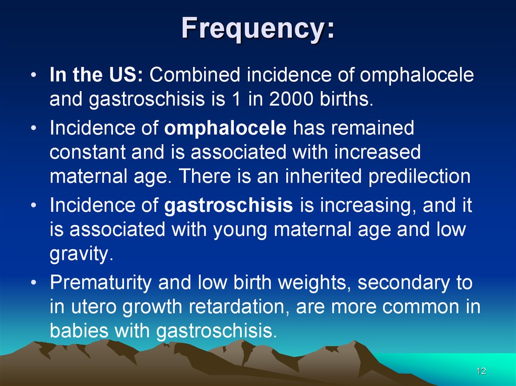
Frequency:• In the US: Combined incidence of omphalocele
and gastroschisis is 1 in 2000 births.
• Incidence of omphalocele has remained
constant and is associated with increased
maternal age. There is an inherited predilection
• Incidence of gastroschisis is increasing, and it
is associated with young maternal age and low
gravity.
• Prematurity and low birth weights, secondary to
in utero growth retardation, are more common in
babies with gastroschisis.
12
13.
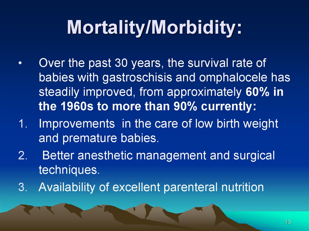
Mortality/Morbidity:Over the past 30 years, the survival rate of
babies with gastroschisis and omphalocele has
steadily improved, from approximately 60% in
the 1960s to more than 90% currently:
1. Improvements in the care of low birth weight
and premature babies.
2. Better anesthetic management and surgical
techniques.
3. Availability of excellent parenteral nutrition
13
14.
Mortality/Morbidity:• Long-term morbidity from gastroschisis is related to
intestinal dysfunction and wound problems.
• Short gut syndrome may be caused by a number of
factors.
1. An antenatal mesenteric vascular accident
2. Constriction of the extruded intestine's mesentery by a
small abdominal wall defect may cause an obstructed,
shortened intestine with diminished absorptive capacity.
3. Gut necrosis may complicate excessively tight closure of
the abdominal wall defect by impeding splanchnic blood
flow with resultant intestinal ischemia and necrotizing
enterocolitis (NEC),
4. or it may occur consequent to closed loop obstruction
caused by adhesions or midgut volvulus.
5. Loss of intestinal length exacerbates the dysfunction
consequent to antenatal exposure of the intestine to
amniotic fluid.
14
15.
Mortality/Morbidity:• Management of babies with short gut syndrome also
has improved significantly as a result of providing
nutrition by parenteral and enteral routes.
• Obtaining venous access and treating catheter sepsis,
and optimizing gut adaptation with innovative surgical
procedures and aggressive treatment of bacterial
overgrowth within stagnant intestinal loops.
• Even so, babies with short gut as a consequence of
gastroschisis comprise a large percentage of children
undergoing intestinal transplantation.
15
16.
Mortality/Morbidity:• Poor healing of the abdominal wound usually
results in a ventral hernia, which may require
secondary surgical repair.
• Paradoxically, babies with small (unimpressive)
omphaloceles are most likely to have associated
abnormalities, including intestinal problems (Meckel
diverticulum, atresia), genetic syndromes (BeckwithWiedemann), and congenital heart disease.
• Babies with giant omphaloceles usually have small,
bell-shaped, thoracic cavities and minimal pulmonary
reserve; reduction and repair of the omphalocele
frequently precipitates respiratory failure, which may
be chronic and require a tracheotomy and long-term
16
ventilator support.
17.
Mortality/Morbidity:• Even with successful repair, which usually
requires a synthetic patch, and good
clinical outcome, the location of the child's
liver is central, directly beneath the patch,
rendering it more vulnerable to trauma.
• Race: No geographic or racial predilection
exists for omphalocele or gastroschisis.
• Sex: The male-to-female ratio is 1.5:1.
17
18.
CLINICAL• Physical:
• Omphalocele
– In babies with omphaloceles, the size of the abdominal
wall defect ranges from 4-12 cm, and the location of the
defect may be central, epigastric, or hypogastric.
– Although the ease of surgical reduction and repair correlate
with the size of the abdominal wall defect, a small
omphalocele is no guarantee of an uncomplicated clinical
course. Associated genetic syndromes involving multiple
organ systems, or abnormalities of the intestine, such as an
atresia or a patent omphalomesenteric duct, are potential
problems.
– With a large omphalocele, dystocia may occur and result in
injury to the baby's liver; hence, a cesarean section may
be indicated.
– The omphalocele sac is usually intact, although it may be
ruptured in 10-20% of cases. Rupture may occur in utero
or during or after delivery.
18
19.
CLINICAL– Babies with the Beckwith-Wiedemann syndrome (ie,
macroglossia, gigantism) have large, rounded facial
features, hypoglycemia from hyperplasia of the pancreatic
islet cells, and visceromegaly. They may have genitourinary
abnormalities, and they are at risk for development of Wilms
tumors, liver tumors (hepatoblastoma), and adrenocortical
neoplasms.
– Pentalogy of Cantrell describes an epigastric omphalocele
associated with a cleft sternum and anterior diaphragmatic
hernia (Morgagni), cardiac defects (eg, ectopia cordis,
ventricular septal defect [VSD]) and an absent pericardium.
– Giant omphaloceles have large central or epigastric defects.
The liver is centrally located and entirely contained within the
omphalocele sac. The abdominal cavity is small and
undeveloped, and operative closure is very difficult. The
thoracic cavity is also small. Associated pulmonary
hypoplasia or restrictive lung disease may be present.
19
20.
Beckwith-Wiedemann syndrome20
21.
Baby with pentalogy of Cantrell21
22.
CLINICAL- Gastroschisis– The defect is fairly uniform in size and location; a 5-cm
vertical opening to the left of the umbilical cord.
– However, the extent of intestinal inflammation and
resultant edema and turgor greatly affect reduction and
closure of the abdomen. Inflammation so distorts the
bowel's appearance that it may be difficult to determine if
associated intestinal atresia is present.
– Once reduction and closure is obtained, inflammation
resolves, and the intestine softens and regains a normal
appearance.
– Correction of associated intestinal atresia is best left until
this time, usually 3 weeks after the first operative
procedure.
– Intestinal dysfunction takes longer to normalize, from 6
weeks to several months.
– If gastroschisis is identified, perform serial examinations
to assess intestinal integrity and amniocentesis to
22
monitor lung maturity.
23.
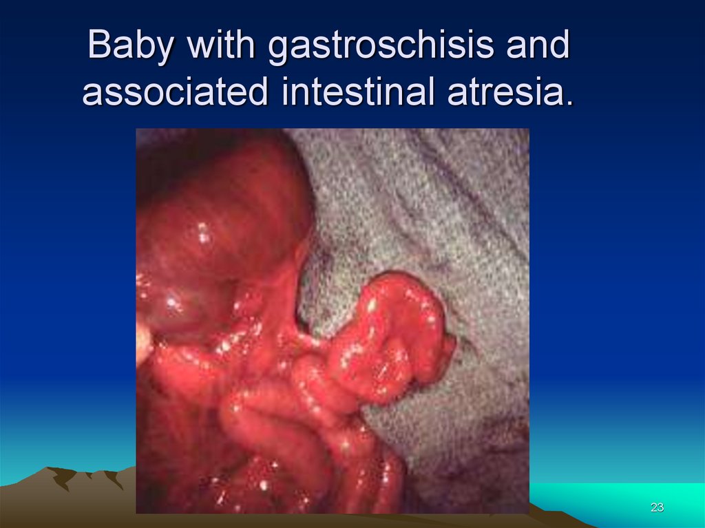
Baby with gastroschisis andassociated intestinal atresia.
23
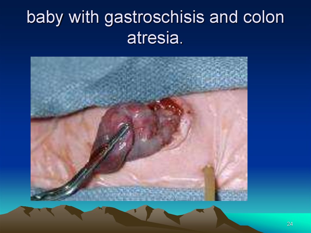
24.
baby with gastroschisis and colonatresia.
24
25.
Causes:• Factors associated with high-risk pregnancies, such as maternal
illness and infection, drug use, smoking, and genetic abnormalities,
also are associated with the birth of babies with omphalocele and
gastroschisis.
• These factors contribute to placental insufficiency and the birth of
small for gestational age (SGA) or premature babies, among whom
gastroschisis and omphalocele most commonly occur.
• Folic acid deficiency, hypoxia, and salicylates have caused
laboratory rats to develop abdominal wall defects, but the clinical
significance of these experiments is conjectural.
• Certainly, elevation of maternal serum alpha-fetoprotein
(MSAFP) warrants investigation by high-resolution sonography to
determine if any structural abnormalities are present in the fetus.
• If such abnormalities are present and associated with an
omphalocele, perform amniocentesis to check for a genetic
abnormality.
• Polyhydramnios suggests fetal intestinal atresia, and this
possibility should be investigated by ultrasound. Ideally, such
information will prompt referral to a tertiary care facility, where the
infant can receive expeditious specialty care.
25
26.
DIFFERENTIALS• Other Problems to be Considered:
• In babies with omphalocele, a 35-80% incidence of other clinical
problems is seen.
• These include congenital heart disease, cleft palate, and
musculoskeletal and dental occlusion abnormalities.
• Patent omphalomesenteric duct and small bowel atresias may
occur in babies with umbilical cord hernias where the size of the
defect is smaller than 4 cm.
Incidence of associated chromosomal abnormalities is 10-40%.
These include trisomies 12, 13, 15, 18, and 21.
Babies with gastroschisis, in which the incidence of chromosomal
anomalies is less than 5 percent, may have gastroesophageal
reflux disease or Hirschsprung disease in addition to abnormal
intestinal absorption and motility.
26
27.
WORKUP• Lab Studies:
• Maternal serum alpha-fetoprotein
– Prenatal diagnosis of abdominal wall defects can be
made by detection of an elevation in MSAPF.
– MSAPF levels are greater in gastroschisis than in
omphalocele.
– MSAPF also is increased in spina bifida, which
additionally demonstrates an increased ratio of
acetylcholinesterase and pseudocholinesterase.
27
28.
WORKUP• Imaging Studies:
• Fetal sonography may detect a genetic abnormality, with
identification of a structural marker of the karyotypic
abnormality.
• Fetal echocardiography also may identify a cardiac
abnormality.
• Confirm positive findings suggestive of a genetic
abnormality by amniocentesis.
• If serial ultrasounds show dilatation and thickening of the
intestine in a baby with gastroschisis, and if lung maturity
can be verified by amniocentesis, delivery is induced.
28
29.
TREATMENT• Medical Care:
• Intestinal inflammation
– Intestinal inflammation may occur with either gastroschisis or ruptured
omphalocele.
– The eviscerated intestine may be either normal or abnormal in structure
and function. The degree of abnormality depends upon the extent of the
inflammatory and ischemic injury, manifested by shortened length and
surface exudate (peel), which is related to the composition and duration
of the intestine’s exposure to the amniotic fluid and fetal urine.
– Inflamed intestine is thick and edematous, the loops of bowel are matted
together, and the mesentery is congested and foreshortened.
– Histologically, atrophy of the myenteric ganglion cells is seen.
– The intestine is dysmotile, with prolonged transit time and decreased
absorption of carbohydrate, fat, and protein. These deleterious effects
remit as the inflammation resolves, usually in 4-6 weeks. During this
time, total parenteral nutrition (TPN) is required.
29
30.
TREATMENT• Intact omphalocele
– Usually, neonates with intact omphalocele are in no
distress, unless associated pulmonary hypoplasia is
present.
– Examine the baby carefully to detect any associated
problems, such as Beckwith-Wiedemann syndrome,
chromosomal abnormalities, congenital heart disease, or
other associated malformations. Give nothing by mouth
(NPO) pending operative repair.
– Administer maintenance IV fluids, and cover the
omphalocele sac with sterile saline-soaked gauze and with
plastic wrap, using sterile technique. As an alternative, the
baby's lower torso may be placed in a bowel bag.
30
31.
TREATMENT– The omphalocele should be supported to
avoid excessive traction to the mesentery.
– Give prophylactic antibiotics preoperatively,
because of the possibility of an associated
intestinal anomaly.
– Closure of a small or moderate size
omphalocele usually is accomplished without
difficulty.
– A ruptured omphalocele is treated like
gastroschisis.
– Closure of a giant omphalocele that contains
the liver can be very challenging.
31
32.
• GastroschisisTREATMENT
– Respiratory distress in neonates with gastroschisis may respond to
gastric decompression, although endotracheal intubation may still be
needed.
– Fluid, electrolyte, and heat losses must be minimized and corrected.
Administer an intravenous fluid bolus (20 mL/kg LR), followed by 5%
dextrose ¼ NS at 2-3 times the baby's maintenance fluid rate.
– The baby should be placed under a radiant heater, and the exposed
intestines should be covered with plastic wrap and supported to avoid
excessive traction on the mesentery. As an alternative, the baby’s lower
torso may be placed in a bowel bag.
– Insert a urinary catheter to monitor urine output and facilitate reduction of
the herniated viscera by avoiding bladder distention.
– Administer antibiotics to prevent infection, since neonates have low
levels of circulating immunoglobulin G (IgG).
– Place a central venous line to provide parenteral nutrition, thereby
minimizing protein loss during the period of gastrointestinal dysfunction.
32
33.
TREATMENT-Surgical Care:• Omphalocele
– Ambroise Pare, the 17th-century French surgeon,
accurately described omphalocele and the dire
consequences of opening the sac to attempt
surgical closure. Certainly, his admonition
encouraged conservative treatment, ie, squeezing
the sac to effect reduction of the herniated viscera
or painting the sac with escharotic agents to
promote epithelization.
– The problem with this approach is that it is slow.
During this time the sac may rupture, resulting in a
wound infection. Even if complications do not occur,
the healing of such a large wound exacts a
significant metabolic and nutritional toll.
33
34.
TREATMENT-Surgical Care:– Healing may be hastened by surgically mobilizing skin flaps
sufficient to cover the omphalocele sac, thereby obtaining
closure of the abdominal wall defect in a way comparable to
closing a burn wound with skin grafts (Gross technique).
This, however, results in the creation of a ventral hernia.
– In 1967, Schuster developed a technique that may be used
in the initial treatment of a baby with a giant omphalocele or
in correcting the ventral hernia created by skin flap closure.
An incision is made along the skin-sac junction of the
abdominal wall defect, which is enlarged in the midline. The
anterior rectus fascia is exposed from the xiphoid to the
pubis, and Teflon sheets are sutured to its medial edge. The
Teflon sheets are then closed over the omphalocele sac and
gradually tightened, approximating the rectus muscles over
the abdominal viscera.
34
35.
TREATMENT-Surgical Care• Gastroschisis
– In 1969, Allen and Wrenn adapted Schuster’s technique for treatment of
gastroschisis.
– Silastic sheets are sutured to the full thickness of the enlarged abdominal
wall defect and closed over the eviscerated intestine, whose reduction is
facilitated by stretching the abdominal musculature, emptying the
stomach and bladder, and manually evacuating the colon.
– A major factor in the reduction of the extruded viscera is resolution of
intestinal inflammation, which results in a change from a rigid, congealed
mass of bowel to soft, pliable loops of intestine, which squeeze into the
abdominal cavity.
– Too tight a closure of the abdominal wall must be avoided, for this limits
excursion of the diaphragm and necessitates increased inspiratory
pressure to compensate for the increase in airway resistance. In general,
peak inspiratory pressures (PIPs) higher than 25 mmHg should be
avoided. High-frequency oscillatory ventilation may be an alternative to
conventional ventilation if intraabdominal pressures are markedly
increased.
35
36.
TREATMENT-Surgical Care– In addition, tight closure of the abdominal cavity
impedes venous return to the heart, compromising
cardiac output and decreasing renal blood flow and
glomerular filtration rate. Renal vein thrombosis and
renal failure may ensue.
– Diminished mesenteric blood flow may facilitate the
development of necrotizing enterocolitis.
– In order to avoid these problems, techniques have
been developed to monitor central venous pressure
(CVP), intraabdominal pressure, intravesicular
pressure, and intragastric pressure (which should not
exceed 20 cm of water).
36
37.
• Consultations:• Neonatologists and pediatric surgeons
usually care for babies with these
anomalies.
• Consult with cardiology, pulmonology,
gastroenterology, and genetics, as
indicated.
37
38.
Diet:• Babies with omphalocele usually do not require
special formulas; their intestines are typically normal,
with the exception of occasional atresias, which, in the
author’s experience, are located in the distal ileum and
are not associated with short gut.
• Babies with gastroschisis, on the other hand,
typically require special elemental, crystalline amino
acid, or protein hydrolysate formulas with nonlactose
carbohydrate and medium-chain triglycerides because
of the associated gut inflammation and resultant
tendency towards substrate malabsorption and
allergy.
• Babies with short gut syndrome absorb medium-chain
triglycerides more readily than long-chain triglycerides;
however, the latter are more valuable with regard to
gut adaptation.
38
39.
Activity:• A child with a repaired giant omphalocele
has an epigastric liver. In this location, the
liver is more vulnerable to trauma.
Avoidance of contact sports is prudent.
39
40.
FOLLOW-UP• Further Inpatient Care:
• Omphalocele
– Babies with omphalocele usually have rapid return of
intestinal function after surgical repair, even if intestinal
atresia occurs concomitantly, because no associated gut
inflammation is present.
– Babies with giant omphaloceles usually have a protracted
hospital course; and overall morbidity and mortality is higher
for these patients. Multiple procedures are necessary to
obtain closure of the abdominal wall defect.
– Respiratory compromise may complicate the repair and
require prolonged support and possibly a tracheotomy.
Ventilator management, tracheotomy care, and, ultimately,
decannulation require close cooperation by the
neonatologist, pulmonologist, and pediatric surgeon.
40
41.
• GastroschisisFOLLOW-UP
– Even if primary closure of the abdominal wall defect is
obtained, a period of several weeks of intestinal dysfunction
(ileus) usually follows, as a result of associated gut
inflammation. In this situation, parenteral nutrition is
essential, followed by the gradual introduction of enteral
feedings. Continuous drip feedings usually are tolerated
optimally.
– If reduction of the herniated intestine requires the use of a
silo, it usually is removed within 5-7 days. The period of
ileus follows, during which the baby requires parenteral
nutrition until the gradual return of intestinal function. If this
expected recovery does not occur within 3-4 weeks,
intestinal obstruction is presumed, and a contrast study is
obtained to document intestinal transit.
– If intestinal obstruction is present, a laparotomy must be
performed.
41
42.
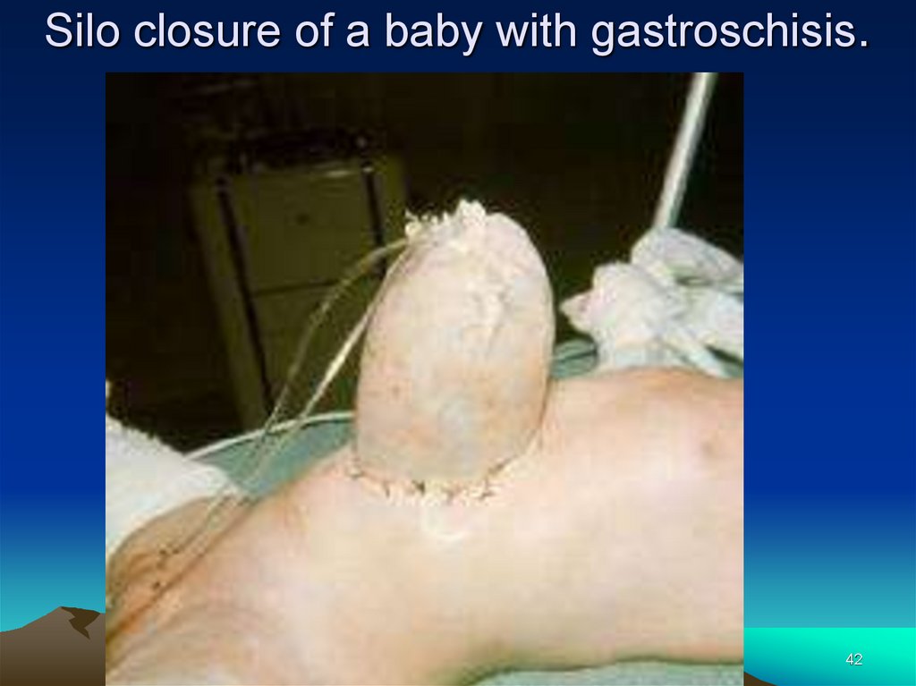
Silo closure of a baby with gastroschisis.42
43.
FOLLOW-UP• Further Outpatient Care:
• After hospital discharge, babies require close
follow-up care to assess growth and weight gain.
• Patients usually have gastroesophageal reflux
and may require medical therapy, but
fundoplication should not be necessary.
• Hirschsprung disease (aganglionic megacolon)
also may occur. Physicians should be alert to a
history of constipation.
43
44.
FOLLOW-UP• Transfer:
• The best way to treat the exposed intestines of a
baby with gastroschisis who is being transported to
a tertiary center includes the application of a moist
lap pad. The moist lap pad is placed over the
intestines and held directly over the abdominal wall
defect with dry Kerlix wrap applied around the
baby's torso including the extruded intestine. This
prevents traction upon the mesentery. A warm, wet,
lap pad placed in a bowel bag with the eviscerated
intestine soon becomes a cold, wet, lap pad.
44
45.
• The patient’s condition improved dramaticallyonce closure of the abdominal cavity was
achieved. Again, the author tried to wean him
from the ventilator, but his copious secretions
and episodes of high fever and drenching
sweats prevented this. Finally, it was determined
that the patient was experiencing narcotic
withdrawal. He had been postoperative for so
long, and narcotics had been used liberally to
provide postoperative pain relief.
45
46.
Prognosis:• Omphalocele
– Prognosis is dependent upon the severity of the
associated problems. Babies with omphalocele
are considerably complex, with involvement of
many other organ systems.
– Even giant omphaloceles can be closed,
although multiple procedures may be necessary.
– The limiting factor for many of these babies,
however, is their diminutive thoracic cavities and
associated pulmonary hypoplasia and resultant
chronic respiratory failure. Even so, lung growth
and development continue well into childhood,
encouraging optimism regarding the ultimate
prognosis.
46
47.
Prognosis• Gastroschisis
– Prognosis is dependent mainly upon severity of
associated problems, including prematurity,
intestinal atresia, short gut, and intestinal
inflammatory dysfunction.
– Many pediatric surgeons believe that prognosis
has improved because of maternal ultrasound
diagnosis and monitoring, which leads to
expeditious delivery of babies at tertiary centers.
– Years ago, obtaining primary closure of a baby
with gastroschisis was unusual. Usually, it was
necessary to use a silo. Now, primary closure is
commonly attained.
47
48.
Patient Education:Instruct parents regarding the significance of
bilious (green) vomiting, since these babies
may develop adhesive small bowel obstruction
or midgut volvulus.
• Inform parents that their child's appendix is
probably in an unusual location and that a CT
scan may be the most reliable way to diagnose
acute appendicitis.
48
49.
Special Concerns:• Prenatal care and planning
– With increased availability of sonography, prenatal
diagnosis is more frequent.
– Diagnosis of omphalocele mandates further workup to
determine if an associated genetic abnormality is
present, in which case appropriate counseling is
necessary.
– When gastroschisis is diagnosed, perform serial
examinations to detect signs of intestinal injury
(decreased peristalsis or distension).
– Provide the baby's parents with information concerning
the anomaly before delivery. Also, optimal management
requires that the obstetrician understands the particular
needs of these babies and ensures that they are
delivered in a facility where neonatal, pediatric
anesthesia, and pediatric surgery services are available.
49
50.
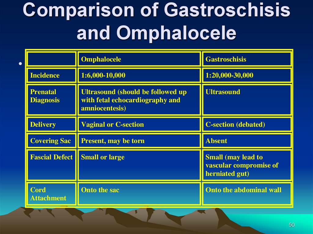
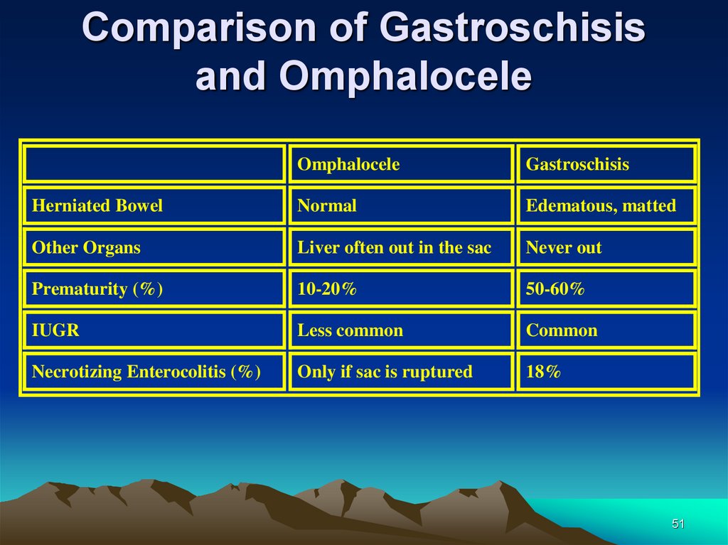
Comparison of Gastroschisisand Omphalocele
Omphalocele
Gastroschisis
Incidence
1:6,000-10,000
1:20,000-30,000
Prenatal
Diagnosis
Ultrasound (should be followed up
with fetal echocardiography and
amniocentesis)
Ultrasound
Delivery
Vaginal or C-section
C-section (debated)
Covering Sac
Present, may be torn
Absent
Fascial Defect
Small or large
Small (may lead to
vascular compromise of
herniated gut)
Cord
Attachment
Onto the sac
Onto the abdominal wall
50
51.
Comparison of Gastroschisisand Omphalocele
Omphalocele
Gastroschisis
Herniated Bowel
Normal
Edematous, matted
Other Organs
Liver often out in the sac
Never out
Prematurity (%)
10-20%
50-60%
IUGR
Less common
Common
Necrotizing Enterocolitis (%)
Only if sac is ruptured
18%
51
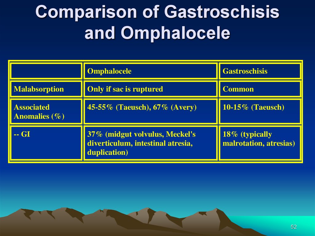
52.
Comparison of Gastroschisisand Omphalocele
Omphalocele
Gastroschisis
Malabsorption
Only if sac is ruptured
Common
Associated
Anomalies (%)
45-55% (Taeusch), 67% (Avery)
10-15% (Taeusch)
-- GI
37% (midgut volvulus, Meckel's
diverticulum, intestinal atresia,
duplication)
18% (typically
malrotation, atresias)
52
53.
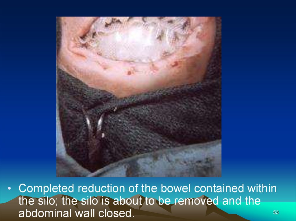
• Completed reduction of the bowel contained withinthe silo; the silo is about to be removed and the
53
abdominal wall closed.
54.
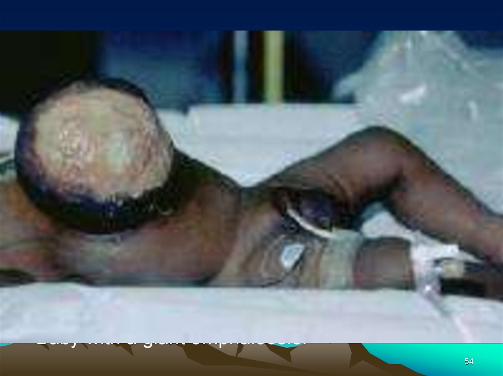
• Baby with a giant omphalocele.54
55.
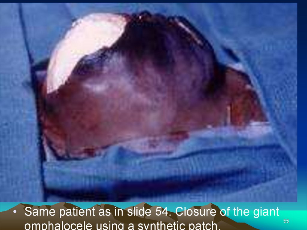
• Same patient as in slide 54. Closure of the giantomphalocele using a synthetic patch.
55
56.
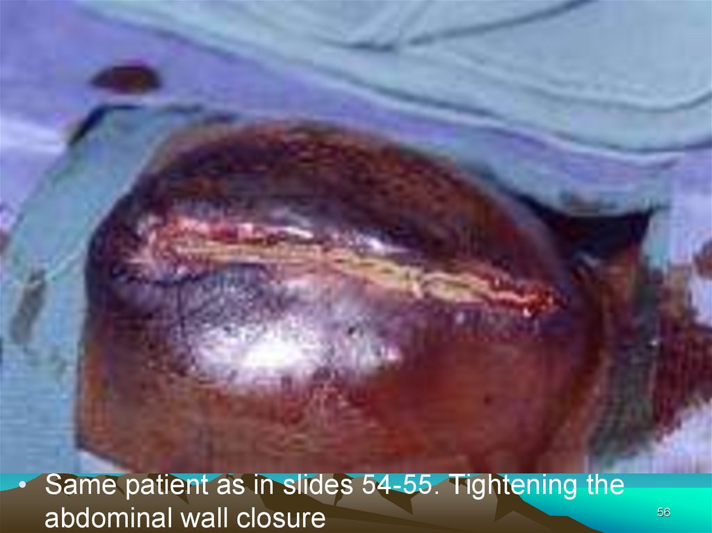
• Same patient as in slides 54-55. Tightening theabdominal wall closure
56
57.
• Same patient as in slides 54-56. Flank flaps wereused to close the giant omphalocele in the baby
whose patch became infected.
57
58.
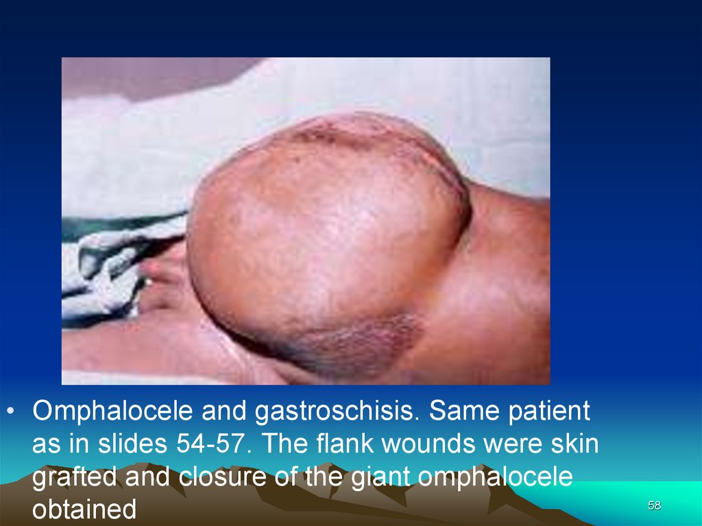
• Omphalocele and gastroschisis. Same patientas in slides 54-57. The flank wounds were skin
grafted and closure of the giant omphalocele
obtained
58