


 Медицина
Похожие презентации:


")


Cirrhosis
1.
CIRRHOSISKOGATAM.LIVINGSTONE
KOPULLA.VIJAY VARDHAN
MOHAMMED. ABDUL SAQIB
SHAIK NAYAB RAHMAN.SADHIK
2.
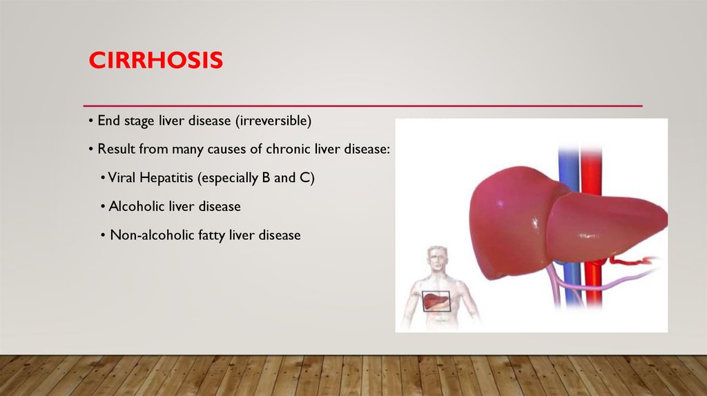
CIRRHOSIS• End stage liver disease (irreversible)
• Result from many causes of chronic liver disease:
• Viral Hepatitis (especially B and C)
• Alcoholic liver disease
• Non-alcoholic fatty liver disease
3.
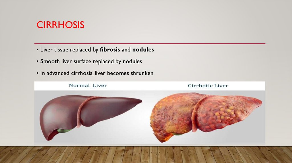
CIRRHOSIS• Liver tissue replaced by fibrosis and nodules
• Smooth liver surface replaced by nodules
• In advanced cirrhosis, liver becomes shrunken
4.
CLINICAL FEATURES• Hyperammonemia
• Asterixis, confusion, coma
5.
HYPERAMMONEMIATREATMENT
• Low protein diet
• Lactulose
• Synthetic disaccharide (laxative)
• Colon breakdown by bacteria to fatty acids
• Lowers colonic pH; favors formation of NH4+ over NH3
• NH4 + not absorbed → trapped in colon
• Result: ↓plasma ammonia concentrations
6.
CIRRHOSISCLINICAL FEATURES
• Jaundice
• Loss of bilirubin metabolism
• Hypoglycemia
• Loss of gluconeogenesis
• Coagulopathy
• Loss of clotting factors
• Elevated PT/PTT
• Hypoalbuminemia
• May cause low oncotic pressure
• Contributes to ascites, edema
7.
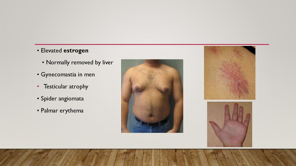
• Elevated estrogen• Normally removed by liver
• Gynecomastia in men
• Testicular atrophy
• Spider angiomata
• Palmar erythema
8.
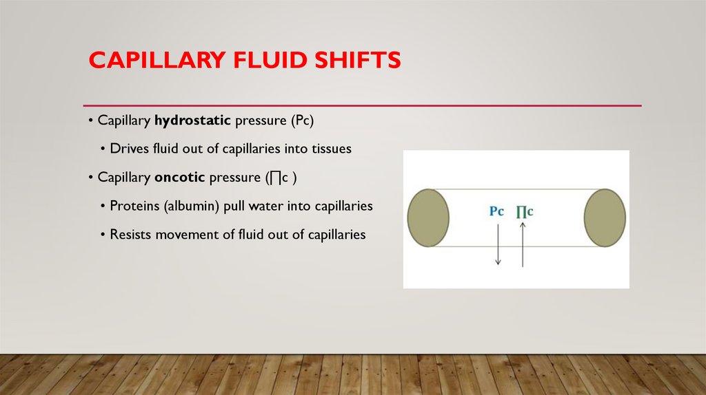
CAPILLARY FLUID SHIFTS• Capillary hydrostatic pressure (Pc)
• Drives fluid out of capillaries into tissues
• Capillary oncotic pressure (∏c )
• Proteins (albumin) pull water into capillaries
• Resists movement of fluid out of capillaries
9.
PORTAL HYPERTENSION• Blood flows portal vein → liver → hepatic vein
• Cirrhosis → obstructed flow through liver
• High pressure in portal vein (“hypertension”)
10.
CIRRHOSIS11.
ASCITES AND EDEMA12.
VENOUS COLLATERALSVENOUS ANASTAMOSES
• High portal pressure opens “venous collaterals”
• Connection between portal-systemic veins
• Normally small, collapsed vessels
• Engorge in portal hypertension
• Key collaterals:
• Umbilicus – physical exam finding: “caput medusa”
• Esophagus – upper gastrointestinal bleeding
• Stomach – upper gastrointestinal bleeding
• Rectum – hemorrhoids which may also bleed
13.
ESOPHAGEAL VARICES14.
GASTRIC VARICES15.
CAPUT MEDUSA16.
INTERNAL HEMORRHOIDS17.
HYPERSPLENISM18.
PORTAL VEIN THROMBOSIS• Rare cause of portal hypertension
• Acute onset abdominal pain
• Splenomegaly (palpable spleen one exam)
• May result in gastric varices with bleeding
• Liver biopsy will be normal
19.
ASCITES• Accumulation of fluid in peritoneal cavity
• In liver disease, from portal hypertension +/- low albumin
20.
SAAGSERUM ASCITES ALBUMIN GRADIENT
• Test of ascitic fluid
• Two reasons for new/worsening ascites
• Portal hypertension
• Malignancy (leaky vasculature)
• Sample of ascitic fluid via paracentesis
• Serum albumin – ascites albumin = SAAG
21.
SAAGSERUM ASCITES ALBUMIN GRADIENT
• SAAG >1.1 g/dL
• Large difference between serum and ascites albumin
• High pressure driving fluid (not albumin) into peritoneum
• Seen in portal hypertension
• SAAG <1.1 g/Dl
• Albumin levels similar between serum and ascites
• Leaky vasculature leading to fluid/albumin into peritoneum
• Seen in malignant ascites (malignant cells in peritoneal cavity)
22.
ASCITES TREATMENT• Sodium restriction
• Spironolactone (drug of choice)
• Potassium-sparing diuretic
• Blocks aldosterone distal tubule
• Most effective drug for ascites
• Loop diuretics (2nd line)
• Large volume paracentesis
• TIPS
23.
TIPSTRANSJUGULAR INTRAHEPATIC PORTOSYSTEMIC
SHUNT
• Transjugular Intrahepatic Portosystemic Shunt
• Treatment of portal hypertension
• Creation of channel in liver
• Connects portal vein to hepatic vein
24.
SBPSPONTANEOUS BACTERIAL PERITONITIS
• Ascitic fluid infection
• Bacteria in gut gain entry into ascitic fluid
• Usually E. coli and Klebsiella; rarely strep/staph
• Fever, abdominal pain/tenderness
• ↑ ascitic absolute PMNs (≥250 cells/mm3)
• Common treatment:
• 3rd generation cephalosporin (cefotaxime)
• Gram positive and gram negative coverage
• Achieves good levels in ascitic fluid
25.
MELD SCOREMODEL FOR END-STAGE LIVER DISEASE
• Scoring system for chronic liver disease or cirrhosis
• Estimates 3-month mortality from liver disease
• Point system using:
• Bilirubin level
• Creatinine level
• INR
• >40 = 71% mortality
• <9 = 2% mortality
26.
CHILD-PUGH CLASSIFICATION• Five variables to predict risk/survival
• Points for encephalopathy, ascites, bilirubin, albumin, PT
• Score ranges from 5 to 15
• 5 or 6: Child-Pugh class A cirrhosis
• 7 to 9: Child-Pugh class B cirrhosis
• 10 to 15: Child-Pugh class C cirrhosis (worst)
27.
CIRRHOSISDIAGNOSIS
• Gold standard is liver biopsy
• Not required if diagnosis is clear from history
• Done only when biopsy will change management
• Imaging (ultrasound, CT, MRI)
• May show small, nodular liver
• Not sensitive or specific for diagnosis
• More helpful for detection of hepatocellular carcinoma
• Clinical diagnosis (common)
• Presence of ascites
• Low platelet count
• Spider angiomata
28.
STELLATE CELLS• Perisinusoidal cell
• Storage site for retinoids (vitamin A metabolites)
• Activated in liver disease
• Secrete TGF-β
• Proliferate and produce fibrous tissue
• Major contributor to cirrhosis