


 Медицина
Похожие презентации:


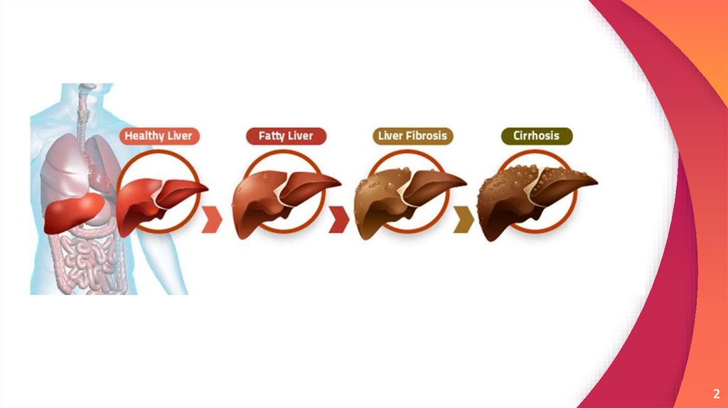
Liver cirrhosis
1.
Liver CirrhosisBY:
GOPAL YOGANANDHAN
LA-2-CO-171(2)
2.
23.
“Liver cirrhosis is a chronic liver
disease accompanied by
irreversible replacement of
parenchymal liver tissue by
fibrous connective tissue.
3
4.
EtiologyAlcohol
Hepatitis B can cause liver inflammation and damage that can
lead to cirrhosis.
Hepatitis C occurs by sexual intercourse or exposure to
infected blood or blood products
Hepatitis D can also cause cirrhosis. It’s often seen in people
who already have hepatitis B.
Autoimmune hepatitis causes inflammation that can lead to
cirrhosis.
4
5.
● Damage to the bile ducts, which function todrain bile: One example of such a condition
is primary biliary cholangitis.
● Disorders that affect the body’s ability to
handle iron and copper: Two examples
are hemochromatosis and Wilson’s disease.
● Medications, including prescription and overthe-counter drugs like acetaminophen, some
antibiotics, and some antidepressants, can
lead to cirrhosis.
5
6.
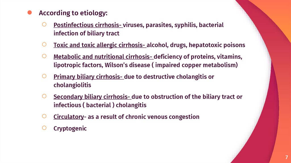
Classification7.
According to etiology:
○
○
○
○
○
○
○
Postinfectious cirrhosis- viruses, parasites, syphilis, bacterial
infection of biliary tract
Toxic and toxic allergic cirrhosis- alcohol, drugs, hepatotoxic poisons
Metabolic and nutritional cirrhosis- deficiency of proteins, vitamins,
lipotropic factors, Wilson’s disease ( impaired copper metabolism)
Primary biliary cirrhosis- due to destructive cholangitis or
cholangiolitis
Secondary biliary cirrhosis- due to obstruction of the biliary tract or
infectious ( bacterial ) cholangitis
Circulatory- as a result of chronic venous congestion
Cryptogenic
7
8.
● According to macroscopic appearance:○ Micronodular cirrhosis
○ Macronodular cirrhosis
● According to microscopic appearance:
○ Monolobular cirrhosis
○ Multilobular cirrhosis
● According to morphogenesis:
○ Portal
○ Post necrotic
○ mixed
8
9.
● According to course:○ Active
○ Inactive
9
10.
Pathogenesis11.
1112.
● Irrespective of the aetiology, cirrhosis ingeneral is initiated by hepatocellular necrosis
● Replacement of BM collagen type iv and vi by
fibrillary collagen type I and iii
● This lead to capillarization with quantitative
and qualitative ECM change
12
13.
● ECM regulates cellular activity and availabilityof growth factors
○ Decorin and biglycan binds TGF-B
○ Fibronectin and laminin binds TNF-alpha
○ Collagen binds PDGF, HGF, IL-2
● Binding of the survival factors to ECM prevents
apoptosis in damage liver and proteolysis
● ECM can modulate the activation of &
proliferation of HSC, angiogenesis GF & MMP
13
14.
● HSC activation represents a critical event inthe fibrosis
● This cell become the primary source of ECM in
liver upon injury
● This is modulated by immune signaling that is
influence by genetic and environmental
factors
14
15.
● Sources of ECM○ HSC
○ Bone marrow derive cells
○ Epithelial mesenchymal transition
○ Portal fibroblast
15
16.
1617.
CYTOKINES AND SIGNALING PATHWAYSIn ammatory cytokines play a key role in brosis, given that persistent
in ammation precedes brosis.
Following liver injury, several cell types can secrete in ammatory cytokines;
Cell types include; KCs, hepatocytes, HSCs, natural killer (NK)cells, lymphocytes,
and dendritic cells.
Ligand + receptor = transduction of extracellular signals into the cell
=modulation of changes in gene expression.
Common form of ligand-receptor interaction=dimerization/trimerization of
receptor molecules
○
○
○
Receptors with intrinsic tyrosine kinase
Receptors lacking intrinsic tyrosine kinase activity .
Seven transmembrane G-protein-coupled receptors (GPCRs). Steroid
hormone receptors.
17
18.
1819.
Systemic SyndromesClinical entity
cause/mechanism
Portal hypertension
Architectural and functional changes in liver resulting in
increased resistance to portal flow leading to portosystemic
collaterals, splanchnic vasodilation, expanded plasma volume
and arterial underfilling
Portopulmonary hypertension
Vasoactive substances not filtered by the damaged liver causing
pulmonary artery vasoconstriction, remodeling of vessels and in
situ thrombosis resulting in increasing pulmonary vasculature
resistance and ultimately right heart failure
Hepatopulmonary syndrome
Intrapulmonary vascular dilatation due to nitric oxide (NO) or
arteriovenous communication (with a predominance at lung
bases) causes ventilation/perfusion mismatch and/or shunting
which worsens when upright due to dependent pooling of blood
at bases
Hepatorenal syndrome
Renal failure caused by intense vasoconstriction of the renal
circulation as a consequence of extreme underfilling of arterial
circulation due to splanchnic vasodilation (due to high NO levels)
and dysregulation of vasocoactive systems in the setting
of advanced liver disease
19
20.
Clinical Features21.
The symptoms of cirrhosis occur because the liver is unable to purify the blood, breakdown toxins, produce clotting proteins, and help with absorption of fats and fatsoluble vitamins. Often, there are no symptoms until the disorder has progressed.
Some of the symptoms include:
decreased appetite
nose bleeds
jaundice (yellow discoloration)
small spider-shaped
veins underneath the skin
weight loss
anorexia
itchy skin
weakness
More serious symptoms include:
confusion and difficulty thinking clearly
abdominal swelling (ascites)
swelling of the legs (edema)
impotence
gynecomastia (when males start to
develop breast tissue)
21
22.
Liver dysfunctionThe following features are a direct consequence of liver cells not functioning.
Spider angiomata or spider nevi are vascular lesions consisting of a central arteriole
surrounded by many smaller vessels (hence the name "spider") and occur due to an increase
in estradiol. One study found that spider angiomata occur in about 1/3 of cases.
Palmar erythema is a reddening of palms at the thenar and hypothenar eminences seen in
about 23% of cirrhosis cases as a result of increased estrogen.
Gynecomastia, or benign increase in breast size in men, is caused by increased estradiol and
can occur in up to 2/3 of cases. This is different from increase in breast fat in overweight
people. A swollen scrotum may also be evident.
Hypogonadism, a decrease in male sex hormones may manifest as impotence, infertility, loss
of sexual drive, and testicular atrophy, and can result from primary gonadal injury or
suppression of hypothalamic/pituitary function. Hypogonadism is associated with cirrhosis
due to alcoholism or iron overload.
Liver size can be enlarged, normal, or shrunken in people with cirrhosis.
Ascites, accumulation of fluid in the peritoneal cavity in the abdomen, gives rise to
bulging flanks.
Jaundice, is yellow discoloration of the skin and mucous membranes notably of the white of
the eyes due to increased levels of bilirubin which may also cause the urine to be dark
colored.
22
23.
Palmnar ErythemaSpider angiomata
23
24.
2425.
2526.
EpitaxisJaundice
26
27.
2728.
Portal hypertensionLiver cirrhosis increases resistance to blood flow and leads to higher pressure in the
portal venous system, resulting in portal hypertension. Effects of portal
hypertension include:
An enlarged spleen is found in 35% to 50% of cases.
Esophageal varices result from collateral portal blood flow through vessels in
the stomach and esophagus (a process called portacaval anastomosis). When
these blood vessels become enlarged, they are called varices and are more
likely to rupture. Variceal rupture often leads to severe bleeding, which can
prove fatal.
Caput medusae are dilated paraumbilical collateral veins due to portal
hypertension. Blood from the portal venous system may be shunted through the
paraumbilical veins and ultimately to the abdominal wall veins, manifesting as
a pattern that may resemble the head of Medusa.
Cruveilhier-Baumgarten bruit is a venous hum heard in the epigastric region (on
examination by stethoscope) due to collateral connections forming between the
portal system and the paraumbilical veins as a result of portal hypertension.
28
29.
2930.
3031.
Advanced diseaseAs the disease progresses, complications may develop. In some people, these may
be the first signs of the disease.
Bruising and bleeding resulting from decreased production of clotting factors.
Hepatic encephalopathy (HE) – occurs when ammonia and related substances
build up in the blood and affect brain function when they are not cleared from
the blood by the liver. This may result in neglect of personal appearance,
unresponsiveness, forgetfulness, trouble concentrating, changes in sleep habits
or psychosis. One classic physical exam findings is asterixis, bilateral
asynchronous flapping of outstretched, dorsiflexed hands. Fetor hepaticus is a
musty breath odor resulting from increased dimethyl sulfide and is a feature of
HE.
Sensitivity to medication caused by decreased metabolism of the active
compounds.
Acute kidney injury (particularly hepatorenal syndrome).
Cachexia associated with muscle wasting and weakness.
31
32.
Cachexicpatient with
jaundice
Bruising
32
33.
Lab findingsThe following findings are typical in cirrhosis:
Thrombocytopenia – typically multifactorial. Due to alcoholic marrow
suppression, sepsis, lack of folate, platelet sequestering in the spleen as well as
decreased thrombopoietin. However, this rarely results in a platelet count < 50
000/mL.
Aminotransferases – AST and ALT are moderately elevated, with AST > ALT.
However, normal aminotransferase levels do not preclude cirrhosis.
Alkaline phosphatase – slightly elevated but less than 2–3 times the upper limit
of normal.
Gamma-glutamyl transferase – correlates with AP levels. Typically much higher
in chronic liver disease from alcohol.
Bilirubin – levels normal when compensated but may elevate as cirrhosis
progresses.
Albumin – levels fall as the synthetic function of the liver declines with
worsening cirrhosis since albumin is exclusively synthesized in the liver
33
34.
Prothrombin time – increases, since the liver synthesizes clotting factors.
Globulins – increased due to shunting of bacterial antigens away from the liver
to lymphoid tissue.
Serum sodium – hyponatremia due to inability to excrete free water resulting
from high levels of ADH and aldosterone.
Leukopenia and neutropenia – due to splenomegaly with splenic margination.
Coagulation defects – the liver produces most of the coagulation factors and
thus coagulopathy correlates with worsening liver disease.
Glucagon – increased in cirrhosis
Vasoactive intestinal peptide – increased as blood is shunted in the intestinal
system because of portal hypertension
Vasodilators – increased (such as nitric oxide and carbon monoxide) reducing
afterload with compensatory increase in cardiac output, mixed venous oxygen
saturation
Renin – increased (as well as sodium retention in kidneys) secondary to fall in
systemic vascular resistance
34
35.
Other laboratory studies performed in newly diagnosed cirrhosis may include:Serology for hepatitis viruses, autoantibodies (ANA, anti-smooth muscle, anti-mitochondria,
anti-LKM)
Ferritin and transferrin saturation: markers of iron overload as in
hemochromatosis, copper and ceruloplasmin: markers of copper overload as in Wilson's
disease
Immunoglobulin levels (IgG, IgM, IgA) – these immunoglobins are non-specific but may help in
distinguishing various causes
Cholesterol and glucose
Alpha 1-antitrypsin
Markers of inflammation and immune cell activation are typically elevated in cirrhotic patients
especially in the decompensated disease stage:
C-reactive protein (CRP)
Procalcitonin (PCT)
Presepsin
soluble CD14
soluble CD163
soluble CD206 (mannose receptor)
soluble TREM-1
35
36.
Liver ultrasound to assess the severity of cirrhosis.
Liver biopsy to identify liver cell changes & alterations in the
lobular structure.
Prolonged prothrombin time (11-12sec)
Rarely are diseases of the bile ducts, such as primary
sclerosing cholangitis, causes of cirrhosis. Imaging of the bile
ducts, such as ERCP or MRCP (MRI of biliary tract and
pancreas) may aid in the diagnosis.
upper endoscopy (to see if esophageal varices are present)
CT scan of the abdomen
36
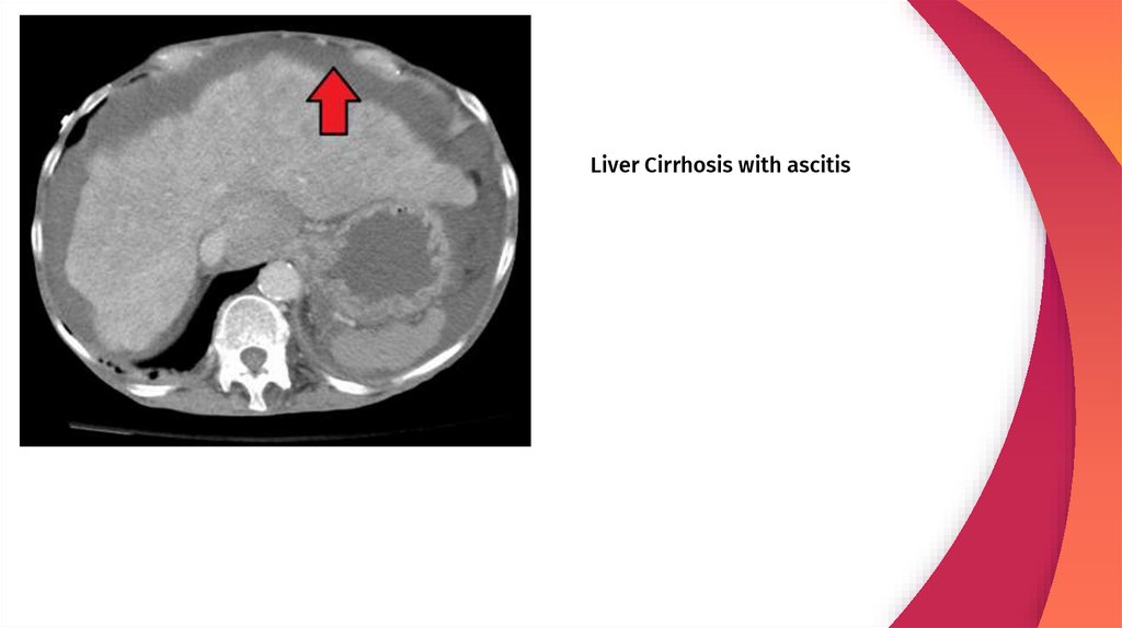
37.
Liver Cirrhosis with ascitis38.
Trichromestain,
showing
cirrhosis as
a nodular
texture
surrounded
by fibrosis
(wherein
collagen is
stained
blue)
39.
Treatment & Prevention40.
Treatment for cirrhosis varies based on what caused it and how far the disorder hasprogressed. Some treatments your doctor might prescribe include:
beta blockers or nitrates (for portal hypertension)
quitting drinking (if the cirrhosis is caused by alcohol)
banding procedures (used to control bleeding from esophageal varices)
intravenous antibiotics (to treat peritonitis that can occur with ascites)
hemodialysis (to purify the blood of those in kidney failure)
lactulose and a low protein diet (to treat encephalopathy)
Liver transplantation is an option of last resort, when other treatments fail.
All patients must stop drinking alcohol. Medications, even over-the-counter
ones, should not be taken without consulting your doctor.
40
41.
PreventionPracticing sex with a barrier method can reduce the risk of getting hepatitis B or
C. The U.S. Centers for Disease Control and Prevention Trusted
Source recommend that all infants and at-risk adults (such as healthcare
providers and rescue personnel) be vaccinated against hepatitis B.
Limiting alcohol intake or avoiding alcohol, eating a balanced diet, and getting
adequate exercise can prevent or slow cirrhosis.
Vaccination of susceptible patients should be considered for hepatitis
A and hepatitis B. Treating the cause of cirrhosis prevents further damage; for
example, giving oral antivirals such as entecavir and tenofovir where cirrhosis is
due to hepatitis B prevents progression of cirrhosis. Similarly, control of weight
and diabetes prevents deterioration in cirrhosis due to non-alcoholic fatty liver
disease.
41
42.
Thankyou
42