


 Медицина
Похожие презентации:
")


Phylum Nemathelminthes (Aschelminthes). Class Nematoda. Lesson 5
1.
Lesson 5Phylum Nemathelminthes
(Aschelminthes)
Class Nematoda.
2.
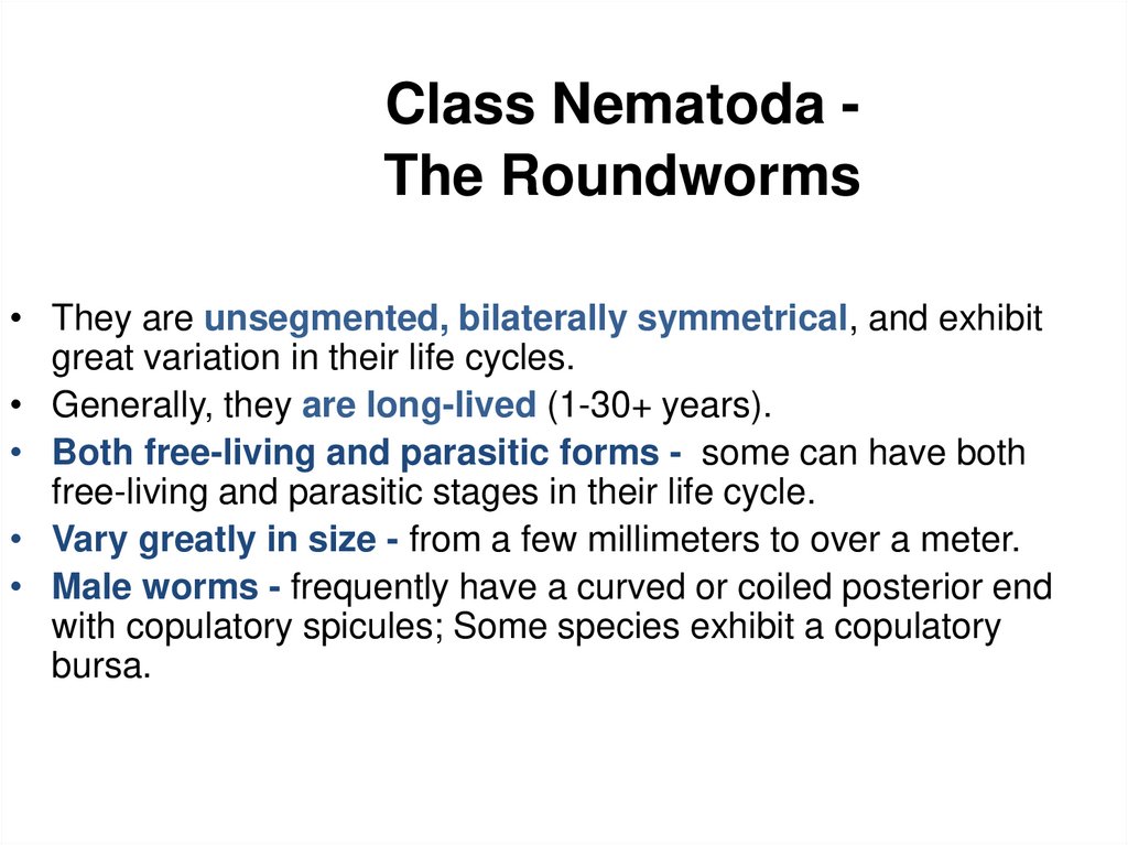
Class Nematoda The Roundworms• They are unsegmented, bilaterally symmetrical, and exhibit
great variation in their life cycles.
• Generally, they are long-lived (1-30+ years).
• Both free-living and parasitic forms - some can have both
free-living and parasitic stages in their life cycle.
• Vary greatly in size - from a few millimeters to over a meter.
• Male worms - frequently have a curved or coiled posterior end
with copulatory spicules; Some species exhibit a copulatory
bursa.
3.
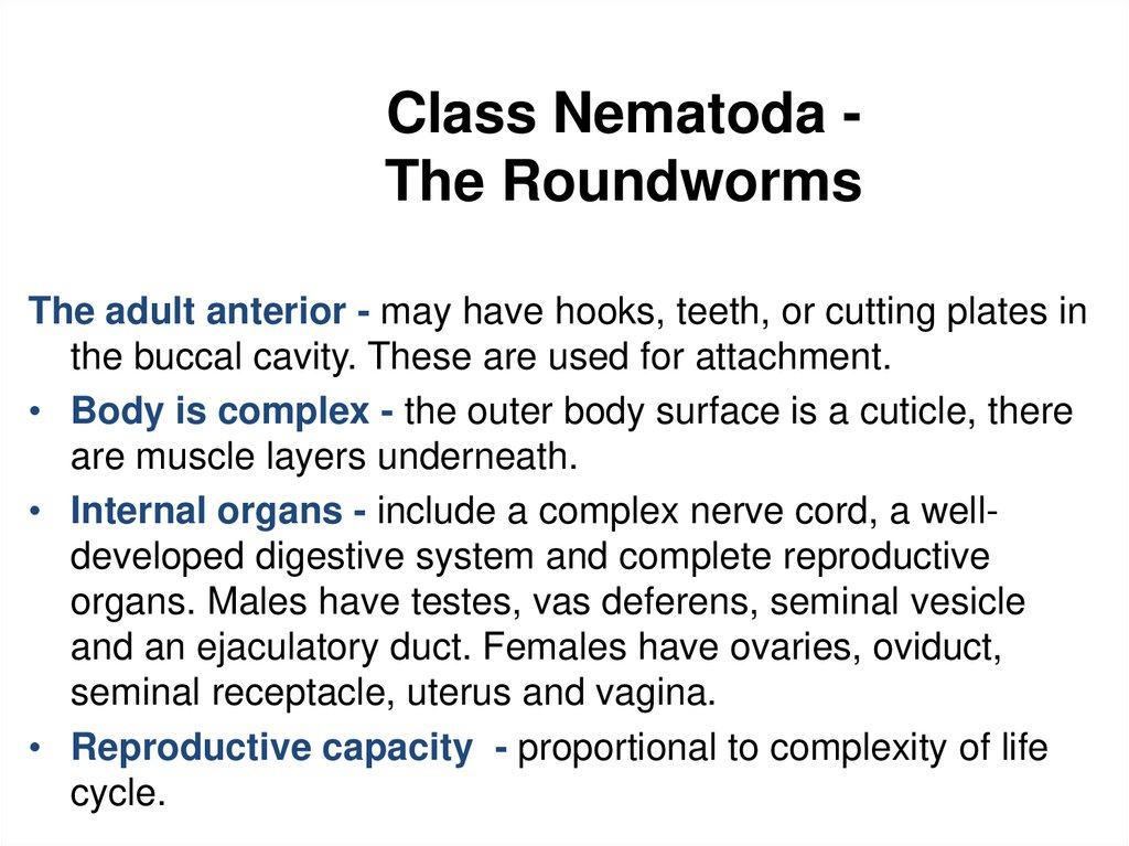
Class Nematoda The RoundwormsThe adult anterior - may have hooks, teeth, or cutting plates in
the buccal cavity. These are used for attachment.
• Body is complex - the outer body surface is a cuticle, there
are muscle layers underneath.
• Internal organs - include a complex nerve cord, a welldeveloped digestive system and complete reproductive
organs. Males have testes, vas deferens, seminal vesicle
and an ejaculatory duct. Females have ovaries, oviduct,
seminal receptacle, uterus and vagina.
• Reproductive capacity - proportional to complexity of life
cycle.
4.
Class Nematoda The Roundworms• Humans are definitive hosts.
• Arthropods may serve as intermediate hosts
and/or vectors. Many nematodes require no
intermediate host.
• The adult female produces fertilized eggs, or
larvae which may be infective to new host.
• Eggs may be immediately infective after ingestion
by humans.
5.
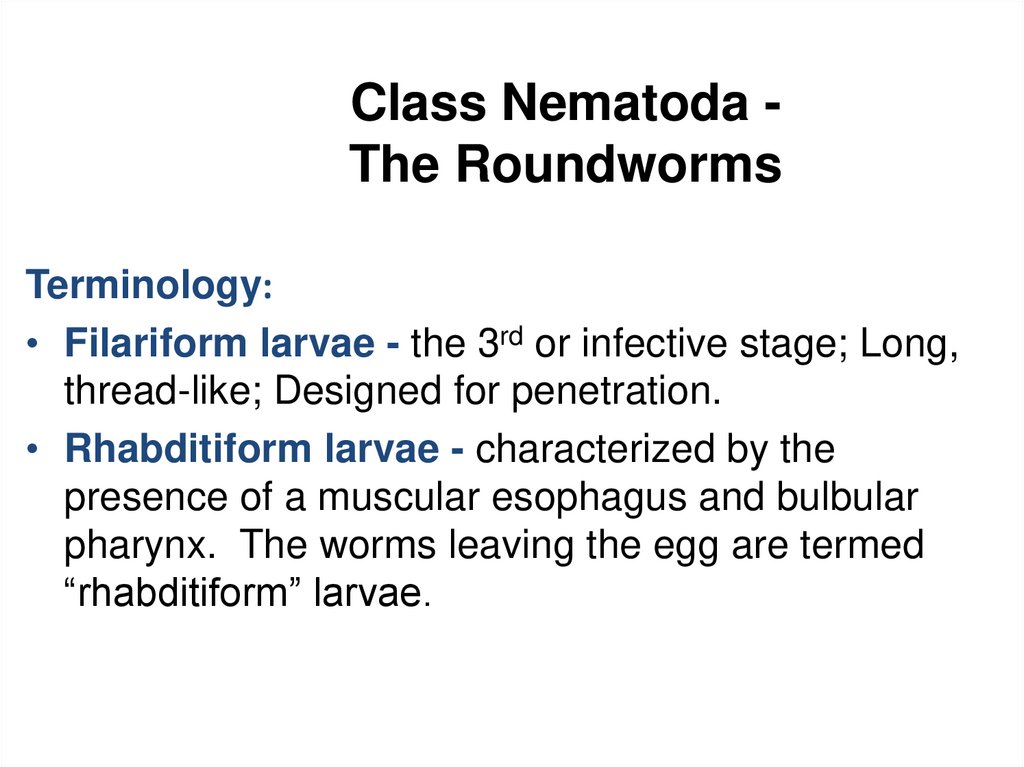
Class Nematoda The RoundwormsTerminology:
• Filariform larvae - the 3rd or infective stage; Long,
thread-like; Designed for penetration.
• Rhabditiform larvae - characterized by the
presence of a muscular esophagus and bulbular
pharynx. The worms leaving the egg are termed
“rhabditiform” larvae.
6.
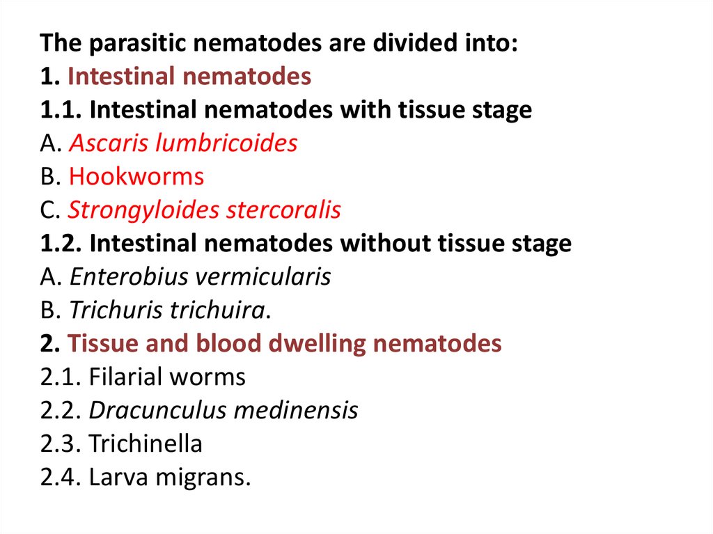
The parasitic nematodes are divided into:1. Intestinal nematodes
1.1. Intestinal nematodes with tissue stage
A. Ascaris lumbricoides
B. Hookworms
C. Strongyloides stercoralis
1.2. Intestinal nematodes without tissue stage
A. Enterobius vermicularis
B. Trichuris trichuira.
2. Tissue and blood dwelling nematodes
2.1. Filarial worms
2.2. Dracunculus medinensis
2.3. Trichinella
2.4. Larva migrans.
7.
INTESTINAL NEMATODES WITHTISSUE STAGE
8.
Class Nematoda The RoundwormsAscaris lumbricoides
Ascaris lumbricoides - Large Intestinal Roundworm
Life cycle: (complex, involves a heart-lung cycle)
• Humans ingest embryonated eggs containing infective larvae.
• Larvae hatch from the eggs in the small intestine, penetrate the
intestine wall, enter the bloodstream, migrate to the liver, travel to
the lung via the blood stream.
• Larvae break out of lung capillaries into alveoli, travel to the
bronchioles, and are coughed up to the pharynx. They are
swallowed and return to the intestine. Two molts to 4th stage
larvae take place in alveoli.
9.
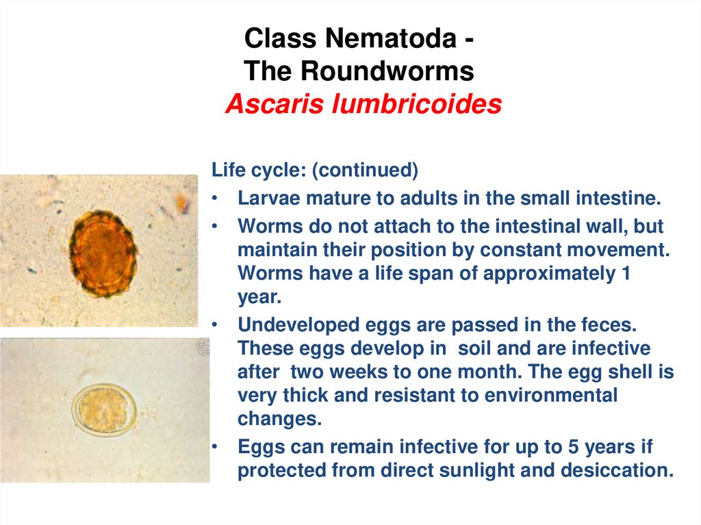
Class Nematoda The RoundwormsAscaris lumbricoides
Life cycle: (continued)
• Larvae mature to adults in the small intestine.
• Worms do not attach to the intestinal wall, but
maintain their position by constant movement.
Worms have a life span of approximately 1
year.
• Undeveloped eggs are passed in the feces.
These eggs develop in soil and are infective
after two weeks to one month. The egg shell is
very thick and resistant to environmental
changes.
• Eggs can remain infective for up to 5 years if
protected from direct sunlight and desiccation.
10.
11.
Class Nematoda The RoundwormsAscaris lumbricoides
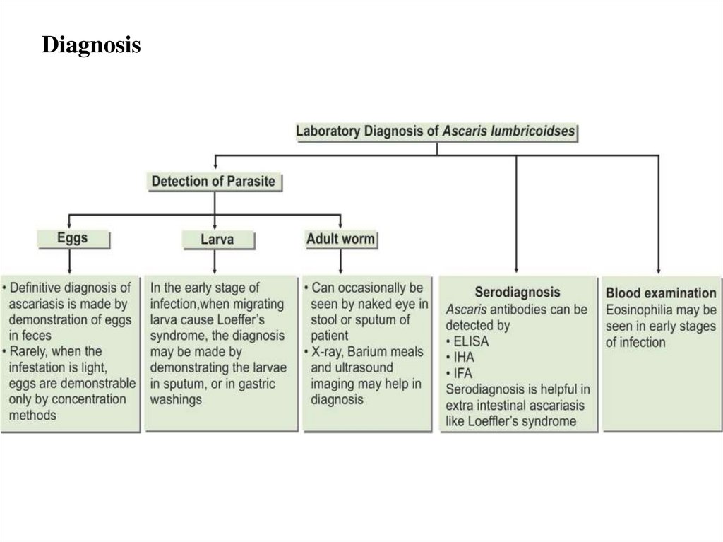
Diagnosis Identification of eggs and/or adults in fecal samples.
Major pathology and symptoms:
• Pneumonia associated with migration of larvae in the lungs.
• Obstruction of the intestines, appendix, or common bile duct.
• Vomiting and abdominal pain.
• May cause malnutrition in children with heavy infections or poor
diet.
• Some infections are asymptomatic.
12.
Diagnosis13.
Class Nematoda The RoundwormsAscaris lumbricoides
Treatment
•pyrantel pamoate 11 mg/kg once; maximum 1 g,
•albendazole 400 mg once,
•mebendazole 100 g twice daily for 3 days or 500 mg once,
•ivermectin 150–200 mg/kg once.
Prophylaxis
• Preventing fecal contamination of soil.
• Treatment of vegetables and other garden crops with water
containing iodine 200 ppm for 15 minutes kills the eggs and
larvae of Ascaris and other helminths.
• Avoid eating raw vegetables.
• Improvement of personal hygiene. Treatment of infected
persons.
14.
HOOK WORMS15.
HOOK WORMSThere are two species of hookworm:
1. Ancylostoma duodenale
2. Necator americanus
The adults are found in the small intestines of man.
Mixed infection is common. Both of the species are
found in Ethiopia, but N. americanus is more common.
16.
17.
18.
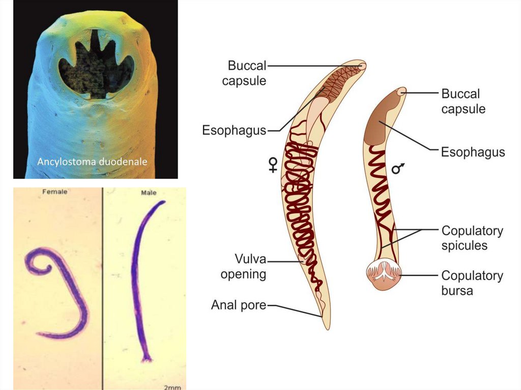
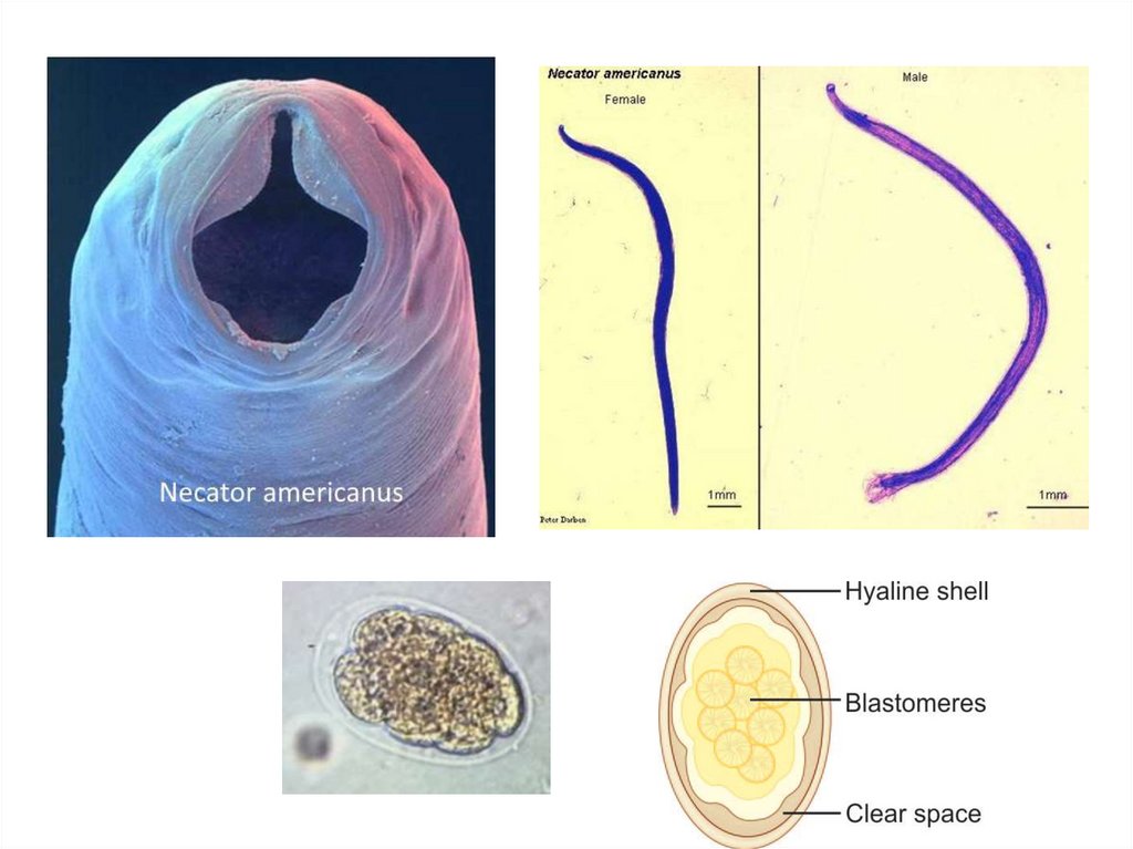
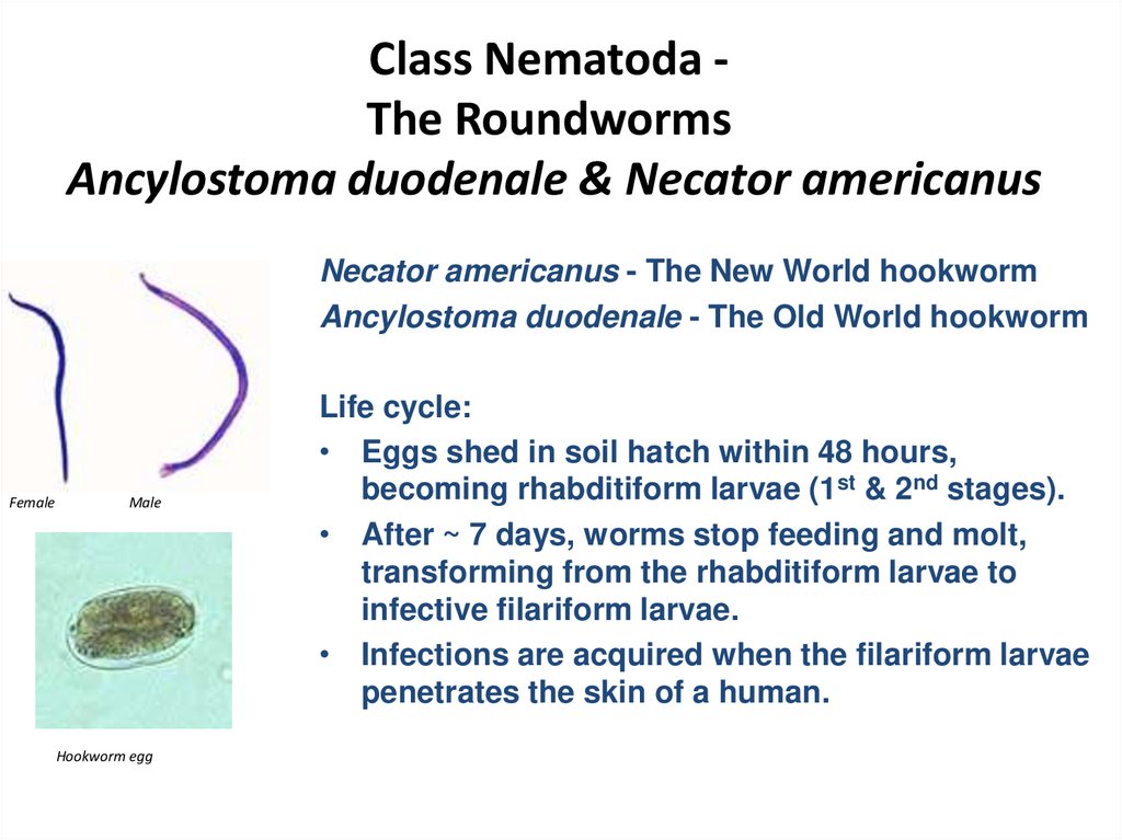
Class Nematoda The RoundwormsAncylostoma duodenale & Necator americanus
Necator americanus - The New World hookworm
Ancylostoma duodenale - The Old World hookworm
Female
Male
Hookworm egg
Life cycle:
• Eggs shed in soil hatch within 48 hours,
becoming rhabditiform larvae (1st & 2nd stages).
• After ~ 7 days, worms stop feeding and molt,
transforming from the rhabditiform larvae to
infective filariform larvae.
• Infections are acquired when the filariform larvae
penetrates the skin of a human.
19.
20.
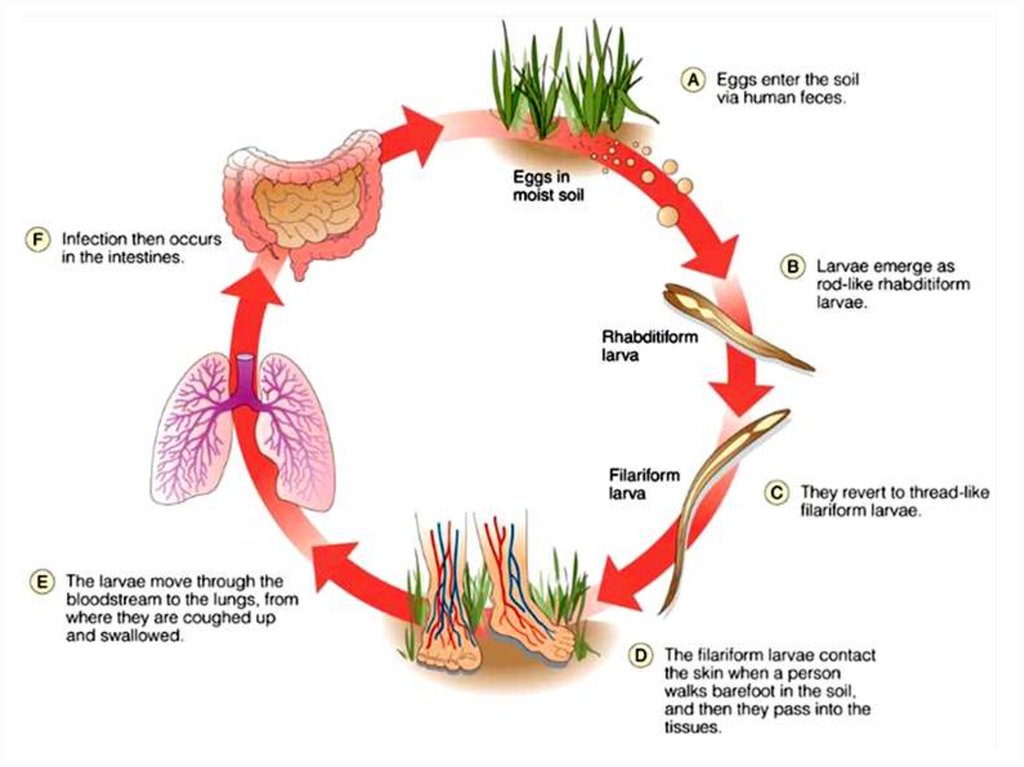
Class Nematoda The RoundwormsAncylostoma duodenale & Necator americanus
Hookworm rhabditiform larva
Hookworm filariform larva
Life cycle: (continued)
• Larvae enter the lymphatic system or
bloodstream, and travel to the lungs. After
maturating in the lungs, they migrate up the
trachea to be swallowed and reach the small
intestine, where they mature to adults.
• Immature adults attach to the intestinal mucosa
by means of their stout mouth parts and suck
blood and tissue juices of the host.
• About five weeks after infection, the worms have
undergone a final molt to become sexually
mature adults. Fertilization occurs, and the
females begin to release eggs. Worm life span is
about 1 year.
21.
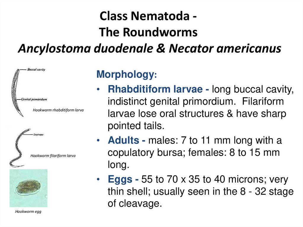
Class Nematoda The RoundwormsAncylostoma duodenale & Necator americanus
Hookworm rhabditiform larva
Hookworm filariform larva
Hookworm egg
Morphology:
• Rhabditiform larvae - long buccal cavity,
indistinct genital primordium. Filariform
larvae lose oral structures & have sharp
pointed tails.
• Adults - males: 7 to 11 mm long with a
copulatory bursa; females: 8 to 15 mm
long.
• Eggs - 55 to 70 x 35 to 40 microns; very
thin shell; usually seen in the 8 - 32 stage
of cleavage.
22.
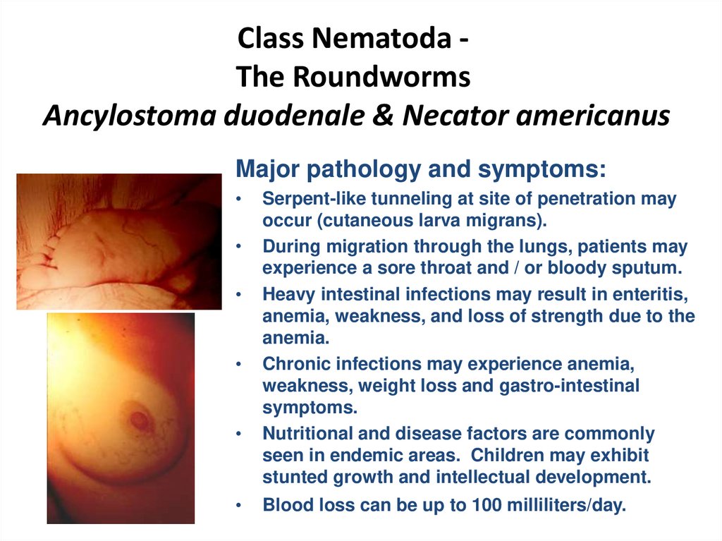
Class Nematoda The RoundwormsAncylostoma duodenale & Necator americanus
Major pathology and symptoms:
Serpent-like tunneling at site of penetration may
occur (cutaneous larva migrans).
During migration through the lungs, patients may
experience a sore throat and / or bloody sputum.
Heavy intestinal infections may result in enteritis,
anemia, weakness, and loss of strength due to the
anemia.
Chronic infections may experience anemia,
weakness, weight loss and gastro-intestinal
symptoms.
Nutritional and disease factors are commonly
seen in endemic areas. Children may exhibit
stunted growth and intellectual development.
Blood loss can be up to 100 milliliters/day.
23.
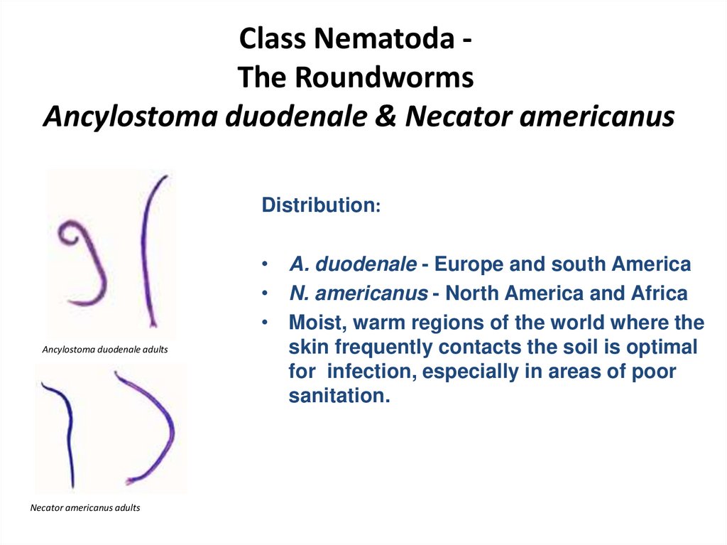
Class Nematoda The RoundwormsAncylostoma duodenale & Necator americanus
Distribution:
Ancylostoma duodenale adults
Necator americanus adults
• A. duodenale - Europe and south America
• N. americanus - North America and Africa
• Moist, warm regions of the world where the
skin frequently contacts the soil is optimal
for infection, especially in areas of poor
sanitation.
24.
Class Nematoda The RoundwormsAncylostoma duodenale & Necator americanus
Symptoms
Adult worms in the intestine feed on blood causing iron
deficiency anemia. The larvae may cause inflammation of the
lungs.
Diagnosis
Examination of stool by direct saline smear to detect the eggs.
Treatment
Mebendazole: 1 tab 2x daily for 3 days.
25.
Strongyloides26.
Strongyloides stercoralisThe worms may be present as parasitic in the host or free living in the soil.
Morphology
Male: The male measures1 mm in length with curved posterior end and carries
two spicules
Female: The female measures 2.5 mm in length with straight posterior end.
Infection: follows skin penetration by filariform larvae
27.
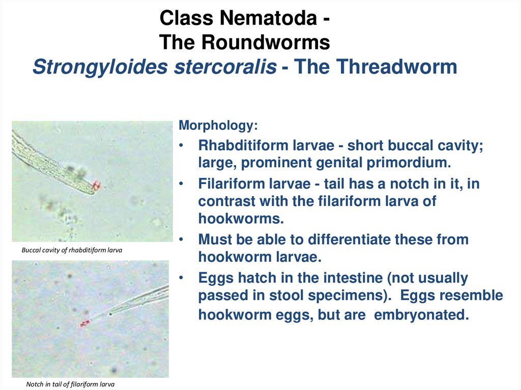
Class Nematoda The RoundwormsStrongyloides stercoralis - The Threadworm
Morphology:
Buccal cavity of rhabditiform larva
Notch in tail of filariform larva
• Rhabditiform larvae - short buccal cavity;
large, prominent genital primordium.
• Filariform larvae - tail has a notch in it, in
contrast with the filariform larva of
hookworms.
• Must be able to differentiate these from
hookworm larvae.
• Eggs hatch in the intestine (not usually
passed in stool specimens). Eggs resemble
hookworm eggs, but are embryonated.
28.
Class Nematoda The RoundwormsStrongyloides stercoralis - The Threadworm
Strongyloides stercoralis rhabditiform larva
Strongyloides stercoralis filariform larva
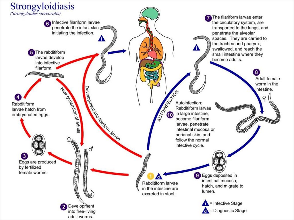
Life cycle: (very complex)
• Infective third stage filariform larvae
penetrate skin, enter the lymphatics or
bloodstream.
• Larvae migrate to the lungs, break out of
lung capillaries into alveoli.
• After maturation, larvae travel to the
pharynx, are swallowed, and return to the
intestine.
• Larvae mature to adults and attach to the
mucosa of the small intestine.
29.
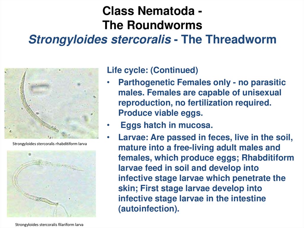
Class Nematoda The RoundwormsStrongyloides stercoralis - The Threadworm
Strongyloides stercoralis rhabditiform larva
Strongyloides stercoralis filariform larva
Life cycle: (Continued)
• Parthogenetic Females only - no parasitic
males. Females are capable of unisexual
reproduction, no fertilization required.
Produce viable eggs.
• Eggs hatch in mucosa.
• Larvae: Are passed in feces, live in the soil,
mature into a free-living adult males and
females, which produce eggs; Rhabditiform
larvae feed in soil and develop into
infective stage larvae which penetrate the
skin; First stage larvae develop into
infective stage larvae in the intestine
(autoinfection).
30.
31.
Class Nematoda The RoundwormsStrongyloides stercoralis - The Threadworm
Symptoms
•Skin – allergic reactions; raised, itchy, red blotches at the site of larval penetration.
•Lungs – pneumonia.
•Intestinal - abdominal pain, diarrhea, vomiting, weight loss, anemia, eosinophilia.
Light infections usually asymptomatic; Heavy infection - bowel becomes
edematous and congested.
•Death occurs in immunosuppressed patients due to heavy autoinfection.
Disseminated strongyloidiasis. Multiplicity of symptoms are present due to
the injury of other organs by the migrating larvae. Organs such as liver, heart
adrenals, pancreas, kidneys, and CNS, etc. may be affected. This is usually seen in
immunocompromized individuals.
32.
Class Nematoda The RoundwormsStrongyloides stercoralis - The Threadworm
Diagnosis:
• Recovery and identification of larvae in the feces.
Recovery and identification of eggs in duodenal drainage.
Treatment
Thiabendazole: 25 mg/kg twice daily for 3 days.
33.
Class Nematoda The RoundwormsStrongyloides stercoralis - The Threadworm
• Distribution - worldwide, similar to hookworm.
• While hookworm infection dies out over a period of years after
the patient has moved from an endemic area, strongyloidiasis
may persist for years, due to autoinfection (internal infection).
• In cases with severe diarrhea, Strongyloides eggs may be present
in stool specimens. These must be differentiated from hookworm
eggs. Strongyloides eggs contain well-developed larvae.
Hookworm eggs do not have well developed larvae until passed
from the body and mature for one to two weeks in the soil.
34.
Intestinal nematodes withouttissue stage
35.
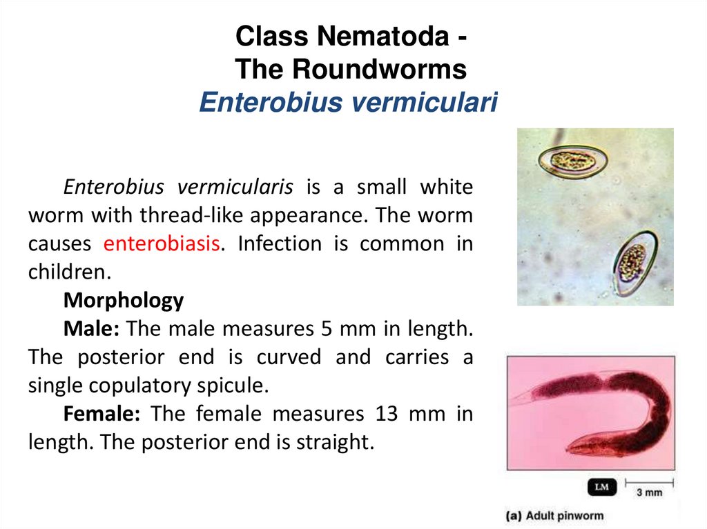
Class Nematoda The RoundwormsEnterobius vermicularis
Enterobius vermicularis is a small white
worm with thread-like appearance. The worm
causes enterobiasis. Infection is common in
children.
Morphology
Male: The male measures 5 mm in length.
The posterior end is curved and carries a
single copulatory spicule.
Female: The female measures 13 mm in
length. The posterior end is straight.
36.
Class Nematoda The RoundwormsEnterobius vermicularis
• Transmission is direct, person-to-person.
• Egg is infective immediately or within hours of being shed by the
female.
• Common worldwide but more prevalent in temperate climates.
• Higher prevalence in Caucasians than in Negroes.
• It is a group infection especially common among children. Very often
associated with low sanitation and hygiene.
• Humans are the only known host. Dogs and cats are not infected.
37.
Class Nematoda The RoundwormsEnterobius vermicularis
Life cycle:
• Eggs are ingested, hatch in
intestine, larvae mature to
adults.
• Gravid females migrate to the
perianal area at night to lay
eggs.
• Eggs develop to infective
stage within 4-6 hours. Eggs
can survive for extended
periods in cool, moist
environment.
• Eggs are found rarely in fecal
samples; Release is most
often external to the intestines.
Dying worms may release
eggs in the bowel.
38.
39.
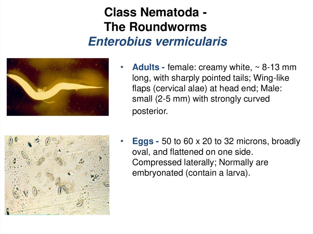
Class Nematoda The RoundwormsEnterobius vermicularis
• Adults - female: creamy white, ~ 8-13 mm
long, with sharply pointed tails; Wing-like
flaps (cervical alae) at head end; Male:
small (2-5 mm) with strongly curved
posterior.
• Eggs - 50 to 60 x 20 to 32 microns, broadly
oval, and flattened on one side.
Compressed laterally; Normally are
embryonated (contain a larva).
40.
Class Nematoda The RoundwormsEnterobius vermicularis
Major pathology and symptoms:
• One third of all cases are asymptomatic.
• Infections rarely cause serious lesions.
• Symptoms associated with the migration of the female out of the anus
to lay her eggs include: perianal itching, nausea or vomiting, loss of
sleep, irritability, irritation of the intestinal mucosa, and vulval irritation
in females due to migrating worms entering the vagina instead of the
re-entering anus.
41.
Class Nematoda The RoundwormsEnterobius vermicularis
Diagnosis:
• Recovery and identification of eggs or adults from the perianal
region utilizing the cellophane tape preparation.
• Specimens must be collected the first thing in the morning upon
waking, especially before bathing or bowel movements.
• Eggs are rarely found in fecal samples because release is usually
external to the intestines.
42.
Class Nematoda The RoundwormsEnterobius vermicularis
Treatment
Pyrantel pamoate 11 mg/kg once, maximum 1 g,
Albendazole 400 mg once
Mebendazole 100 mg once
Prophylaxis
Maintainance of personal and community hygiene such as
frequent hand washing, _ nger nail cleaning, and regular bathing.
Frequent washing of night clothes and bed linen.
43.
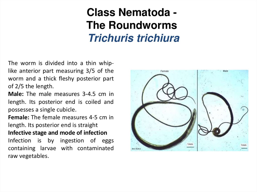
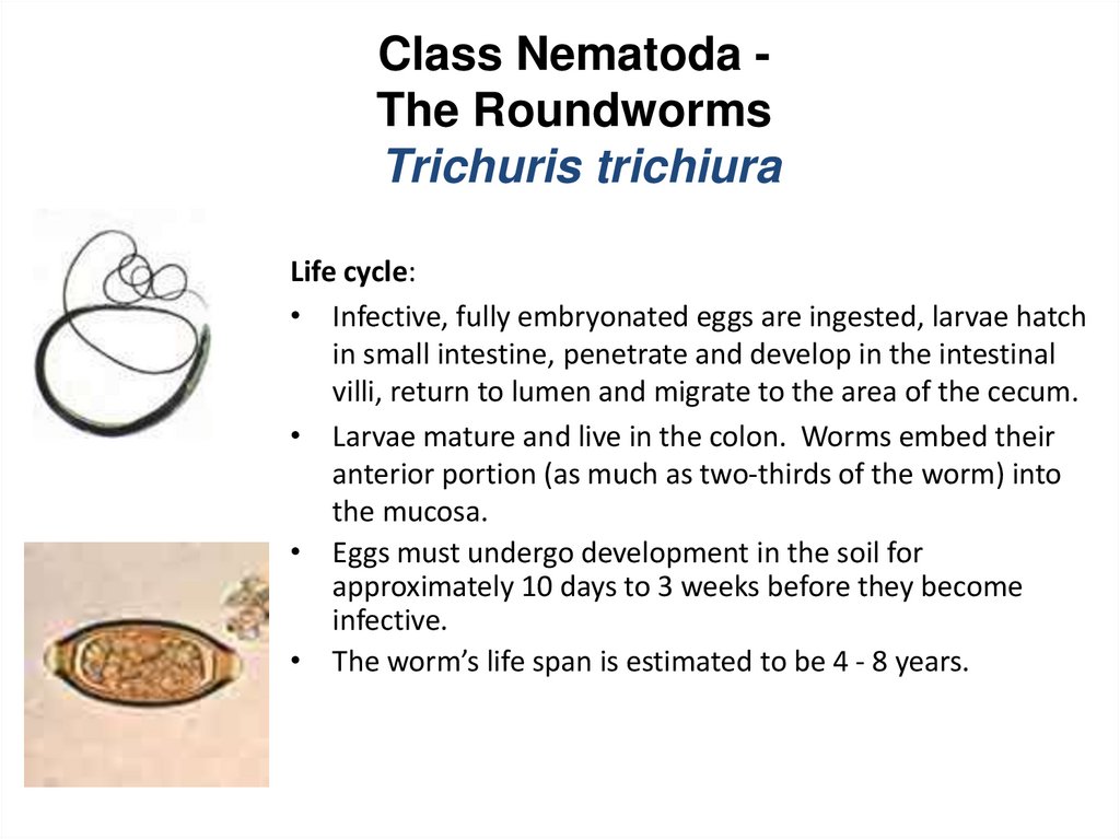
Class Nematoda The RoundwormsTrichuris trichiura
The worm is divided into a thin whiplike anterior part measuring 3/5 of the
worm and a thick fleshy posterior part
of 2/5 the length.
Male: The male measures 3-4.5 cm in
length. Its posterior end is coiled and
possesses a single cubicle.
Female: The female measures 4-5 cm in
length. Its posterior end is straight
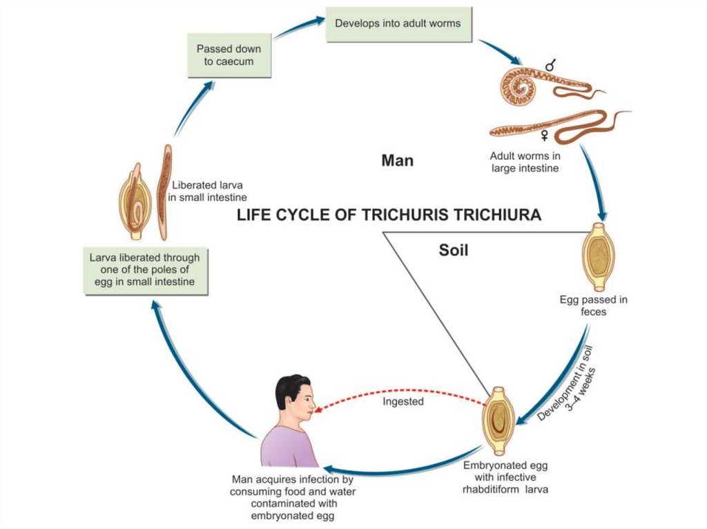
Infective stage and mode of infection
Infection is by ingestion of eggs
containing larvae with contaminated
raw vegetables.
44.
Class Nematoda The RoundwormsTrichuris trichiura
Morphology:
• Adults - females: 35 to 50 mm long, anterior two-thirds is long
and threadlike, expanding into a broader posterior; males: 30
to 45 mm long, similar to female but exhibiting a strong
curvature of tail.
• Eggs - 50 to 55 x 22 to 25 microns, barrel-shaped, with clear
polar plug at each end.
45.
Class Nematoda The RoundwormsTrichuris trichiura
Life cycle:
• Infective, fully embryonated eggs are ingested, larvae hatch
in small intestine, penetrate and develop in the intestinal
villi, return to lumen and migrate to the area of the cecum.
• Larvae mature and live in the colon. Worms embed their
anterior portion (as much as two-thirds of the worm) into
the mucosa.
• Eggs must undergo development in the soil for
approximately 10 days to 3 weeks before they become
infective.
• The worm’s life span is estimated to be 4 - 8 years.
46.
47.
Class Nematoda The RoundwormsTrichuris trichiura
• Diagnosis - recovery and identification of eggs in the
feces.
Major pathology and symptoms:
• Slight infections - usually asymptomatic.
• Heavy infections - surface of colon is matted with worms
which causes bloody or mucous diarrhea.
• weight loss and weakness - infections with 200 or more
worms in children may cause a chronic dysentery,
profound anemia and growth retardation.
48.
SymptomsThe patient complains of dysentery (blood and mucus in stool together
with tenesmus). Rectal prolapse is also possible.
Treatment
Mebendazole: 1 tablet twice daily for 2 days.
Egg of Trichuris trichiura