


 Медицина
Похожие презентации:


")


")


Medical Helmintology
1.
MEDICAL HELMINTOLOGYPhylum Platyhelminthes
Trematodes (Flukes)
2.
Medical helminthology is concerned with the study of helminthes or parasitic worms.Helminthes are trophoblastic metazoa (multi-cellular organisms).
Helminthes are among the common parasitic causes of human suffering. They are the
cause of high morbidity and mortality of people worldwide. They cause different
diseases in humans, but few helminthic infections cause life- threatening diseases. They
cause anemia and malnutrition. In children they cause a reduction in academic
performance. Helminthes also cause economic loss as a result of infections of domestic
animals.
There is age dependent distribution of infections from geohelminthes and
schistosomes. As a result of predisposing behavioral and immunological status, children
disproportionately carry the burden of schistosomes and geo-helminthes.
3.
Transmission of helmintes:The sources of the parasites are different. Exposure of humans to the parasites may
occur in one of the following ways:
1. Contaminated soil (Geo-helminthes), water (cercariae of blood flukes) and food (Taenia
in raw meat).
2. Blood sucking insects or arthropods (as in filarial worms).
3. Domestic or wild animals harboring the parasite (as in echinococcus in dogs).
4. Person to person (as in Enterobius vermicularis, Hymenolopis nana).
5. Oneself (auto-infection) as in Enterobius vermicularis.
They enter the body through different routes including: mouth, skin and the respiratory
tract by means of inhalation of airborne eggs. The Trematodes and Cestodes are groups of
flat worms.
4.
MEDICALLY IMPORTANTTREMATODES (FLUKES)
1. BLOOD FLUKES
5.
Schistosomiasis. Shistosoma spp.It is estimated that about 600 million people in 79 countries suffer from schistosomiasis. The schistosomes
cause intestinal, hepatosplenic, pulmonary, urogenital, cerebral and other forms of schistosomiasis. Schistosome
is the only fluke with separate sexes. The female worm lies in the gynecophoral canal of the male. This condition
is important for transportation.
There are five medically important species:
1. Schistosoma mansoni: causes intestinal schistosomiasis.
2. Schistosoma haematobium: causes vesical (urinary) schistosomiasis.
3. Schistosoma japonicum: causes intestinal schistosomiasis.
4. Schistosoma intercalatum: causes intestinal schistosomiasis.
5. Schistosoma mekongi: causes intestinal schistosomiasis. This seems to cause milder disease in man. It causes
disease in other vertebrate hosts. The first two schistosomes (S. mansoni and S. haematobium) are prevalent in
Ethiopia.
6.
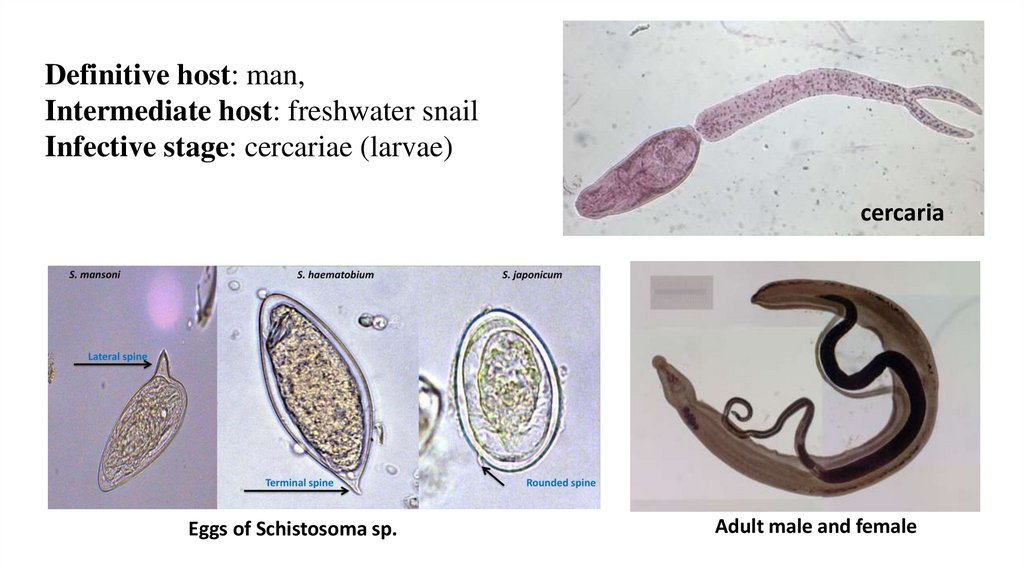
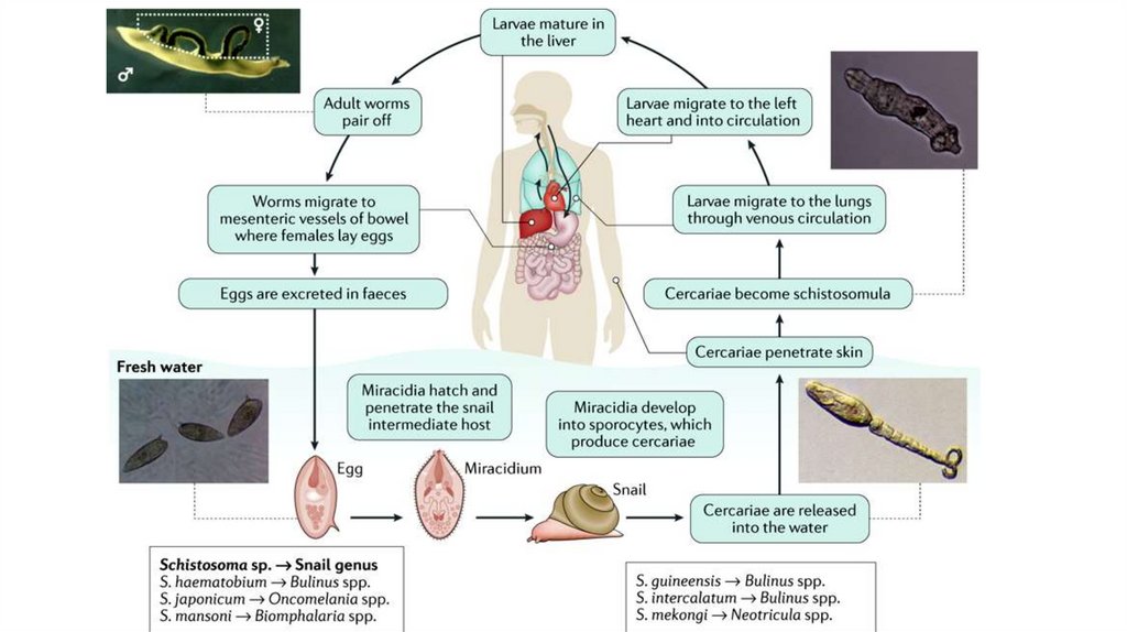
Definitive host: man,Intermediate host: freshwater snail
Infective stage: cercariae (larvae)
cercaria
Eggs of Schistosoma sp.
Adult male and female
7.
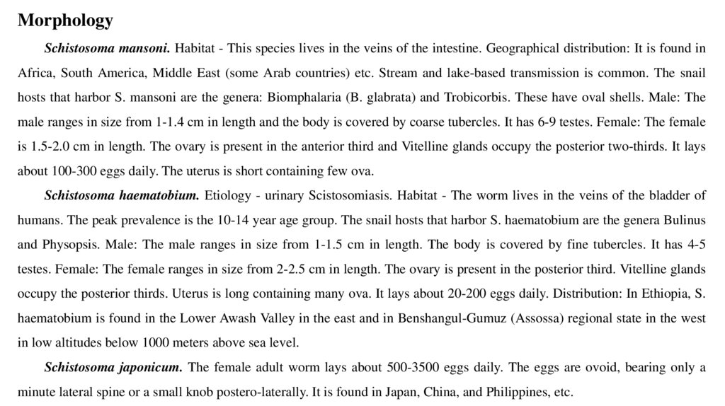
MorphologySchistosoma mansoni. Habitat - This species lives in the veins of the intestine. Geographical distribution: It is found in
Africa, South America, Middle East (some Arab countries) etc. Stream and lake-based transmission is common. The snail
hosts that harbor S. mansoni are the genera: Biomphalaria (B. glabrata) and Trobicorbis. These have oval shells. Male: The
male ranges in size from 1-1.4 cm in length and the body is covered by coarse tubercles. It has 6-9 testes. Female: The female
is 1.5-2.0 cm in length. The ovary is present in the anterior third and Vitelline glands occupy the posterior two-thirds. It lays
about 100-300 eggs daily. The uterus is short containing few ova.
Schistosoma haematobium. Etiology - urinary Scistosomiasis. Habitat - The worm lives in the veins of the bladder of
humans. The peak prevalence is the 10-14 year age group. The snail hosts that harbor S. haematobium are the genera Bulinus
and Physopsis. Male: The male ranges in size from 1-1.5 cm in length. The body is covered by fine tubercles. It has 4-5
testes. Female: The female ranges in size from 2-2.5 cm in length. The ovary is present in the posterior third. Vitelline glands
occupy the posterior thirds. Uterus is long containing many ova. It lays about 20-200 eggs daily. Distribution: In Ethiopia, S.
haematobium is found in the Lower Awash Valley in the east and in Benshangul-Gumuz (Assossa) regional state in the west
in low altitudes below 1000 meters above sea level.
Schistosoma japonicum. The female adult worm lays about 500-3500 eggs daily. The eggs are ovoid, bearing only a
minute lateral spine or a small knob postero-laterally. It is found in Japan, China, and Philippines, etc.
8.
9.
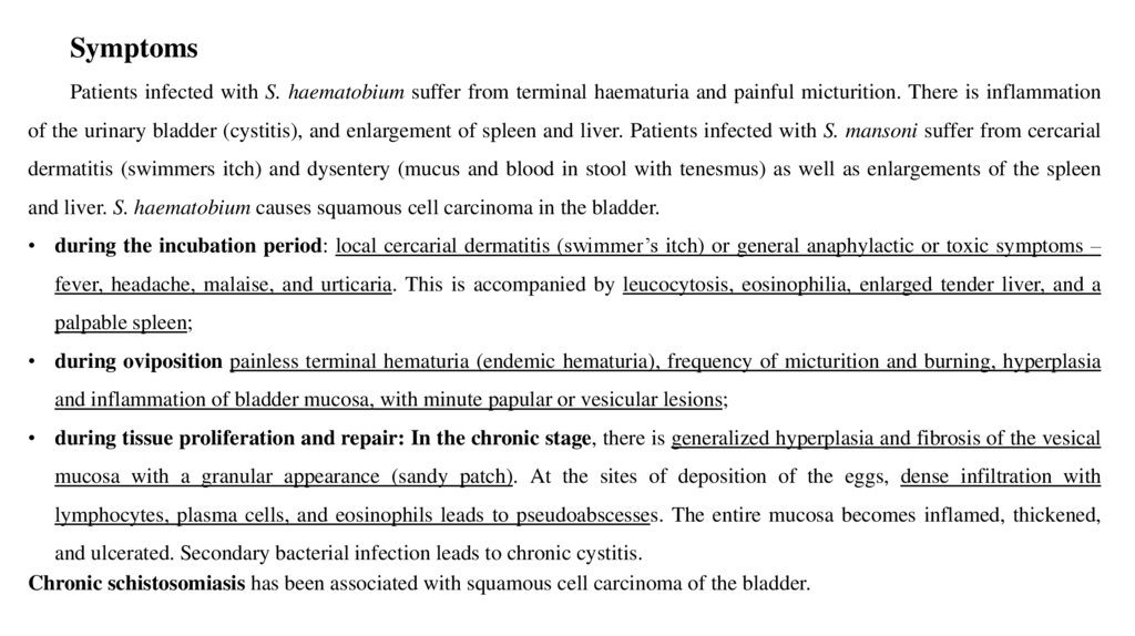
SymptomsPatients infected with S. haematobium suffer from terminal haematuria and painful micturition. There is inflammation
of the urinary bladder (cystitis), and enlargement of spleen and liver. Patients infected with S. mansoni suffer from cercarial
dermatitis (swimmers itch) and dysentery (mucus and blood in stool with tenesmus) as well as enlargements of the spleen
and liver. S. haematobium causes squamous cell carcinoma in the bladder.
• during the incubation period: local cercarial dermatitis (swimmer’s itch) or general anaphylactic or toxic symptoms –
fever, headache, malaise, and urticaria. This is accompanied by leucocytosis, eosinophilia, enlarged tender liver, and a
palpable spleen;
• during oviposition painless terminal hematuria (endemic hematuria), frequency of micturition and burning, hyperplasia
and inflammation of bladder mucosa, with minute papular or vesicular lesions;
• during tissue proliferation and repair: In the chronic stage, there is generalized hyperplasia and fibrosis of the vesical
mucosa with a granular appearance (sandy patch). At the sites of deposition of the eggs, dense infiltration with
lymphocytes, plasma cells, and eosinophils leads to pseudoabscesses. The entire mucosa becomes inflamed, thickened,
and ulcerated. Secondary bacterial infection leads to chronic cystitis.
Chronic schistosomiasis has been associated with squamous cell carcinoma of the bladder.
10.
DiagnosisS. mansoni:
♦ Microscopic examination of the stool for eggs after concentration by sedimentation method. The egg has
characteristic lateral spine.
♦ Rectal snip
S. haematobium:
♦ Urine Microscopy. Examination of the urine after allowing it to sediment in a conical urinalysis glass. A drop
from the sediment is taken and examined for eggs. Egg has terminal spine.
♦ Biopsy from bladder
♦ Detection of specific schistosome antigens in serum or urine.
♦ Serological tests
♦ Intradermal skin tests
♦ Imaging (X-ray, cytoscopy, Ultrasonography (USG), Intravenous pyelogram (IVP))
11.
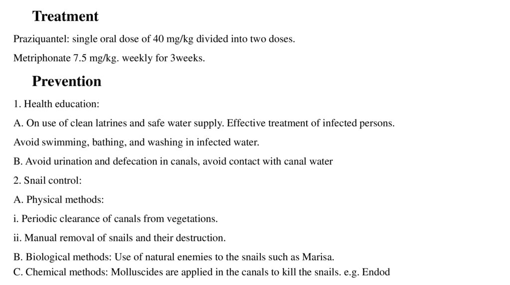
TreatmentPraziquantel: single oral dose of 40 mg/kg divided into two doses.
Metriphonate 7.5 mg/kg. weekly for 3weeks.
Prevention
1. Health education:
A. On use of clean latrines and safe water supply. Effective treatment of infected persons.
Avoid swimming, bathing, and washing in infected water.
B. Avoid urination and defecation in canals, avoid contact with canal water
2. Snail control:
A. Physical methods:
i. Periodic clearance of canals from vegetations.
ii. Manual removal of snails and their destruction.
B. Biological methods: Use of natural enemies to the snails such as Marisa.
C. Chemical methods: Molluscides are applied in the canals to kill the snails. e.g. Endod
12.
2. LIVER FLUKES13.
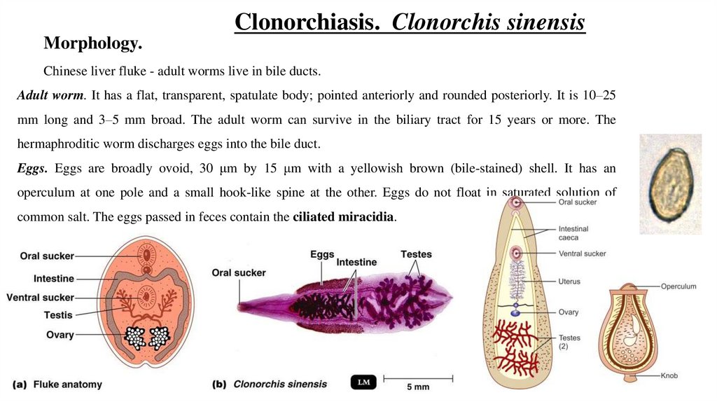
Clonorchiasis. Clonorchis sinensisMorphology.
Chinese liver fluke - adult worms live in bile ducts.
Adult worm. It has a flat, transparent, spatulate body; pointed anteriorly and rounded posteriorly. It is 10–25
mm long and 3–5 mm broad. The adult worm can survive in the biliary tract for 15 years or more. The
hermaphroditic worm discharges eggs into the bile duct.
Eggs. Eggs are broadly ovoid, 30 μm by 15 μm with a yellowish brown (bile-stained) shell. It has an
operculum at one pole and a small hook-like spine at the other. Eggs do not float in saturated solution of
common salt. The eggs passed in feces contain the ciliated miracidia.
14.
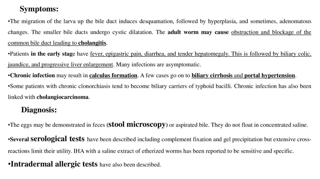
Symptoms:•The migration of the larva up the bile duct induces desquamation, followed by hyperplasia, and sometimes, adenomatous
changes. The smaller bile ducts undergo cystic dilatation. The adult worm may cause obstruction and blockage of the
common bile duct leading to cholangitis.
•Patients in the early stage have fever, epigastric pain, diarrhea, and tender hepatomegaly. This is followed by biliary colic,
jaundice, and progressive liver enlargement. Many infections are asymptomatic.
•Chronic infection may result in calculus formation. A few cases go on to biliary cirrhosis and portal hypertension.
•Some patients with chronic clonorchiasis tend to become biliary carriers of typhoid bacilli. Chronic infection has also been
linked with cholangiocarcinoma.
Diagnosis:
•The eggs may be demonstrated in feces (stool
•Several serological
tests
microscopy) or aspirated bile. They do not float in concentrated saline.
have been described including complement fixation and gel precipitation but extensive cross-
reactions limit their utility. IHA with a saline extract of etherized worms has been reported to be sensitive and specific.
•Intradermal allergic tests have also been described.
15.
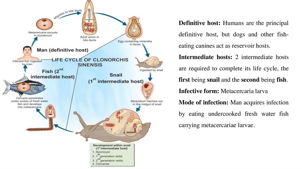
Definitive host: Humans are the principaldefinitive host, but dogs and other fisheating canines act as reservoir hosts.
Intermediate hosts: 2 intermediate hosts
are required to complete its life cycle, the
first being snail and the second being fish.
Infective form: Metacercaria larva
Mode of infection: Man acquires infection
by eating undercooked fresh water fish
carrying metacercariae larvae.
16.
Treatment:• Drug of choice is Praziquantel 25 mg/kg, 3 doses in 1 day.
• Surgical intervention may become necessary in cases with obstructive jaundice.
Prophylaxis:
•Proper cooking of fish
•Proper disposal of feces
•Control of snails.
17.
Opisthorchiasis. Opisthorchis felineusOpisthorchis felineus is found mainly in Italy, Germany, Belarus, Russia, Kazakhstan, and Ukraine.
Adults of Opisthorchis spp. are similar to, but often smaller than, Clonorchis sinensis. Adults
measure approximately 7 mm long by 1.5 mm wide in the human host (adults are slightly
smaller in feline hosts). Adults of Opisthorchis spp. differ from adults of Clonorchis in the
shape of the testes. The distribution of the vitelline glands is also different. Both genera are
similar, however, in having a ventral sucker (acetabulum) smaller than the oral sucker. Adults
reside in the bile ducts of the definitive host.
Definitive host: Humans are the principal definitive host, but dogs and other
fish-eating canines act as reservoir hosts.
Intermediate hosts: 2 intermediate hosts are required to complete its life
cycle, the first being snail and the second being fish.
Infective form: Metacercaria larva
Mode of infection: Man acquires infection by eating undercooked fresh water
fish carrying metacercariae larvae.
18.
19.
Diagnosis:•Microscopic identification of eggs in stool specimens.
•The adult fluke can also be recovered at surgery.
•Serologic testing
Symptoms:
Most infections are asymptomatic. Most pathologic manifestations result from
inflammation and intermittent obstruction of the biliary ducts. In mild cases, manifestations
include dyspepsia, abdominal pain, diarrhea, or constipation. With infections of longer
duration, the symptoms can be more severe, and hepatomegaly and malnutrition may be
present. In rare cases, cholangitis, cholecystitis, and chlolangiocarcinoma may develop. In
addition, fever, facial edema, lymphadenopathy, arthralgias, rash, and eosinophilia.
20.
Diagnosis:• stool examinations
• Imaging (ultrasound, CT, MRI).
• Serologic testing
Treatment:
• Praziquantel, adults, 75mg/kg/day orally, three doses per day for 2 days; the pediatric
dosage is the same. Praziquantel should be taken with liquids during meals.
• Albendazole, the dosage is 10mg/kg/day for 7 days. The pediatric dosage is the same.
Albendazole should be taken with food; a fatty meal increases the bioavailability.
Prophylaxis:
•Proper cooking of fish
•Proper disposal of feces
•Control of snails.
21.
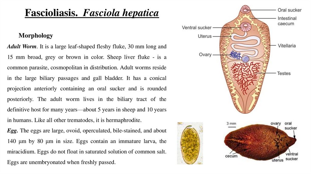
Fascioliasis. Fasciola hepaticaMorphology
Adult Worm. It is a large leaf-shaped fleshy fluke, 30 mm long and
15 mm broad, grey or brown in color. Sheep liver fluke - is a
common parasite, cosmopolitan in distribution. Adult worms reside
in the large biliary passages and gall bladder. It has a conical
projection anteriorly containing an oral sucker and is rounded
posteriorly. The adult worm lives in the biliary tract of the
definitive host for many years—about 5 years in sheep and 10 years
in humans. Like all other trematodes, it is hermaphrodite.
Egg. The eggs are large, ovoid, operculated, bile-stained, and about
140 μm by 80 μm in size. Eggs contain an immature larva, the
miracidium. Eggs do not float in saturated solution of common salt.
Eggs are unembryonated when freshly passed.
22.
Symptoms:•In traversing the liver tissue, it causes parenchymal injury.
•As humans are not its primary host, it causes more severe inflammatory response. Some larvae penetrate
right through the liver and diaphragm ending up in the lung.
•In acute phase during the migration of the larva, patients present with fever, right upper quadrant pain,
eosinophilia, and tender hepatomegaly. The symptoms subside as parasites reach their final destination.
•In chronic phase, patients may develop biliary obstruction, biliary cirrhosis, obstructive jaundice,
cholelithiasis, and anemia. No association to hepatic malignancy has been ascribed to fascioliasis.
Occasionally, ingestion of raw liver of infected sheep results in a condition called halzoun (meaning
suffocation).
•The adult worms in the liver attach to the pharyngeal mucosa, causing edematous congestion of the
pharynx and surrounding areas, leading to dyspnea, acute dysphagia, deafness, and rarely, asphyxiation.
23.
Diagnosis:•Stool Microscopy
•Blood Picture
•Serodiagnosis
•Imaging (USG, CT scan, Endoscopic Retrograde Choangiopancreatography (ERCP) and
percutaneous cholangiography.
Treatment:
•triclabendazole (10 mg/kg once)
•bithionol (30–50 mg for 10–15 days)
•Prednisolone at a dose of 10–20 mg/kg is used to control toxemia.
Prophylaxis:
•Health education
•Preventing pollution of water courses with sheep, cattle, and human feces
•Proper disinfection of watercresses and other water vegetations before consumption.
24.
Life CycleF. hepatica passes its life cycle in 1
definitive host and 2 intermediate hosts.
Definitive host: Sheep, goat, cattle, and
man.
Intermediate host: Snails of the genus
Lymnaea
and
Succinea.
Encystment
occurs on aquatic plants, which act as
second intermediate host.
Mode of infection: The definitive host,
sheep and man, get infection by ingestion
of metacerceriae encysted on aquatic
vegetation.
25.
Trematodes (Flukes)Name of disease
Latin name of
parasite
Forms of parasites
Definitive host
Intermediate host
Infective stage
Transmission
Symptoms
Diagnosis
Treatment
Prevention
Schstosomiasis Clonorchiasis
Opisthorchiasis Fascioliasis