 Медицина
Похожие презентации:


")

. Class Nematoda. Lesson 5")


Medical helmintology. Cestodes
1.
Medical helmintology.Cestodes
2.
CESTODES (TAPEWORMS)The tapeworms are hermaphroditic and require an
intermediate host. The adult tapeworms found in humans
have flat body, white or grayish in color.
They consist of an anterior attachment organ or scolex
and a chain of segments (proglottids) also called strobilla.
The strobilla is the entire body except the scolex. The
scolex has suckers or grooves. It has rosetellum, which
has 1 or 2 rows of hooks situated on the center of the
scolex.
Adult tapeworms inhabit the small intestine, where
they live attached to the mucosa. Tapeworms do not have
a digestive system. Their food is absorbed from the host’s
intestine.
3.
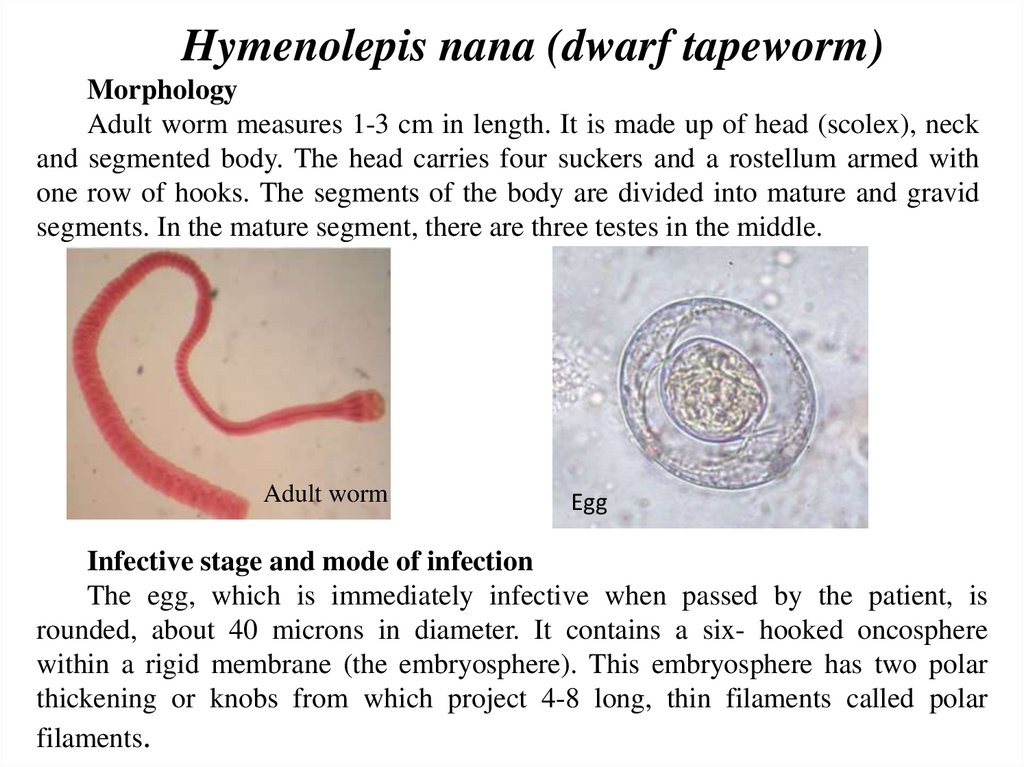
Hymenolepis nana (dwarf tapeworm)Morphology
Adult worm measures 1-3 cm in length. It is made up of head (scolex), neck
and segmented body. The head carries four suckers and a rostellum armed with
one row of hooks. The segments of the body are divided into mature and gravid
segments. In the mature segment, there are three testes in the middle.
Adult worm
Egg
Infective stage and mode of infection
The egg, which is immediately infective when passed by the patient, is
rounded, about 40 microns in diameter. It contains a six- hooked oncosphere
within a rigid membrane (the embryosphere). This embryosphere has two polar
thickening or knobs from which project 4-8 long, thin filaments called polar
filaments.
4.
5.
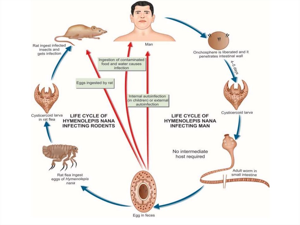
Infection takes place by:1. Ingestion of egg with contaminated raw vegetables.
2. Direct infection from a patient
3. Auto infection: the eggs of H. nana are infective as soon as
they are
passed with feces by the patient. If the hands of the patient are
contaminated by these eggs, she/he infects herself/himself again
and
again.
Symptoms
Light infections produce no symptoms. In fairly heavy infections,
children may
show lack of appetite, abdominal pain and diarrhea.
Treatment
Niclosamide: 4 tablets chewed in a single dose daily for 5 days.
6.
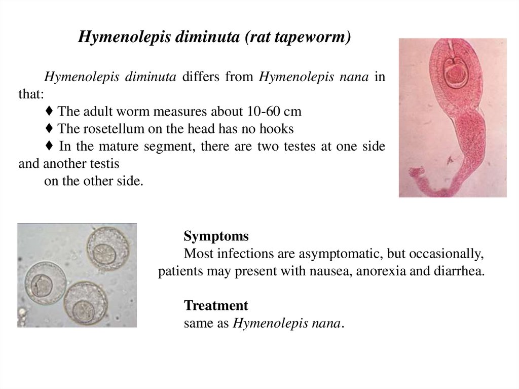
Hymenolepis diminuta (rat tapeworm)Hymenolepis diminuta differs from Hymenolepis nana in
that:
♦ The adult worm measures about 10-60 cm
♦ The rosetellum on the head has no hooks
♦ In the mature segment, there are two testes at one side
and another testis
on the other side.
Symptoms
Most infections are asymptomatic, but occasionally,
patients may present with nausea, anorexia and diarrhea.
Treatment
same as Hymenolepis nana.
7.
8.
Echinococcus granulosus (dog tape worm)Responsible for most cases of echinococcosis. Echinococcosis is caused by
larval tapeworms. The disease is common in East Africa (the highest prevalence
is seen in Kenya: 10-15%).
Morphology
The adult worm measures 3-6 mm in length (up to 1 cm). It has scolex, neck
and strobilla. The scolex is pyriform, with 4 suckers and a prominent rostellum
bearing 2 circular rows of hooklets Adult worms live in small intestine of
definitive host (dog). Man is an intermediate host - carrying the hydatid cyst
(larva). Man contracts infection by swallowing eggs in excreta of definitive host.
The neck is short than the rest of the worm (3 - 6 mm). The strobila is composed
of only 3 proglottids, the anterior immature, the middle mature, and the posterior
gravid segment. The terminal proglottid is longer and wider than the rest of the
worm and contains a branched uterus filled with eggs. The adult worm lives for
6–30 months.
9.
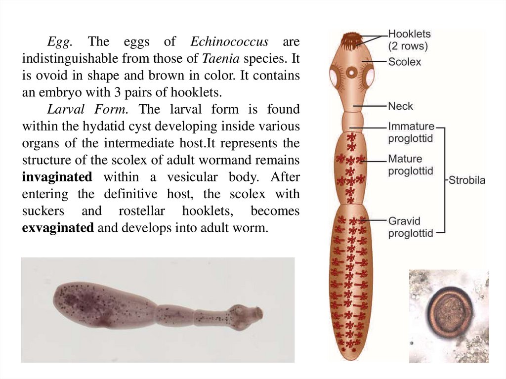
Egg. The eggs of Echinococcus areindistinguishable from those of Taenia species. It
is ovoid in shape and brown in color. It contains
an embryo with 3 pairs of hooklets.
Larval Form. The larval form is found
within the hydatid cyst developing inside various
organs of the intermediate host.It represents the
structure of the scolex of adult wormand remains
invaginated within a vesicular body. After
entering the definitive host, the scolex with
suckers and rostellar hooklets, becomes
exvaginated and develops into adult worm.
10.
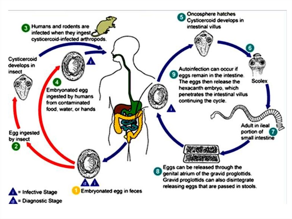
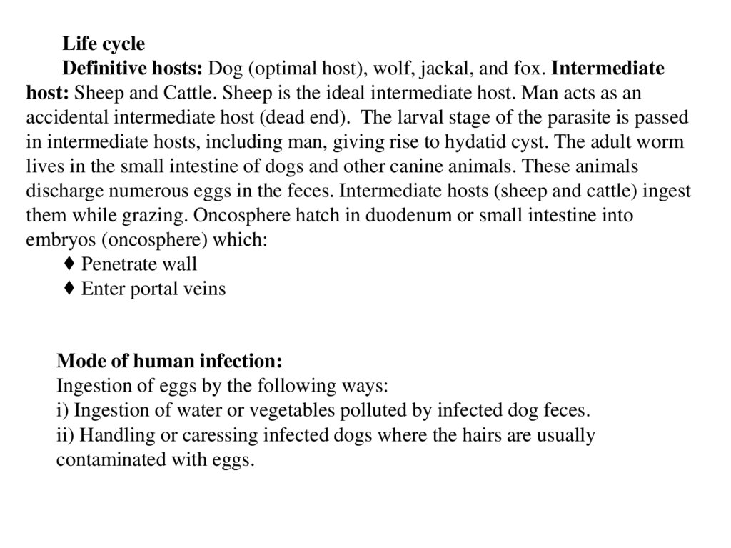
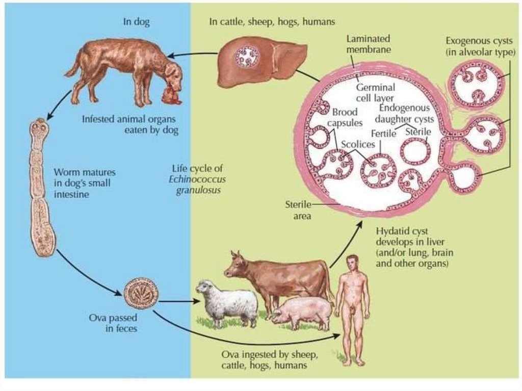
Life cycleDefinitive hosts: Dog (optimal host), wolf, jackal, and fox. Intermediate
host: Sheep and Cattle. Sheep is the ideal intermediate host. Man acts as an
accidental intermediate host (dead end). The larval stage of the parasite is passed
in intermediate hosts, including man, giving rise to hydatid cyst. The adult worm
lives in the small intestine of dogs and other canine animals. These animals
discharge numerous eggs in the feces. Intermediate hosts (sheep and cattle) ingest
them while grazing. Oncosphere hatch in duodenum or small intestine into
embryos (oncosphere) which:
♦ Penetrate wall
♦ Enter portal veins
Mode of human infection:
Ingestion of eggs by the following ways:
i) Ingestion of water or vegetables polluted by infected dog feces.
ii) Handling or caressing infected dogs where the hairs are usually
contaminated with eggs.
11.
12.
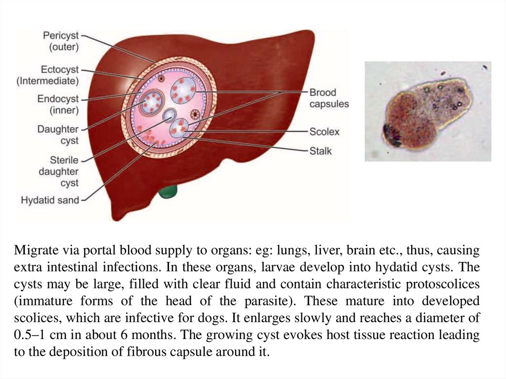
Migrate via portal blood supply to organs: eg: lungs, liver, brain etc., thus, causingextra intestinal infections. In these organs, larvae develop into hydatid cysts. The
cysts may be large, filled with clear fluid and contain characteristic protoscolices
(immature forms of the head of the parasite). These mature into developed
scolices, which are infective for dogs. It enlarges slowly and reaches a diameter of
0.5–1 cm in about 6 months. The growing cyst evokes host tissue reaction leading
to the deposition of fibrous capsule around it.
13.
SymptomsAsymptomatic infection is common, but in symptomatic patients
It may cause cough - with hemoptysis in lung hydatid disease.
Hepatomegaly - with abdominal pain and discomfort
Pressure -from expanding cyst
Rupture of cyst - severe allergic reaction – anaphylaxis, hypersensitivity
to the echinococcal antigen.
Clinical disease develops only when the hydatid cyst has grown big
enough to cause obstructive symptoms. Disease results mainly from pressure
effects caused by the enlarging cysts. In about half the cases, the primary
hydatid cyst occurs in liver (63%), mostly in the right lobe. Hepatomegaly,
pain, and obstructive jaundice are the usual mainfestations.
The next common site is the lung (25%) (most common being the lower
lobe of the right lung). Cough, hemoptysis, chest pain, pneumothorax, and
dyspnea constitute the clinical picture.
In the kidney (2%), hydatid cyst causes pain and hematuria.
Other sites affected include spleen (1%), brain (1%), pelvic organs, orbit,
and bones (3%).
14.
DiagnosisX-ray or ultrasonography (USG), CT scan, and MRI
Demonstration of protoscolices in cyst after operation
Serology
Blood Examination (It may reveal a generalized eosinophilia of 20–25%).
PCR
Casoni’s Intradermal Test (It is an immediate hypersensitivity skin test)
Treatment
Surgery
Albendazole 400 mg twice a day for one to eight periods of 28 days
each,separated by drug-free rest intervals of 14 to 28 days.
Prophylaxis
Ensuring pet dogs do not eat animal carcass or offal.
Periodical deworming of pet dogs.
Destruction of stray and infected dogs.
Mantaining personal hygiene such as washing of hands after touching dogs
and avoidance of kissing pet dogs.
15.
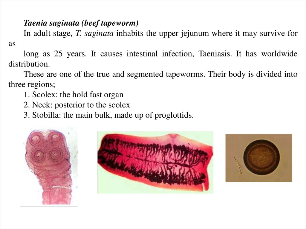
Taenia saginata (beef tapeworm)In adult stage, T. saginata inhabits the upper jejunum where it may survive for
as
long as 25 years. It causes intestinal infection, Taeniasis. It has worldwide
distribution.
These are one of the true and segmented tapeworms. Their body is divided into
three regions;
1. Scolex: the hold fast organ
2. Neck: posterior to the scolex
3. Stobilla: the main bulk, made up of proglottids.
16.
MorphologyAdult worm is opalescent white in color, ribbon-like, dorsoventrally
flattended, and segmented measures 5-10 meters in length. The scolex (head) of T.
saginata is about 1–2 mm in diameter, quadrate in cross-section, bearing 4
hemispherical suckers situated at its four angles. They may be pigmented. The
scolex has no rostellum or hooklets. The suckers serve as the sole organ for
attachment. The neck is long and narrow. The strobila (trunk) consists of 1000 to
2000 proglottides or segments—immature, mature and gravid. The gravid segments
are nearly four times as they are broad, about 20 mm long and 5 mm broad. The
segment contains male and female reproductive structures. The mature segments
have irregularly alternate lateral genital pores. Each of the terminal segments
contains only a uterus made up of a median stem with 15-30 lateral branches.
Eggs of both species are indistinguishable. The egg is spherical, measuring
30–40 μm in diameter. It has a thin hyaline embryonic membrane around it, which
soon disappears after release. The inner embryophore is radially striated and is
yellow-brown due to bile staining. In the center is a fully-developed embryo
(oncosphere) with 3 pairs of hooklets (hexacanth embryo). The eggs do not float
in saturated salt solution. The eggs of T. saginata are infective only to cattle and not
to humans, whereas the eggs of T. solium are infective to pigs and humans too.
The larval stage of Taenia is called as cysticercus.
17.
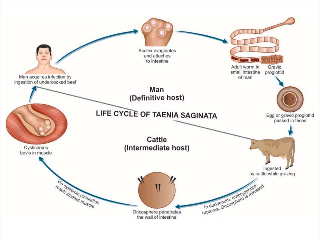
Life cycleThe adult worm lives in the small intestine of man. Gravid
segments pass out in the stool and become disintegrated and eggs
come out to the soil. The gravid proglottid uterus contains about
100,000 eggs. The egg of T. saginata is round, about 40 microns in
diameter. The 6-hooked embryo is enclosed in a radially striated
embryophore. Eggs are ingested by an intermediate host, cattle. The
6-hooked embryo escapes from its shell, penetrates through the
intestinal wall into the blood vessels and is carried to the muscles
where it develops into a larval stage, cysticercus bovis (made up of
an invaginated /inverted head and spherical body). Infection to man
takes place by the ingestion of raw or insufficiently cooked beef. In
the small intestine of man, the head of the cysticercus gets
invaginated and the body becomes segmented.
18.
19.
SymptomsInfected persons may complain of epigastric pain, abdominal discomfort,
indigestion, diarrhea, nausea, weight loss, hunger sensation, vomiting, etc. Occasional
cases of acute intestinal obstruction, acute appendicitis, and pancreatitis have also been
reported.
Diagnosis
Microscopy of stool
Eggs can also be detected by cellophane swab method (NIH Swab) in 85–95%
patients.
Detection of Taenia Antigen in feces: Antigen capture enzyme-linked
immunosorbent assay (ELISA)
Serodiagnosis (Specific antibodies in serum can be demonstrated by ELISA,
indirect immunofluroscence test and indirect hemagglutination (IHA) test
Polymerase chain reaction (PCR)
Treatment
Praziquantel (10–20 mg/kg)
Niclosamide: Four tablets chewed in a single dose.
Mebendazole 100mg twice daily for three days
Prevention
♦ Thorough cooking of meat (above 570C).
♦ Proper disposal of human excret
♦ Maintainence of clean personal habits and general sanitary measures.
20.
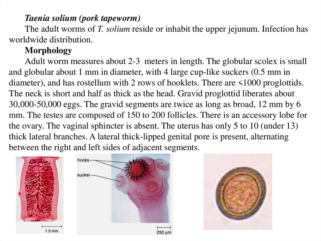
Taenia solium (pork tapeworm)The adult worms of T. solium reside or inhabit the upper jejunum. Infection has
worldwide distribution.
Morphology
Adult worm measures about 2-3 meters in length. The globular scolex is small
and globular about 1 mm in diameter, with 4 large cup-like suckers (0.5 mm in
diameter), and has rostellum with 2 rows of hooklets. There are <1000 proglottids.
The neck is short and half as thick as the head. Gravid proglottid liberates about
30,000-50,000 eggs. The gravid segments are twice as long as broad, 12 mm by 6
mm. The testes are composed of 150 to 200 follicles. There is an accessory lobe for
the ovary. The vaginal sphincter is absent. The uterus has only 5 to 10 (under 13)
thick lateral branches. A lateral thick-lipped genital pore is present, alternating
between the right and left sides of adjacent segments.
21.
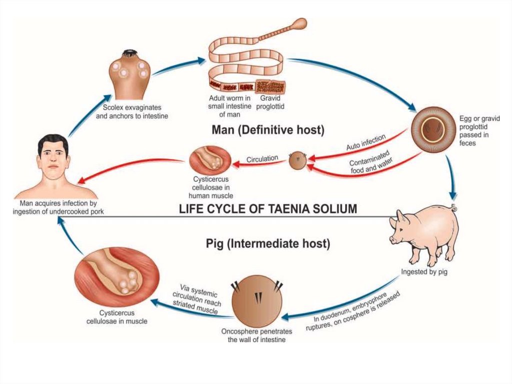
Life cycleDefinitive host: Man
Intermediate host: Pig, occasionally man (in case of cysticercosis).
Mode of infection: Undercooked (measly) pork containing cysticercus
cellulosae; autoinfection and egg in contaminated vegetable, food, and water. Eggs
are infective to human.
Embryonated eggs passed with stool are ingested by pig and the embryo is
released. It penetrates the intestinal wall and is carried by vascular channels to all
parts of the body. After a period of 2-3 months of development the encysted larval
stage called cysticerci or bladder worm occurs in the striated muscles of the
tongue, neck, trunk brain, eye, and the nervous system. The cysticercus survives
for 5 years. Humans become infected by eating pork containing larvae, cysticercus
cellulosae. When improperly cooked cysticercus infected meat is eaten by man, the
scolex remains undigested and attaches itself to the intestinal wall and chain of
proglottids begin to grow to adult worm.
22.
23.
SymptomsInfected persons may complain of epigastric pain, abdominal discomfort,
indigestion, diarrhea, nausea, weight loss, hunger sensation, vomiting, etc.
Cysticercosis. It is caused by larval stage (cysticecus cellulosae) of T. solium.
Cysticercus cellulosae may be solitary or more often multiple. Any organ or tissue
may be involved, the most common being subcutaneous tissues and muscles. It may
also affect the eyes, brain, and less often the heart, liver, lungs, abdominal cavity,
and spinal cord. The cysticercus is surrounded by a fibrous capsule except in the
eye and ventricles of the brain. The larvae evoke a cellular reaction starting with
infiltration of neutrophils, eosinophils, lymphocytes, plasma cells, and at times,
giant cells. This is followed by fibrosis and death of the larva with eventual
calcification. The clinical features depend on the site affected: Subcutaneous
nodules are mostly asymptomatic, Muscular cysticerosis may cause acute
myositis, Neurocysticerosis (cysticercosis of brain) is the most common and most
serious form of cysticercosis. About 70% of adult-onset epilepsy is due to
neurocysticercosis.
24.
DiagnosisStool Examination: Microscopy of stool specimen, cellophane swab method
(NIH Swab), Detection of Taenia Antigen in feces: Antigen capture enzymelinked immunosorbent assay (ELISA),
Serodiagnosis
Polymerase chain reaction (PCR)
Biopsy of the lesion and its microscopic examination
Imaging (X-ray, CT-scan, MRI scan)
Treatment
Praziquantel 50 mg/kg in 3 divided doses for 20–30 days
Niclosamide: 2 gm PO stat
Albendazole 400 mg twice daily for 30 days
Prevention
♦ Treatment of infected persons.
♦ Thorough cooking of pork and proper processing, avoidance of eating
undercooked pork and raw vegetables
♦ Proper disposal of human excreta (good hygiene/sanitation).
25.
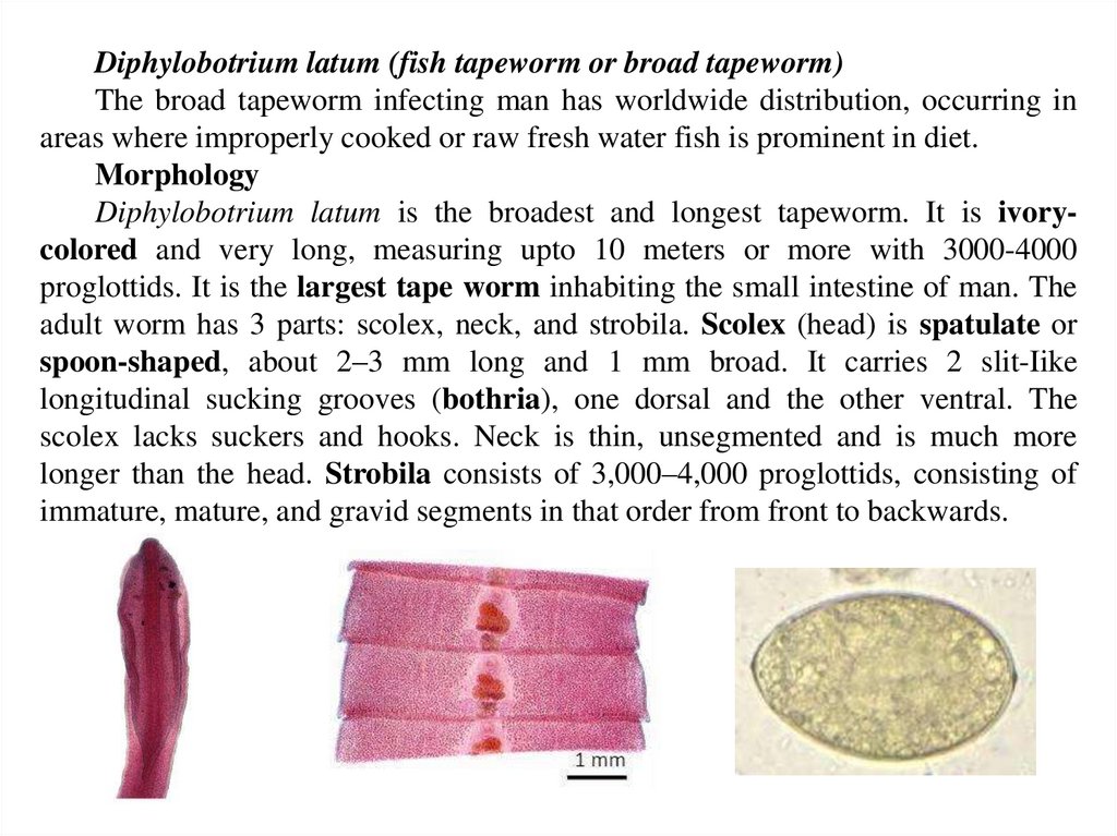
Diphylobotrium latum (fish tapeworm or broad tapeworm)The broad tapeworm infecting man has worldwide distribution, occurring in
areas where improperly cooked or raw fresh water fish is prominent in diet.
Morphology
Diphylobotrium latum is the broadest and longest tapeworm. It is ivorycolored and very long, measuring upto 10 meters or more with 3000-4000
proglottids. It is the largest tape worm inhabiting the small intestine of man. The
adult worm has 3 parts: scolex, neck, and strobila. Scolex (head) is spatulate or
spoon-shaped, about 2–3 mm long and 1 mm broad. It carries 2 slit-Iike
longitudinal sucking grooves (bothria), one dorsal and the other ventral. The
scolex lacks suckers and hooks. Neck is thin, unsegmented and is much more
longer than the head. Strobila consists of 3,000–4,000 proglottids, consisting of
immature, mature, and gravid segments in that order from front to backwards.
26.
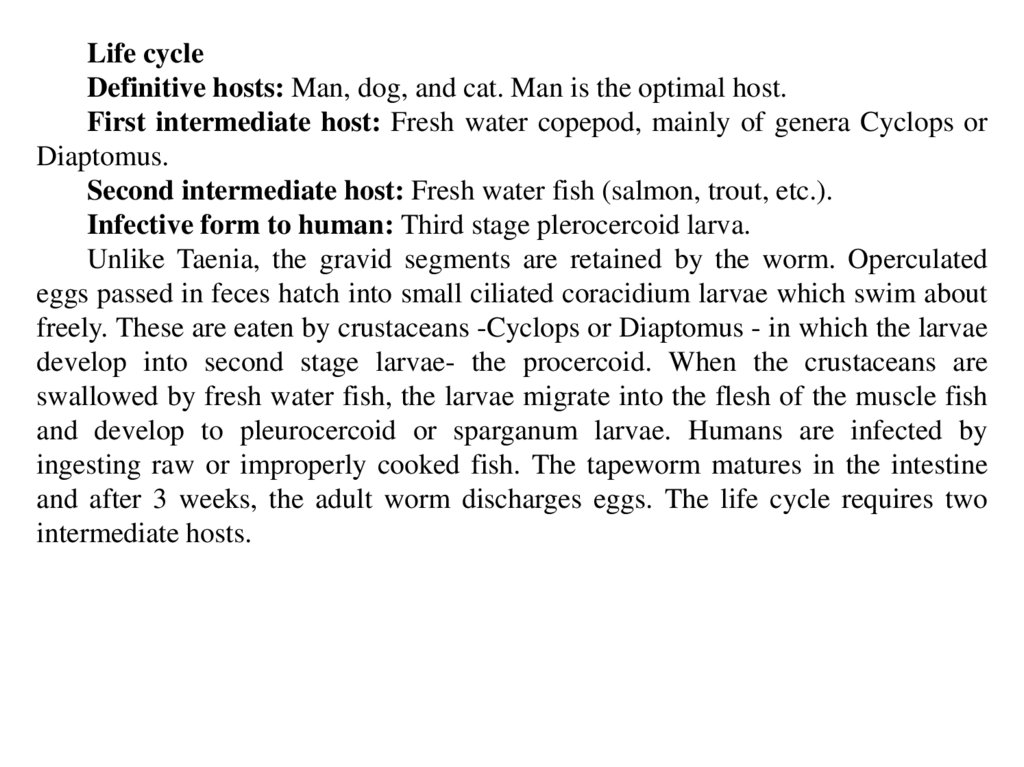
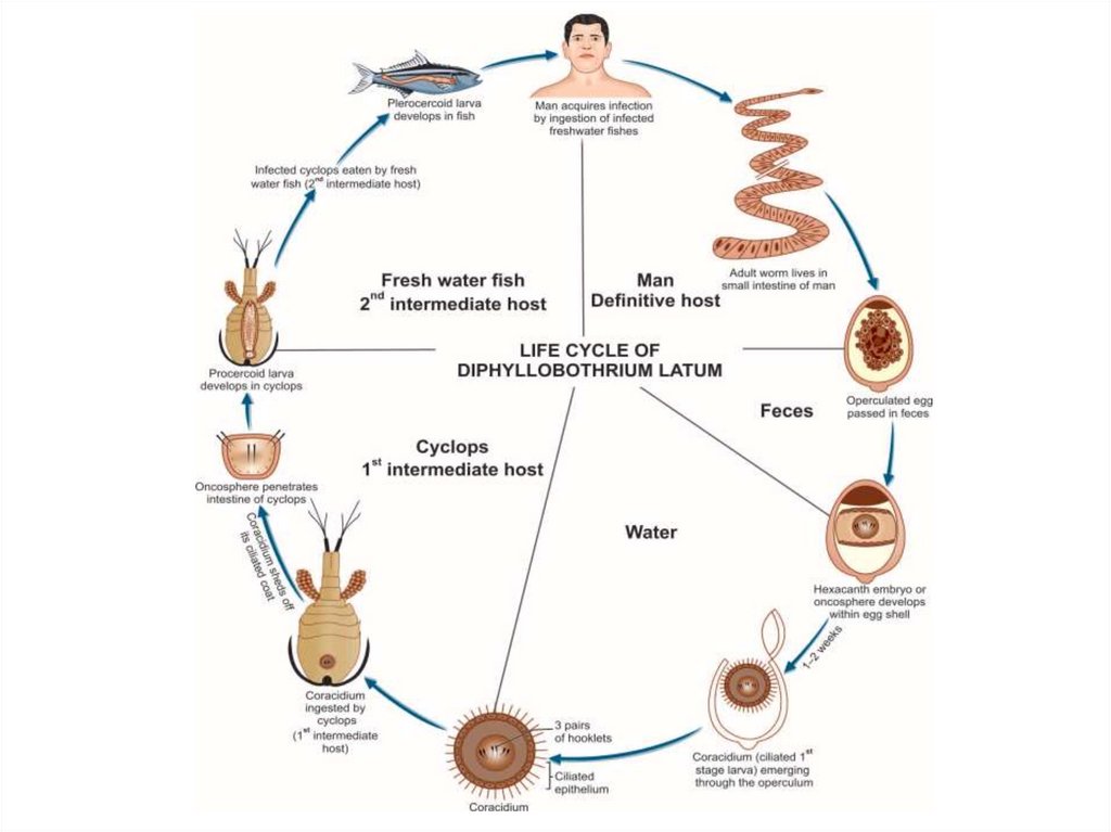
Life cycleDefinitive hosts: Man, dog, and cat. Man is the optimal host.
First intermediate host: Fresh water copepod, mainly of genera Cyclops or
Diaptomus.
Second intermediate host: Fresh water fish (salmon, trout, etc.).
Infective form to human: Third stage plerocercoid larva.
Unlike Taenia, the gravid segments are retained by the worm. Operculated
eggs passed in feces hatch into small ciliated coracidium larvae which swim about
freely. These are eaten by crustaceans -Cyclops or Diaptomus - in which the larvae
develop into second stage larvae- the procercoid. When the crustaceans are
swallowed by fresh water fish, the larvae migrate into the flesh of the muscle fish
and develop to pleurocercoid or sparganum larvae. Humans are infected by
ingesting raw or improperly cooked fish. The tapeworm matures in the intestine
and after 3 weeks, the adult worm discharges eggs. The life cycle requires two
intermediate hosts.
27.
28.
SymptomsMost infections are asymptomatic. Rarely, it causes severe cramping,
mechanical Obstruction, abdominal discomfort, vomiting, diarrhea, nausea,
anemia, weakness and weight loss. Pernicious anemia can also result, due to
interference of vitamin B12 absorption in jejunum.
Diagnosis
Eggs in stool: Single shell with operculum at one end and a knob on the other.
Treatment
Praziquantel in a single dose of 10 mg/kg is effective.
Parenteral vit B12 should be given, if B12 deficiency is present.
Niclosamide: 2 mg after light breakfast.
Prevention
Proper cooking of fish
Deep freezing (–10°C for 24–48 hours ) of fish, if it is to be cosumed raw
Prevention of fecal pollution of natural waters
Periodical deworming of pet dogs and cats.
29.
HOME work. CestodesLatin name of
parasite
Forms of
parasites
Definitive host
Intermediate
host
Infective stage
Transmission
Way of infection
Symptoms
Diagnosis
Treatment
Prevention
Dyphilobothrium
latum
Echinococcus
granulosus
Taenia
solium
Taenia
saginatum