

 Медицина
Похожие презентации:
. Class Nematoda. Lesson 5")


")
Nematodes (round worms)
1.
NEMATODES (ROUND WORMS)Phylum Nemathelminthes
(Aschelminthes)
Class Nematoda.
2.
All the important human parasites of the PhylumNemathelminthes (Aschelminthes) belong to the Class Nematoda.
They are un-segmented, elongated and cylindrical.
They have separate sexes with separate appearances.
They have a tough protective covering or cuticle.
They have a complete digestive tract with both oral and anal
openings.
The nematodes are free living (Majority) or parasites of
humans, plants or animals.
The parasitic nematodes:
The nematodes are generally light cream-white colored. Their
life cycle includes: egg, larvae and adult.
3.
The parasitic nematodes are divided into:1. Intestinal nematodes
1.1. Intestinal nematodes with tissue stage
A. Ascaris lumbricoides
B. Hookworms
C. Strongyloides stercoralis
1.2. Intestinal nematodes without tissue stage
A. Enterobius vermicularis
B. Trichuris trichuira.
2. Tissue and blood dwelling nematodes
2.1. Filarial worms
2.2. Dracunculus medinensis
2.3. Trichinella
2.4. Larva migrans.
4.
INTESTINAL NEMATODESWITH TISSUE STAGE
5.
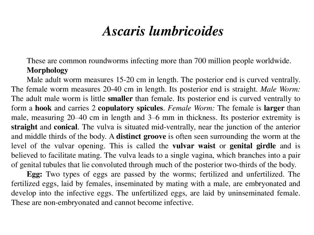
Ascaris lumbricoidesThese are common roundworms infecting more than 700 million people worldwide.
Morphology
Male adult worm measures 15-20 cm in length. The posterior end is curved ventrally.
The female worm measures 20-40 cm in length. Its posterior end is straight. Male Worm:
The adult male worm is little smaller than female. Its posterior end is curved ventrally to
form a hook and carries 2 copulatory spicules. Female Worm: The female is larger than
male, measuring 20–40 cm in length and 3–6 mm in thickness. Its posterior extremity is
straight and conical. The vulva is situated mid-ventrally, near the junction of the anterior
and middle thirds of the body. A distinct groove is often seen surrounding the worm at the
level of the vulvar opening. This is called the vulvar waist or genital girdle and is
believed to facilitate mating. The vulva leads to a single vagina, which branches into a pair
of genital tubules that lie convoluted through much of the posterior two-thirds of the body.
Egg: Two types of eggs are passed by the worms; fertilized and unfertilized. The
fertilized eggs, laid by females, inseminated by mating with a male, are embryonated and
develop into the infective eggs. The unfertilized eggs, are laid by uninseminated female.
These are non-embryonated and cannot become infective.
6.
7.
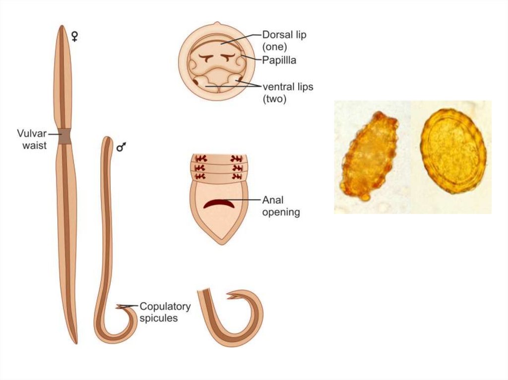
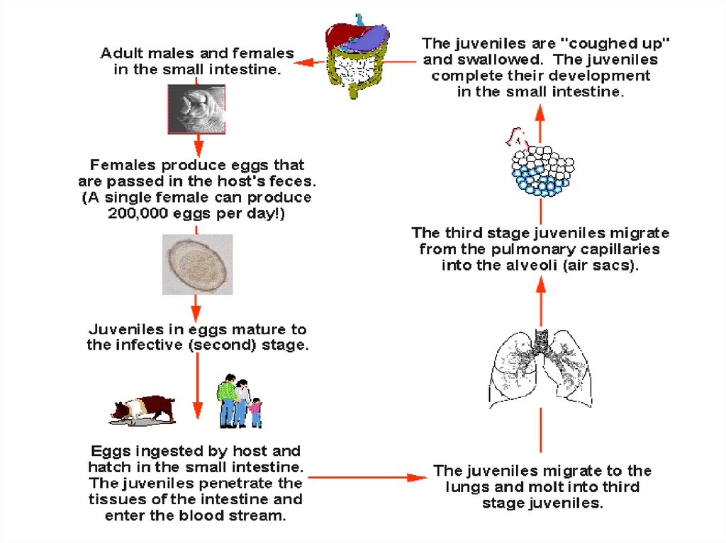
Life cycleLife cycle of Ascaris involves only 1 host. Natural host: Man.
There is no intermediate host. Infective form: Embryonated eggs.
Mode of transmission: Infection occurs when the egg containing
the infective rhabditiform larva is swallowed. A frequent mode
of transmission is through fresh vegetables grown in fields
manured with human feces (‘night soil’). Infection may also be
transmitted through contaminated drinking water.
Ingested eggs hatch in the duodenum. The larvae penetrate the
intestinal wall and circulate in the blood. From the heart they
migrate to the lungs, ascend to the trachea, descend to the
esophagus and finally reach the small intestine to become adult.
The female pass immature eggs which pass to the soil and mature
in 2 weeks.
8.
9.
SymptomsFever, urticaria, angioneurotic edema, wheezing, and
conjunctivitis.
Acute biliary obstruction or pancreatitis, obstructive
appendicitis.
Spoliative action–protein and vitamin A deficiency.
Toxic action–utricaria and angioneurotic edema.
Mechanical action–intestinal obstruction, intussusception,
volvulus, intestinal perforation.
In Lungs–Ascaris can cause pneumonia (Loeffler’s syndrome).
10.
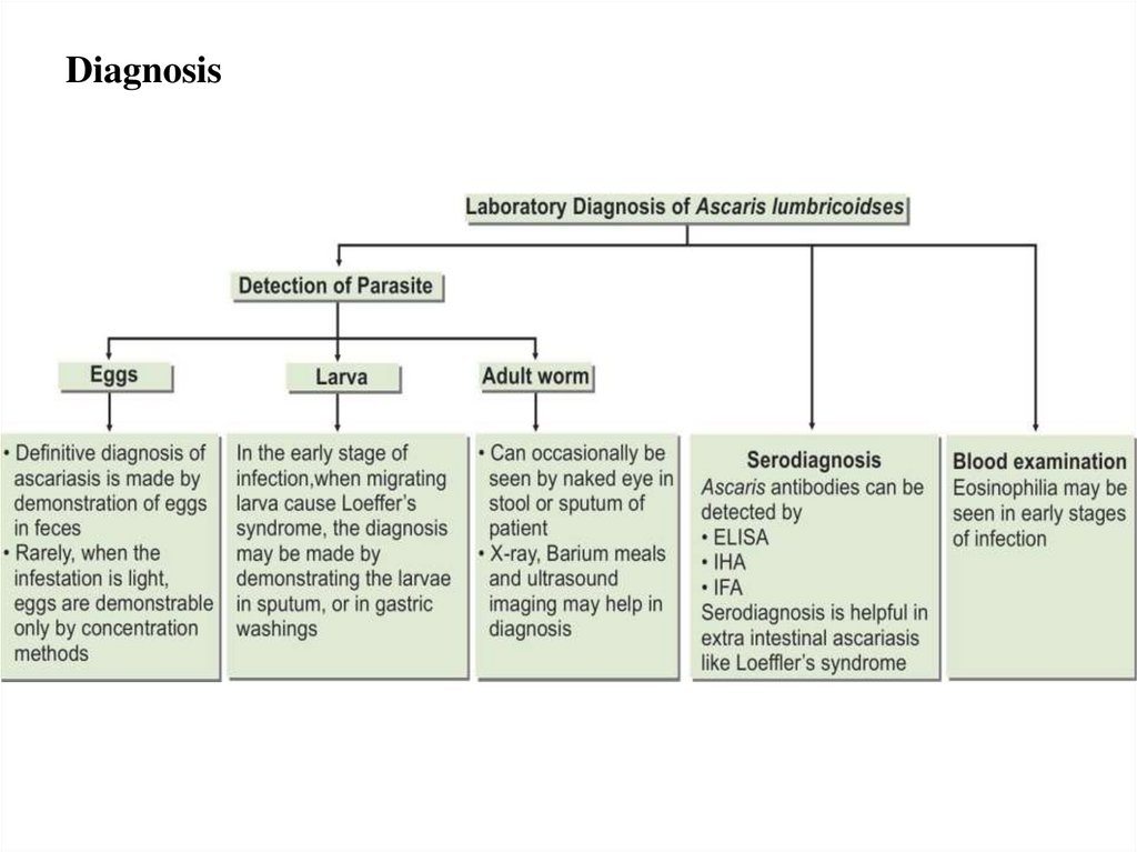
Diagnosis11.
Treatmentpyrantel pamoate 11 mg/kg once; maximum 1 g,
albendazole 400 mg once,
mebendazole 100 g twice daily for 3 days or 500 mg once,
ivermectin 150–200 mg/kg once.
Prophylaxis
Preventing fecal contamination of soil.
Treatment of vegetables and other garden crops with water
containing iodine 200 ppm for 15 minutes kills the eggs and larvae of
Ascaris and other helminths.
Avoid eating raw vegetables.
Improvement of personal hygiene. Treatment of infected persons.
12.
HOOK WORMS13.
HOOK WORMSThere are two species of hookworm:
1. Ancylostoma duodenale
2. Necator americanus
The adults are found in the small intestines of man.
Mixed infection is common. Both of the species are
found in Ethiopia, but N. americanus is more common.
14.
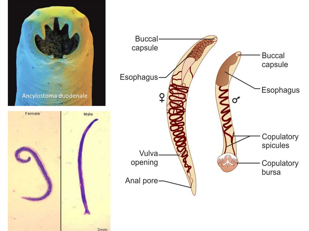
Ancylostoma duodenaleGrayish-white in color. The body is slightly ventrally curved.
The anterior end follows the body curvature. The buccal cavity is
provided ventrally with pairs of teeth and dorsally with a notched
dental plate.
Distribution: This species is found in the northern part of the
world including China, Japan, Europe, North Africa and Ethiopia.
Morphology
Male: The male measures 10 cm in length. The posterior end is
broadened into a membraneous copulatory bursa that is provided
with two long spicules.
Female: The female measures 12 cm in length. The posterior
end is straight.
15.
16.
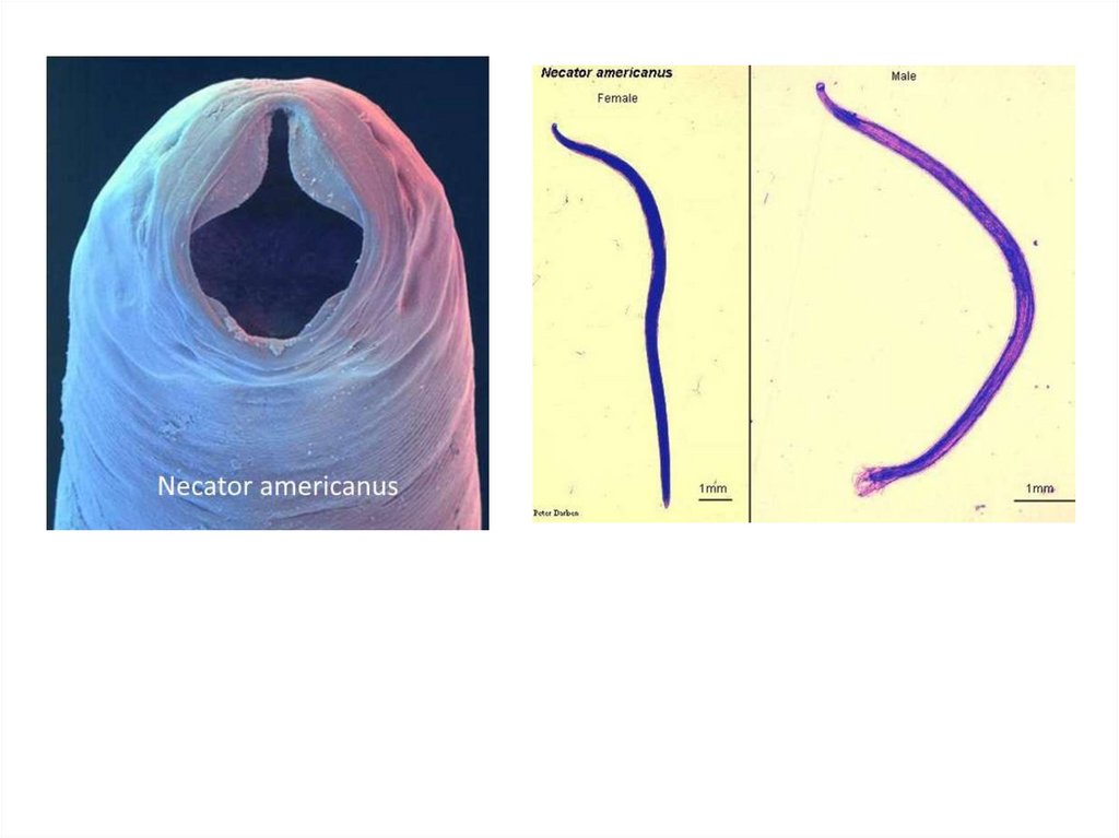
Necator americanusThis species, so called American hookworm, is found in
predominantly the tropics. The anterior end is hooked against the
body curvature. The mouth is provided ventrally and dorsally with
cutting plate.
Morphology
Male: The male measures 8 cm in length. The posterior end is
broadened into a membraneous copulatory bursa, which is
provided with two long spicules fused distally.
Female: The female measures 10 cm in length. The posterior
end is straight
Infective stage and methods of infection: The filariform
larva infects by skin penetration.
17.
18.
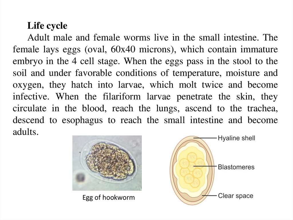
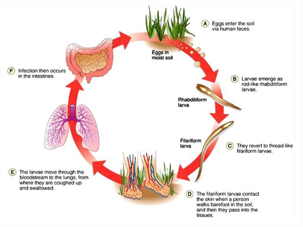
Life cycleAdult male and female worms live in the small intestine. The
female lays eggs (oval, 60x40 microns), which contain immature
embryo in the 4 cell stage. When the eggs pass in the stool to the
soil and under favorable conditions of temperature, moisture and
oxygen, they hatch into larvae, which molt twice and become
infective. When the filariform larvae penetrate the skin, they
circulate in the blood, reach the lungs, ascend to the trachea,
descend to esophagus to reach the small intestine and become
adults.
Egg of hookworm
19.
20.
SymptomsAdult worms in the intestine feed on blood causing iron
deficiency anemia. The larvae may cause inflammation of the
lungs.
Diagnosis
Examination of stool by direct saline smear to detect the eggs.
Treatment
Mebendazole: 1 tab 2x daily for 3 days.
21.
LARVA MIGRANS22.
There are three types of larva migrans:a. Cutaneous larva migrans (Creeping eruption)
Various animals harbor hookworms. Two species of dogs and cats are
important.
1. Ancylostoma braziliens: infects both dogs and cats.
2. Ancylostoma caninum: infects only dogs.
Both of these are common in the tropics and subtropical regions where
human hookworms can best complete their life cycles. If man comes in contact
with infective larvae, penetration of the skin may take place; but the larvae are
then unable to complete their migratory cycle. Trapped larvae may survive for
weeks or even months, migrating through the subcutaneous tissues. They may
evoke a fairly severe reaction - pruritus and dermatitis. The dermatitis leads to
scratching and then bacterial superinfection.
Treatment
Thiabendazole: Applied topically.
b. Visceral larva migrans
A syndrome caused by the migration of parasitic larvae in the viscera of a
host for months or years. It may be caused by transient larval migration in the
life cycles of several parasites such as hookworm, Ascaris lumbricoides, T.
spiralis, S. strecoralis and other filarial worms.
23.
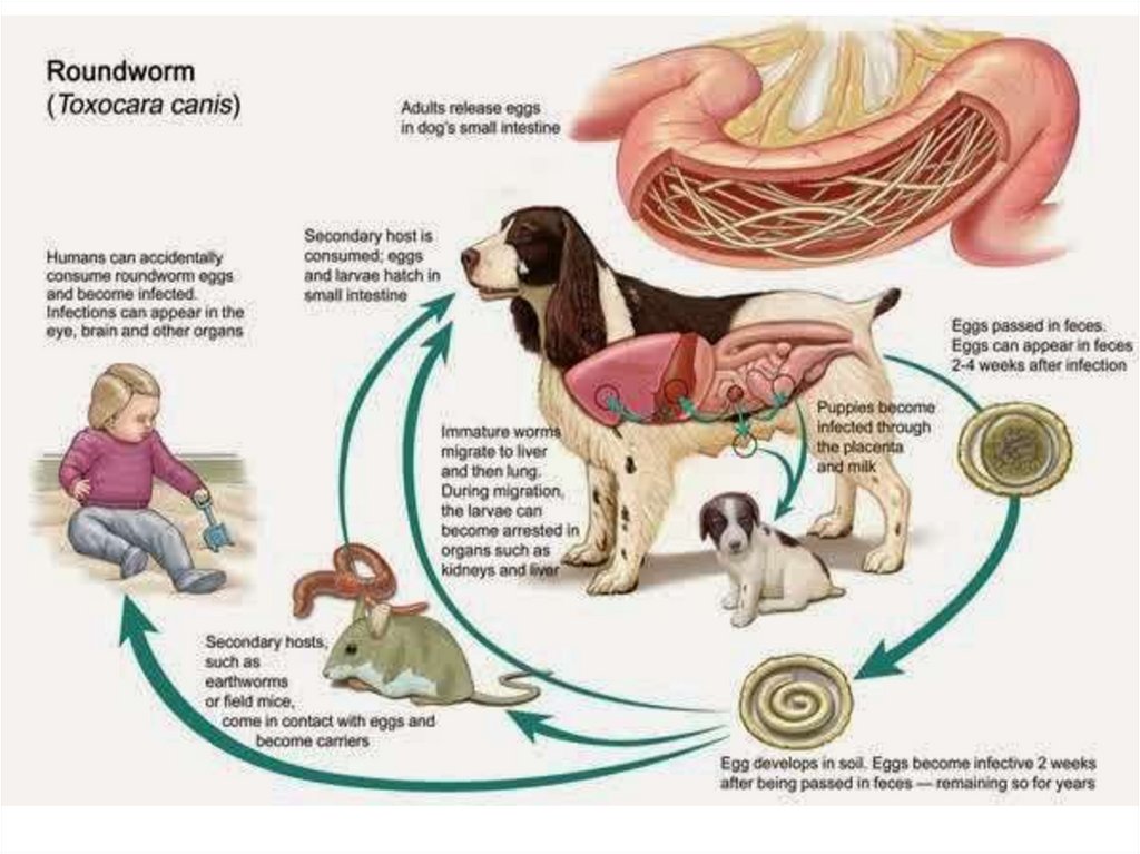
ToxocariasisThis is a kind of visceral larva migrans caused by
♦ Toxocara canis (Dog ascarid) and
♦ Toxocara catis (Cat ascarid).
These cause persistent larval migration and thus the visceral larva migrans is called
toxocariasis.
Morphology
♦ The larvae of Toxocara canis and Toxocara catis measure about 400 μm in
length.
♦ The life cycle of these parasites in their respective hosts is similar to that of
A.lumbricoides in humans.
24.
25.
Epidemiology. Visceral larva migrans is cosmopolitan indistribution.
Transmission: Ingestion of eggs of Toxocara species in
contaminated food or soil or direct contact with infected patients.
Children are more at risk.
Symptoms
♦ Majority are asymptomatic.
♦ Eosinophilia
♦ Cerebral, myocardial and pulmonary involvement may cause
death.
Diagnosis
Identification of larvae in tissue.
Treatment
Thiabendazole: 25 mg/kg twice daily for 5 days.
26.
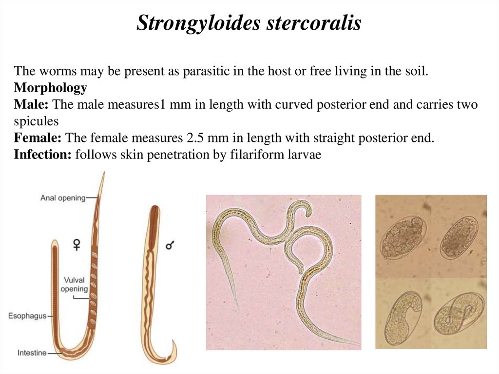
Strongyloides stercoralisThe worms may be present as parasitic in the host or free living in the soil.
Morphology
Male: The male measures1 mm in length with curved posterior end and carries two
spicules
Female: The female measures 2.5 mm in length with straight posterior end.
Infection: follows skin penetration by filariform larvae
27.
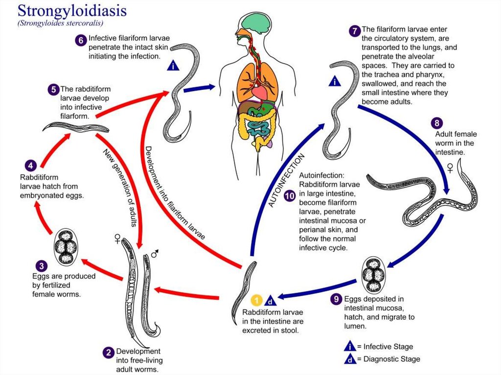
Life cycleAdult male and female worms live in the small intestine. After
fertilization, the female penetrates the mucosa of the small intestine
and lay eggs in the submucosa. The eggs hatch and the larvae
penetrate the mucosa back to the lumen. If the environmental
conditions are favorable, the larvae will come out with the stool to
the soil. They transform into adults, which lay eggs, and hatching
larvae get transformed to adults and so on. If the environmental
conditions are not favorable, the larvae in the stool will moult and
transform into infective filariform larvae, which pierce the intestine
(auto-infection). Larvae penetrating the skin from the soil or by
autoinfection are carried by the blood to the lungs, ascend to the
trachea, descend to the esophagus and mature in the small intestine.
28.
29.
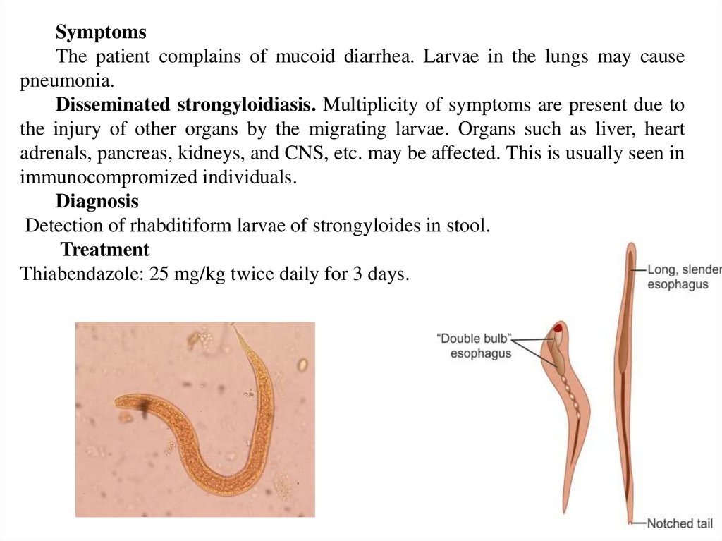
SymptomsThe patient complains of mucoid diarrhea. Larvae in the lungs may cause
pneumonia.
Disseminated strongyloidiasis. Multiplicity of symptoms are present due to
the injury of other organs by the migrating larvae. Organs such as liver, heart
adrenals, pancreas, kidneys, and CNS, etc. may be affected. This is usually seen in
immunocompromized individuals.
Diagnosis
Detection of rhabditiform larvae of strongyloides in stool.
Treatment
Thiabendazole: 25 mg/kg twice daily for 3 days.
30.
INTESTINAL NEMATODESWITHOUT TISSUE STAGE
31.
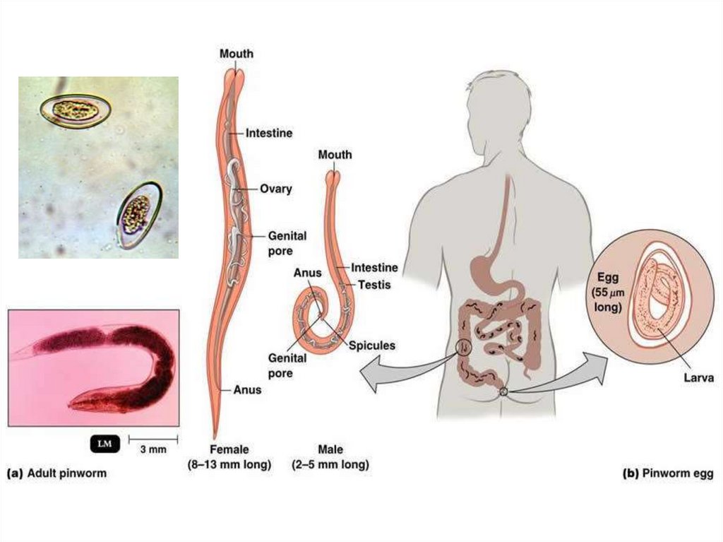
Enterobius vermicularis(pin worm or thread worm)
Enterobius vermicularis is a small white worm with thread-like
appearance. The worm causes enterobiasis. Infection is common in
children.
Morphology
Male: The male measures 5 mm in length. The posterior end is
curved and carries a single copulatory spicule. Female: The female
measures 13 mm in length. The posterior end is straight.
32.
33.
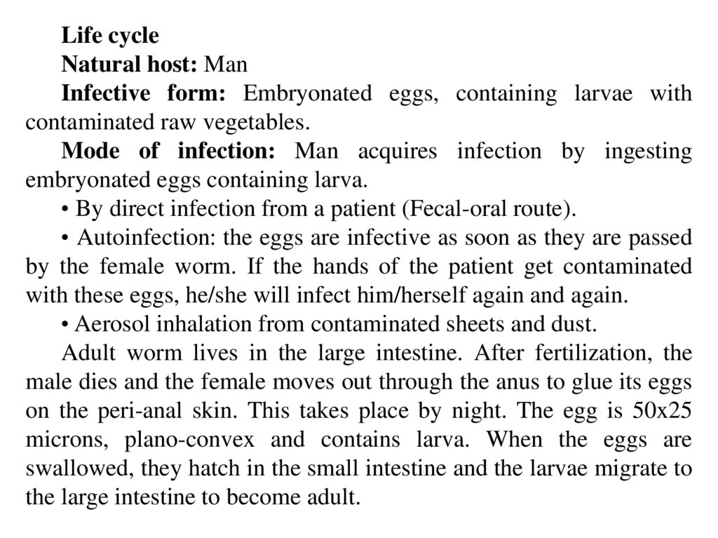
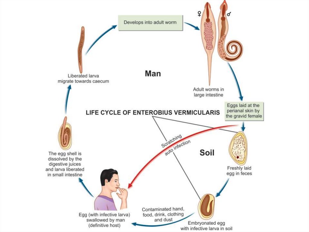
Life cycleNatural host: Man
Infective form: Embryonated eggs, containing larvae with
contaminated raw vegetables.
Mode of infection: Man acquires infection by ingesting
embryonated eggs containing larva.
• By direct infection from a patient (Fecal-oral route).
• Autoinfection: the eggs are infective as soon as they are passed
by the female worm. If the hands of the patient get contaminated
with these eggs, he/she will infect him/herself again and again.
• Aerosol inhalation from contaminated sheets and dust.
Adult worm lives in the large intestine. After fertilization, the
male dies and the female moves out through the anus to glue its eggs
on the peri-anal skin. This takes place by night. The egg is 50x25
microns, plano-convex and contains larva. When the eggs are
swallowed, they hatch in the small intestine and the larvae migrate to
the large intestine to become adult.
34.
35.
SymptomsEnterobiasis occurs mostly in children. It is more common in
females than in males. About one-third of infections are
asymptomatic. The worm produces intense irritation and pruritus of
the perianal and perineal area (pruritis ani), when it crawls out of
the anus to lay eggs. This leads to scratching and excoriation of the
skin around the anus. As the worm migrates out at night, it disturbs
sleep. Nocturnal enuresis is sometimes seen. The worm crawling
into the vulva and vagina causes irritation and a mucoid discharge.
It may migrate upto the uterus, fallopian tubes and into the
peritoneum. This may cause symptoms of chronic salpingitis,
cervicitis, peritiontis, and recurrent urinary tract infections. The
worm is sometimes found in surgically removed appendix and has
been claimed to be responsible for appendicitis.
36.
Diagnosis♦ Detection of eggs by NIH swab and cellophane scotch tape
method. Detection of eggs under finger nail Detection of adult
worm and eggs in stool.
Treatment
Pyrantel pamoate 11 mg/kg once, maximum 1 g,
Albendazole 400 mg once
Mebendazole 100 mg once
Prophylaxis
Maintainance of personal and community hygiene such as frequent
hand washing, _ nger nail cleaning, and regular bathing.
Frequent washing of night clothes and bed linen.
37.
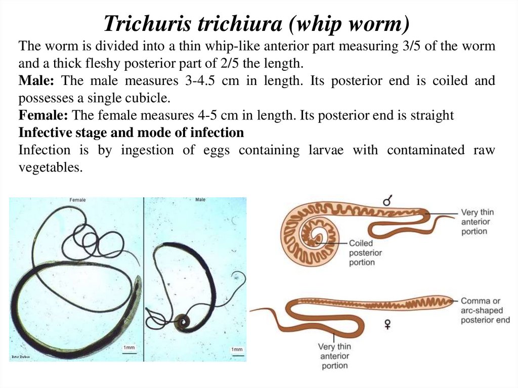
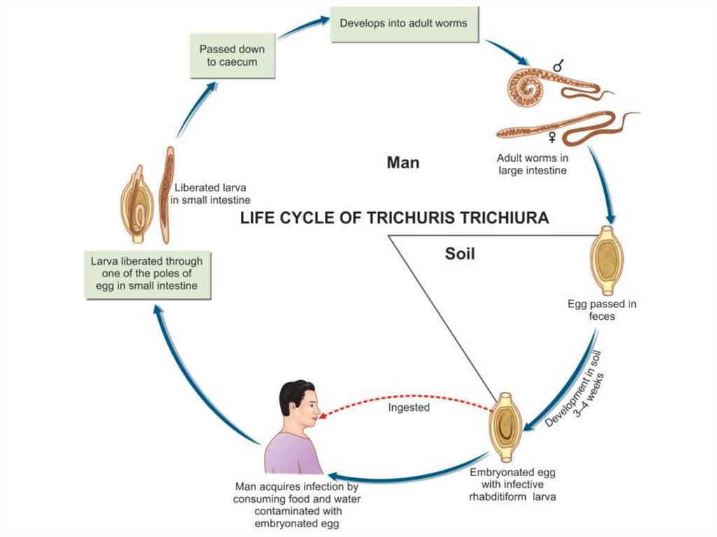
Trichuris trichiura (whip worm)The worm is divided into a thin whip-like anterior part measuring 3/5 of the worm
and a thick fleshy posterior part of 2/5 the length.
Male: The male measures 3-4.5 cm in length. Its posterior end is coiled and
possesses a single cubicle.
Female: The female measures 4-5 cm in length. Its posterior end is straight
Infective stage and mode of infection
Infection is by ingestion of eggs containing larvae with contaminated raw
vegetables.
38.
39.
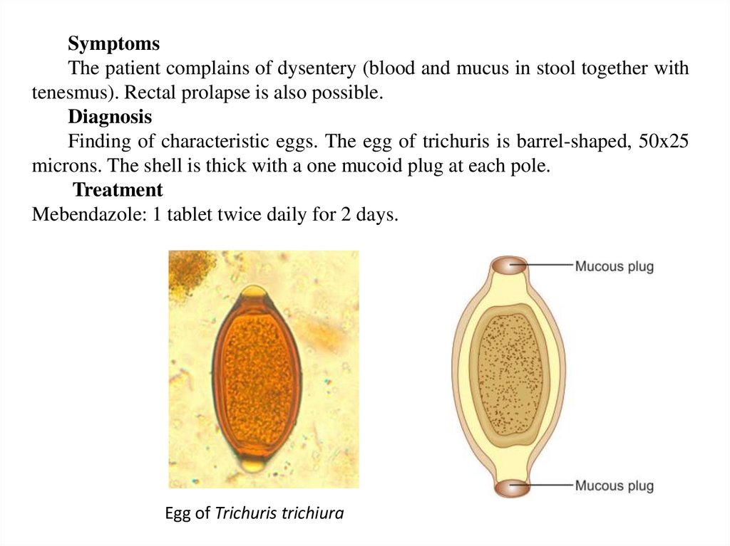
SymptomsThe patient complains of dysentery (blood and mucus in stool together with
tenesmus). Rectal prolapse is also possible.
Diagnosis
Finding of characteristic eggs. The egg of trichuris is barrel-shaped, 50x25
microns. The shell is thick with a one mucoid plug at each pole.
Treatment
Mebendazole: 1 tablet twice daily for 2 days.
Egg of Trichuris trichiura
40.
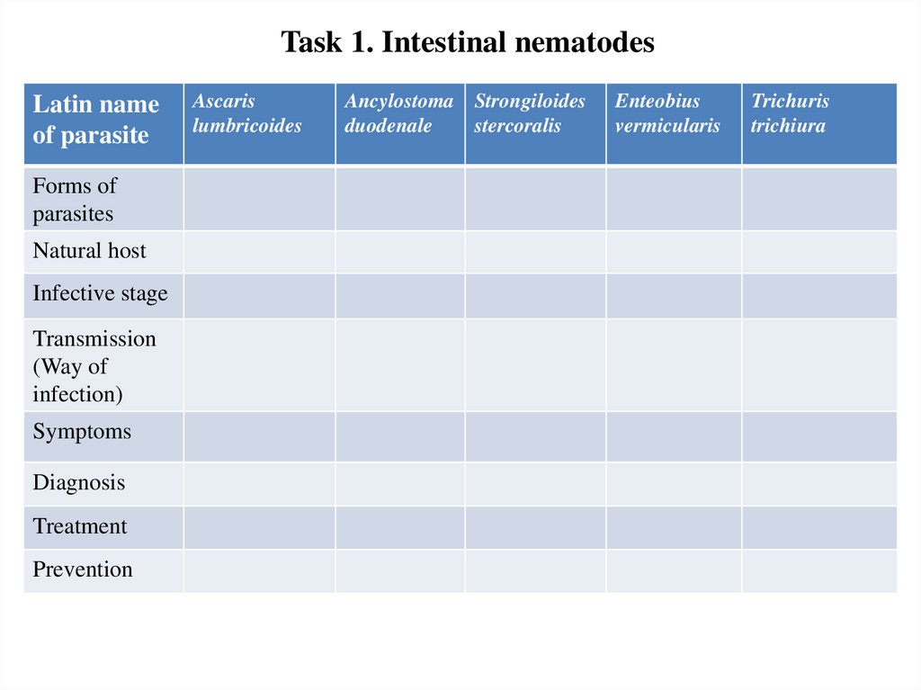
Task 1. Intestinal nematodesLatin name
of parasite
Forms of
parasites
Natural host
Infective stage
Transmission
(Way of
infection)
Symptoms
Diagnosis
Treatment
Prevention
Ascaris
lumbricoides
Ancylostoma Strongiloides
duodenale
stercoralis
Enteobius
vermicularis
Trichuris
trichiura
41.
TISSUE NEMATODES.FILARIAL WORMS
42.
Filarial wormsThis group includes the filarial worms, the guinea worm
(Dranculuculus medinensis) and Trichinella spiralis.
The filarial worms have complex life cycles involving a
developmental stage in an insect vector. They require an arthropod
vector for their transmission. The worms inhabit either the
lymphatic system or the subcutaneous tissues of man. The female
worm gives rise to a young worm called microfilaria. The
microfilariae, when taken by the arthropod intermediate host during
biting, develop into filariform larvae, which are the infective stages.
Humans get infected when bitten by the infected arthropod
intermediate host.
43.
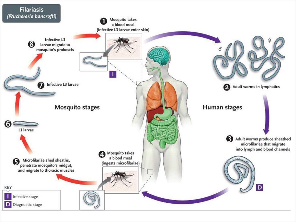
Wuchereria bancroftiThis is a parasite of lymph nodes and lymphatic vessels- causing lymphatic
filariasis. This filarial worm is transmitted by the bite of various species of
mosquitoes. It is believed that over 100 million people are infected. The
microfilariae are nocturnal – seen in greatest numbers in peripheral blood in the
night between 10 PM-2 AM.
44.
Mode of transmission and pathogenesisThe filariform larvae are introduced through the skin by the bite
of the arthropod intermediate host. The larvae invade the
lymphatics, usually the lower limb, where they develop into adult
worms. The microfilariae are librated into the blood stream. They
remain in the pulmonary circulation during day, emerging into the
peripheral circulation only during night, to coincide with the biting
habit of the vector. Presence of the adult worms causes lymphatic
blockage and gross lymphedema, which sometimes lead to
elephantiasis.
Epidemiology: W. bancrofti infection is not reported in higher
altitudes of Ethiopia, but limited to lowlands of Gambella. The
epidemic area covers a long distance along the Baro River.
45.
46.
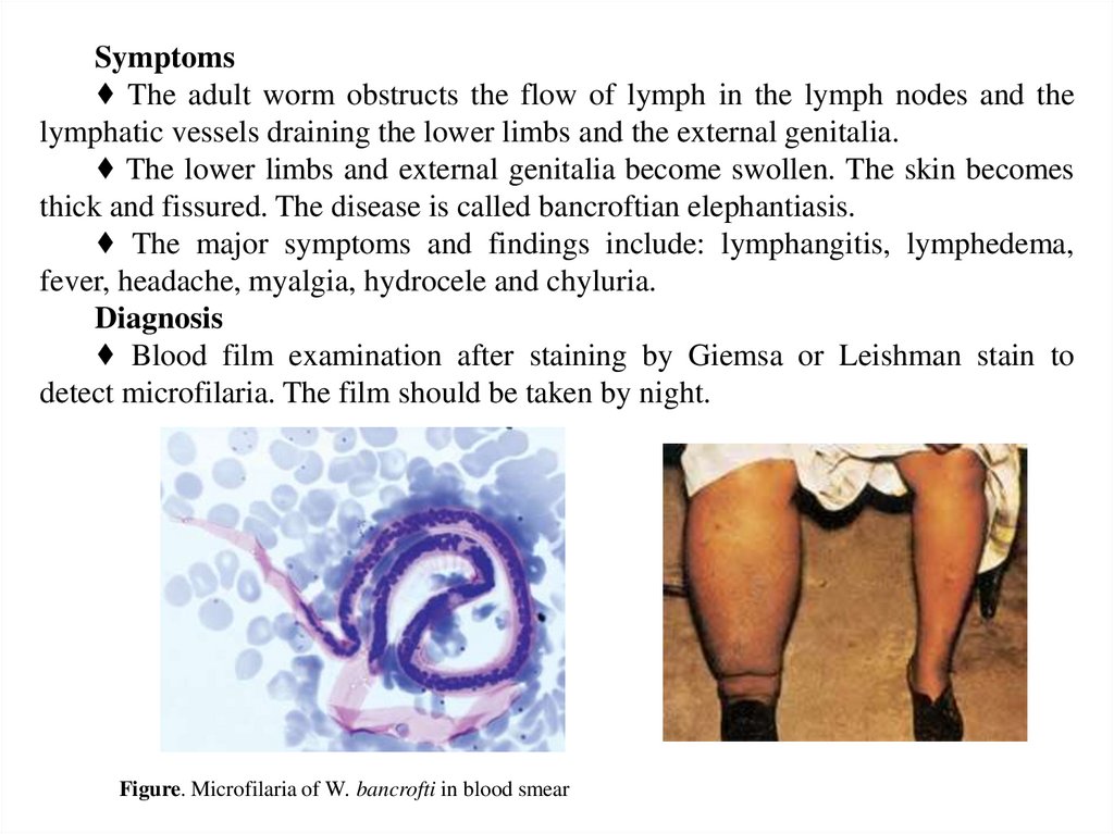
Symptoms♦ The adult worm obstructs the flow of lymph in the lymph nodes and the
lymphatic vessels draining the lower limbs and the external genitalia.
♦ The lower limbs and external genitalia become swollen. The skin becomes
thick and fissured. The disease is called bancroftian elephantiasis.
♦ The major symptoms and findings include: lymphangitis, lymphedema,
fever, headache, myalgia, hydrocele and chyluria.
Diagnosis
♦ Blood film examination after staining by Giemsa or Leishman stain to
detect microfilaria. The film should be taken by night.
Figure. Microfilaria of W. bancrofti in blood smear
47.
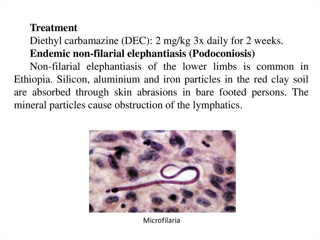
TreatmentDiethyl carbamazine (DEC): 2 mg/kg 3x daily for 2 weeks.
Endemic non-filarial elephantiasis (Podoconiosis)
Non-filarial elephantiasis of the lower limbs is common in
Ethiopia. Silicon, aluminium and iron particles in the red clay soil
are absorbed through skin abrasions in bare footed persons. The
mineral particles cause obstruction of the lymphatics.
Microfilaria
48.
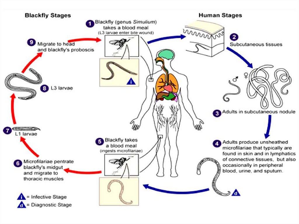
Onchocerca volvulusInfection by this filarial worm is common in Ethiopia. Endemic
foci are found in Bebeka, Gojeb valley, Dedessa valley, Agaro,
Metekel, and in Northwestern Ethiopia around Gondar.
Morphology
Male: Similar to that of Wuchereria bancrofti.
Female: The female measures 30-50 cm in length. It is present
inside of a fibrous nodule (onchocercomata or onchocerca tumor).
Intermediate Host and vector: Female Simulium, (Simulium
damnosum), Black fly, found around plantations following rivers or
river basins.
Microfilaria. Measures 300 microns in length. It is nonsheathed microfilaria. It is present in the subcutaneous tissue fluids
and not in blood.
49.
50.
Infective stage and mode of infection: microfilaria.Symptoms
The disease, onchocerciasis or river blindness includes:
• Skin fibrous nodules (onchocercomata) enclosing female worms. The nodules are
common in neck, iliac crest and the coccyx.
• Skin hypo- or hyper- pigmentation. Dermatitis is present. In advanced cases, the skin
becomes thickened and wrinkled, showing lizard or leopard skin appearance.
• Elephantiasis of the external genitalia and corneal opacity and optic atrophy may
finally cause blindness.
Diagnosis
Superficial biopsy (skin snip) is taken from the skin using sharp razor blade. The
specimen is allowed to stand for 30 minutes in saline before it is examined microscopically
for microfilariae.
Treatment
Ivermectin: 50 mg/kg bodyweight, given every 6 or 12 months. Because it kills
microfilariae but not adult worms, retreatment is necessary over a period of years.
Prevention
• Vector control
• Mass treatment
• Establishment of villages away from Simulium breeding places.
• Use of repellents
• Protective clothing
51.
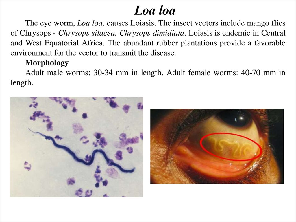
Loa loaThe eye worm, Loa loa, causes Loiasis. The insect vectors include mango flies
of Chrysops - Chrysops silacea, Chrysops dimidiata. Loiasis is endemic in Central
and West Equatorial Africa. The abundant rubber plantations provide a favorable
environment for the vector to transmit the disease.
Morphology
Adult male worms: 30-34 mm in length. Adult female worms: 40-70 mm in
length.
52.
53.
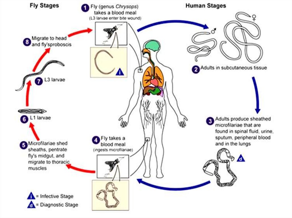
SymptomsThe microfilaria have a sheath. Their diurnal periodicity
corresponds to the feeding pattern of the insect vector, which bites
humans from 10:00 AM to 4:00 PM. Incubation period is about
one year. It causes calabar swelling beneath the skin due to
parasites. There is fever, pain, pruritus, urticaria, allergic reactions,
retinopathy, glomerulonephritis, meningo-encephalitis etc.
Diagnosis
• Detection of microfilaria in peripheral blood, urine, sputum,
CSF - stained with Giemsa or unstained
• Eosinophilia
Treatment
DEC, 6 to 10 mg per kilogram per day for 2 to 3 weeks: but
has side effects - allergic reactions
54.
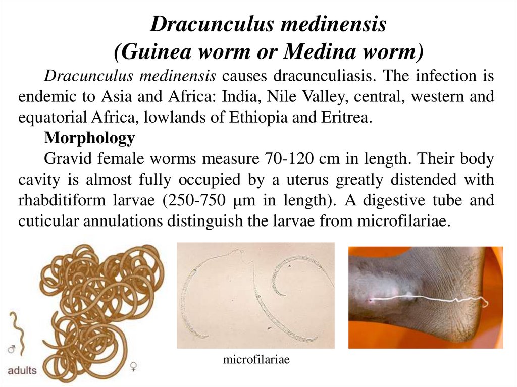
Dracunculus medinensis(Guinea worm or Medina worm)
Dracunculus medinensis causes dracunculiasis. The infection is
endemic to Asia and Africa: India, Nile Valley, central, western and
equatorial Africa, lowlands of Ethiopia and Eritrea.
Morphology
Gravid female worms measure 70-120 cm in length. Their body
cavity is almost fully occupied by a uterus greatly distended with
rhabditiform larvae (250-750 μm in length). A digestive tube and
cuticular annulations distinguish the larvae from microfilariae.
microfilariae
55.
Life cycleDefinitive host: Man. No animal host or reservoir is known for
W. bancrofti.
Intermediate host: Female mosquito, of different species acts
as vectors in different geographic areas. The major vector in India
and most other parts of Asia is Culex quinquefasciatus (C. fatigans).
Infective form: Actively motile third-stage filariform larva is
infective to man.
Mode of transmission: Humans get infection by bite of
mosquito carrying filariform larva.
56.
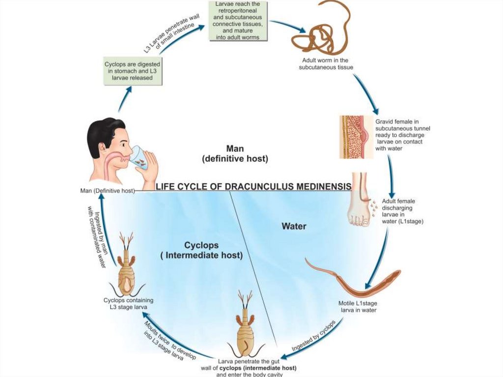
Infection is acquired by drinking unfiltered or not boiled water that containsCyclops species. The larvae are released in the stomach, penetrate the intestinal
wall and find their way to the subcutaneous tissue. Mating takes place in the
axillary or inguinal regions 3 months after infection. The male worms then die in
the tissue and the female worms move down to the limbs within 10 months. In
about 1 year, female worms in the subcutaneous tissue provoke the formation of a
burning blister in the skin of the legs. When in water, the blister bursts, and about
5 cm of the worm is extruded from the resulting ulcer - thus releasing many
thousands of first stage larvae. The larvae swim in water and are ingested by the
intermediate host - Cyclops species- within about 4 days. Inside the Cyclops, the
larvae molt twice and become infective in 2 weeks.
57.
58.
SymptomsThe female parasites in the subcutaneous tissue release toxic byproducts of
histamine-like nature, which cause systemic allergic reactions, like erythema,
urticaria, pruritus, fainting, asthma, dyspnea, etc. This is followed by the
appearance of a blister on the legs, which ruptures on contact with water releasing
larvae into the water by the female worm. The wound may ulcerate. The worms
migrate into other tissues and may cause arthritis, pericarditis, abscesses etc. It
occasionally penetrates the eyeball and causes loss of the eye.
Early stage–fever, malaise, urticaria, fugitive swelling, lymphangitis. Chronic
stage–lymphadenitis,
lymphangiovarix, chyluria, hydrocele, and elephantiasis. Tropical pulmonary
eosinophilia occurs due to hypersensitivity reaction to fi larial antigen.
Diagnosis
Examination of blood (eosonophilia)
Microscopy of peripheral blood (microfilaria)
Demonstration of adult worm in biopsy
X-ray.
Serological tests.
PCR
59.
TreatmentSurgical excision when the worm is in the leg
Niridazole (Ambilhar) or DEC
Prophylaxis
Eradication of the vector mosquito
Detection and treatment of carriers.
60.
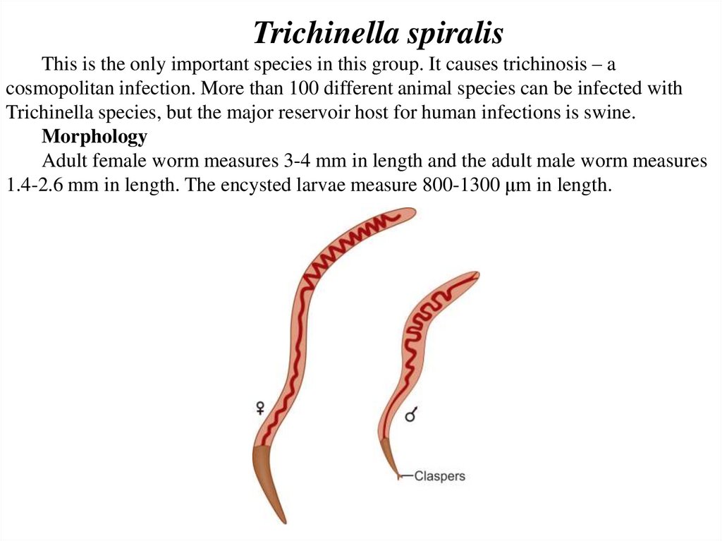
Trichinella spiralisThis is the only important species in this group. It causes trichinosis – a
cosmopolitan infection. More than 100 different animal species can be infected with
Trichinella species, but the major reservoir host for human infections is swine.
Morphology
Adult female worm measures 3-4 mm in length and the adult male worm measures
1.4-2.6 mm in length. The encysted larvae measure 800-1300 μm in length.
61.
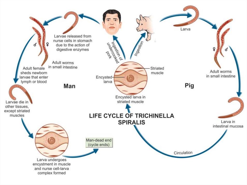
Life cycleAfter ingesting infected meat, the capsule of the encysted larvae
is digested by gastric juice, and the larvae are released in the
duodenum or jejunum where they molt four times to become adult
worm. After mating, the male worm dies and the female worm
begins to deliver the embryos 4-7 days after the infection. The
larvae penetrate the intestinal wall and migrate through the
lymphatic vessels to the blood stream, which carries them to various
organs. Skeletal muscles and diaphragm are most frequently
parasitized. Others include the tongue, masseter and ocular muscles.
62.
63.
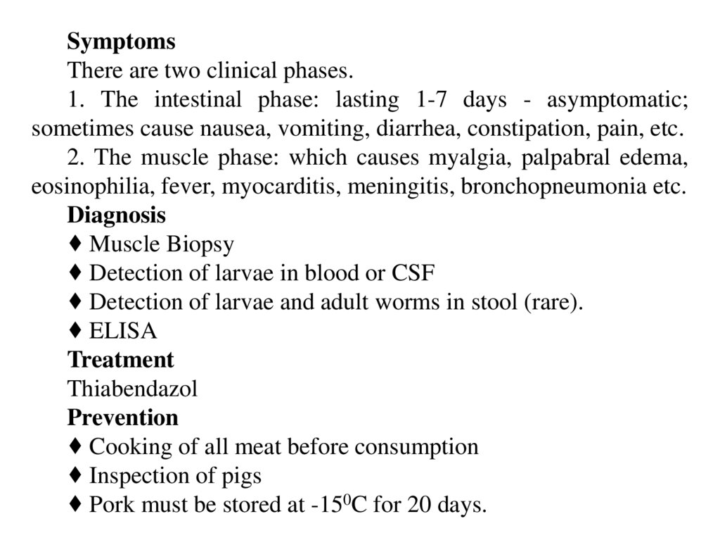
SymptomsThere are two clinical phases.
1. The intestinal phase: lasting 1-7 days - asymptomatic;
sometimes cause nausea, vomiting, diarrhea, constipation, pain, etc.
2. The muscle phase: which causes myalgia, palpabral edema,
eosinophilia, fever, myocarditis, meningitis, bronchopneumonia etc.
Diagnosis
♦ Muscle Biopsy
♦ Detection of larvae in blood or CSF
♦ Detection of larvae and adult worms in stool (rare).
♦ ELISA
Treatment
Thiabendazol
Prevention
♦ Cooking of all meat before consumption
♦ Inspection of pigs
♦ Pork must be stored at -150C for 20 days.
64.
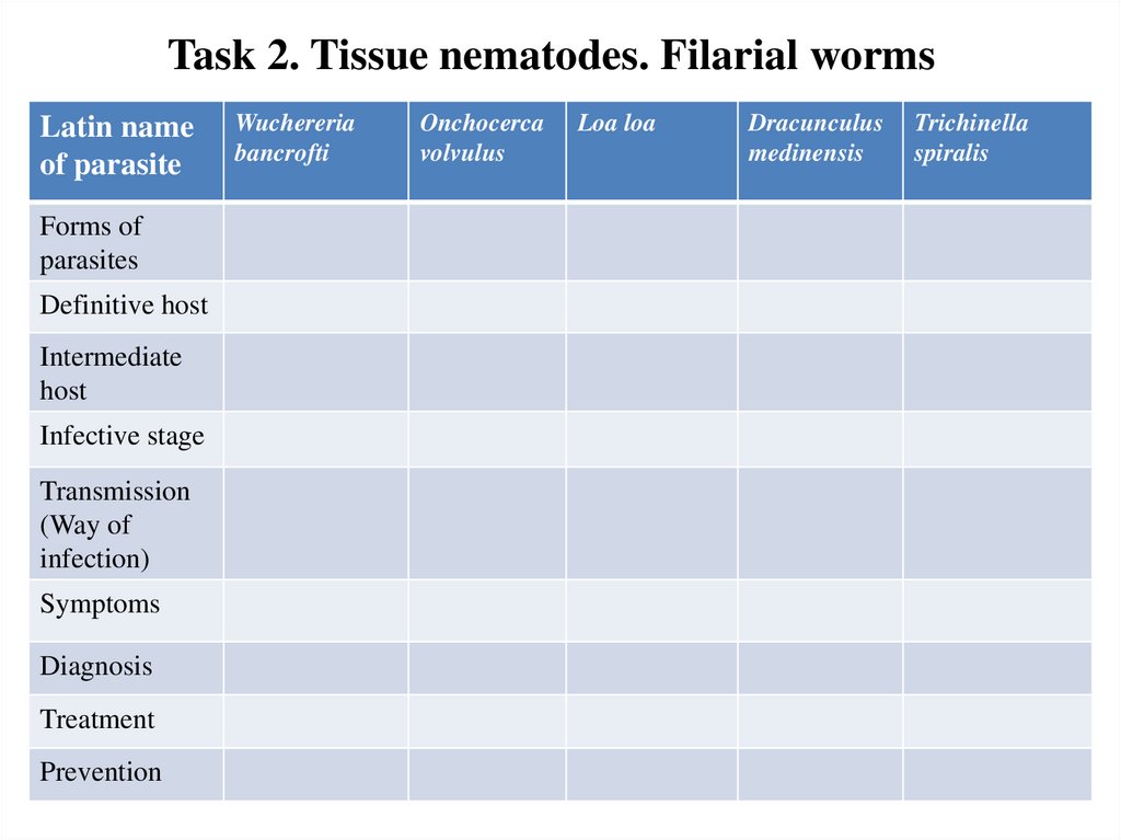
Task 2. Tissue nematodes. Filarial wormsLatin name
of parasite
Forms of
parasites
Definitive host
Intermediate
host
Infective stage
Transmission
(Way of
infection)
Symptoms
Diagnosis
Treatment
Prevention
Wuchereria
bancrofti
Onchocerca
volvulus
Loa loa
Dracunculus
medinensis
Trichinella
spiralis