


 Медицина
Похожие презентации:


Ultrasound Elastography in the Evaluation of Venous Thrombosis - try to apply in clinical practice
1. Ultrasound Elastography in the Evaluation of Venous Thrombosis - try to apply in clinical practice
VARADY’S 30TH INTERNATIONAL WORKSHOP FOR PHLEBOLOGY, LYMPHOLOGY AND ANGIOLOGYFRANKFURT 17-18.04.2015
Ultrasound Elastography in the Evaluation
of Venous Thrombosis
- try to apply in clinical practice
TA D E U S Z W I L KO S Z , T O M A S Z D R Ą Ż K I E W I C Z , G R Z E G O R Z L E W I Ń S K I
J O H N PA U L I I H O S P I TA L , C R A C O W - P O L A N D
2.
What is Elastography?Elastography is an imaging technique to measure the stiffness of tissues
Images are acquired before and after soft compression of tissues and the
deformation is evaluated
Initially elastography used manual compression and was only qualitative,
now some methods appears to apply a non operator dependant
compression
3.
Elastography was developed first in the US fieldThree step approach:
1. Organs mechanically stressed by either external or internal forces.
2. Measurement of tissues movement induced
3. Qualitative or quantitative evaluation of tissue elastic properties from
the measured displacement of tissues.
4.
Several Approaches1. Manual compression by operator using the transducer (static
elastography - strain elastography)
2. Organ compression by heartbeat or vascular pulsations
3. Push pulse waves compression
4. Supersonic shear waves
5. Sonoelastography
• Strain elastography (manual mechanical compression of transducer)is a ultrasonic (US) technique invented by Hitachi Medical and
Tsukuba University in Japan
• Dynamic Elastography ( Shear Wave Elastography) introduced by
SuperSonic Imagine in France
6.
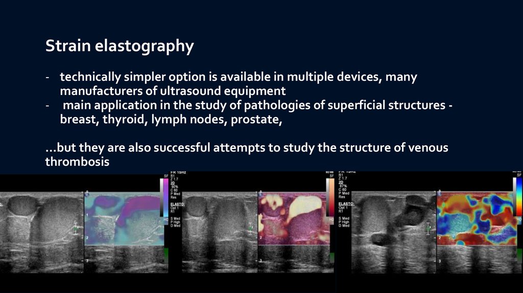
Strain elastography- technically simpler option is available in multiple devices, many
manufacturers of ultrasound equipment
- main application in the study of pathologies of superficial structures breast, thyroid, lymph nodes, prostate,
…but they are also successful attempts to study the structure of venous
thrombosis
7.
STRAIN (STATIC) ELASTOGRAPHYAxial and lateral deformations
after an axial constraint
B-mode
Elastography mode
8.
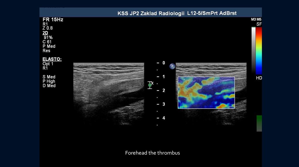
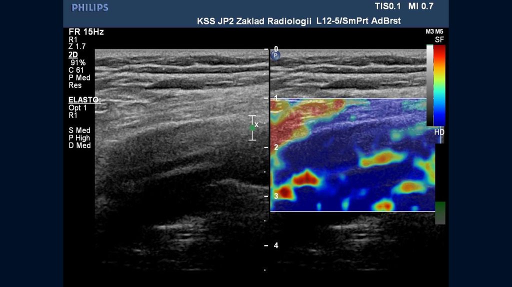
Application of vein elastographyWhat is the age of the thrombus?
Whether there are features of recanalization?
9.
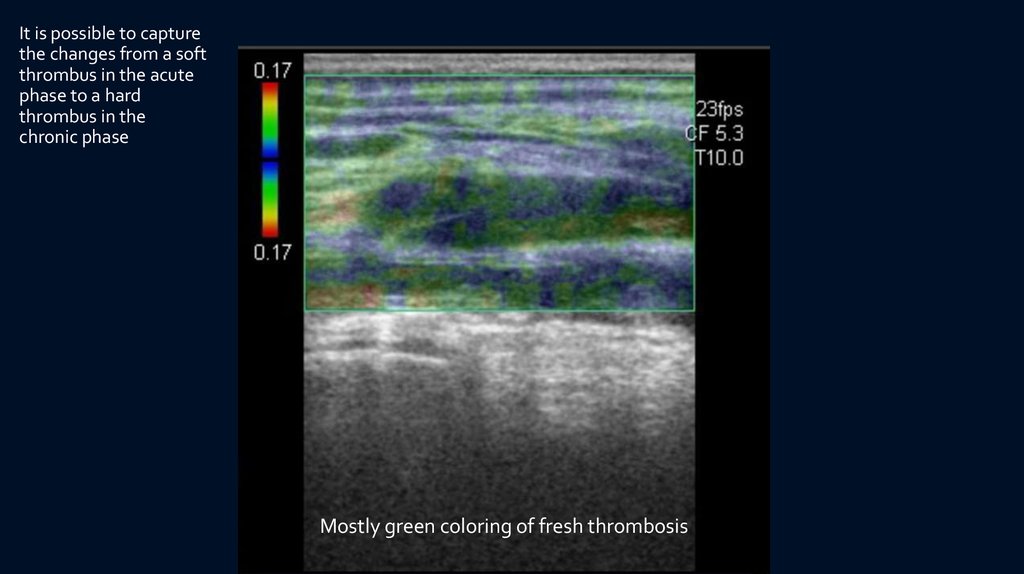
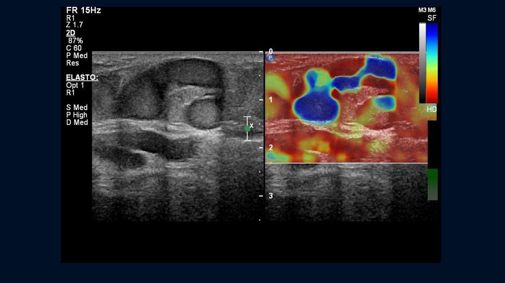
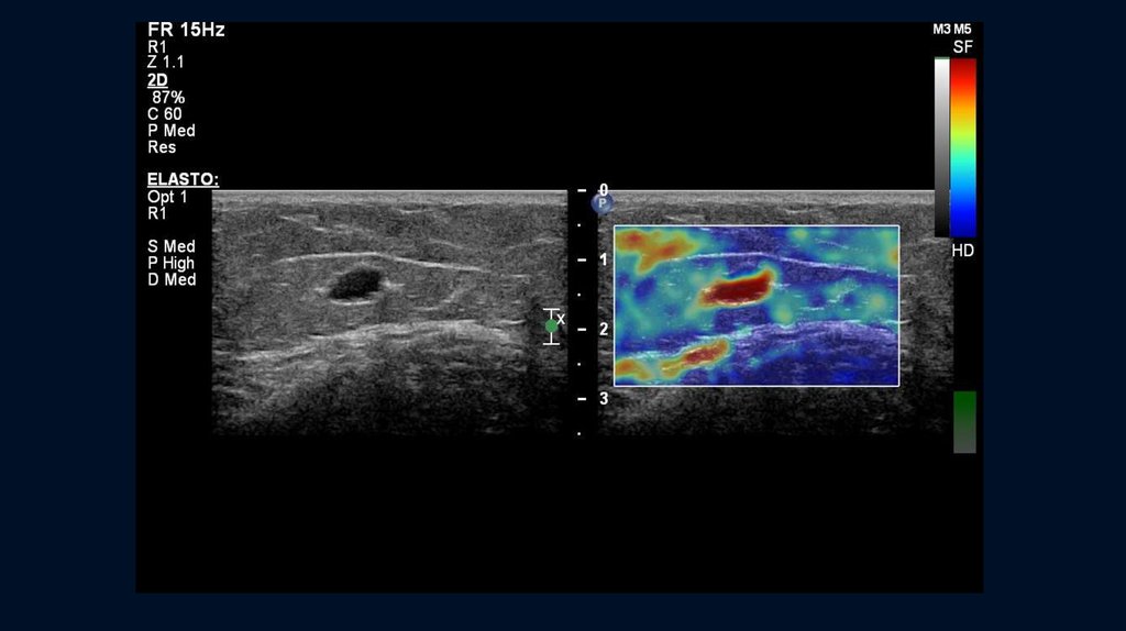
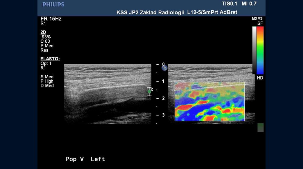
It is possible to capturethe changes from a soft
thrombus in the acute
phase to a hard
thrombus in the
chronic phase
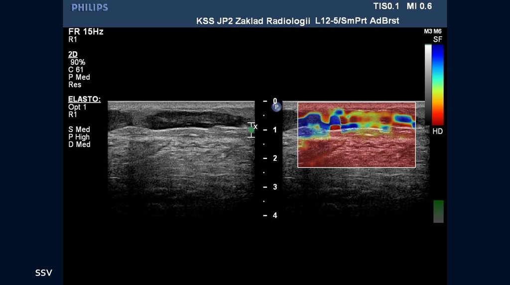
Mostly green coloring of fresh thrombosis
10.
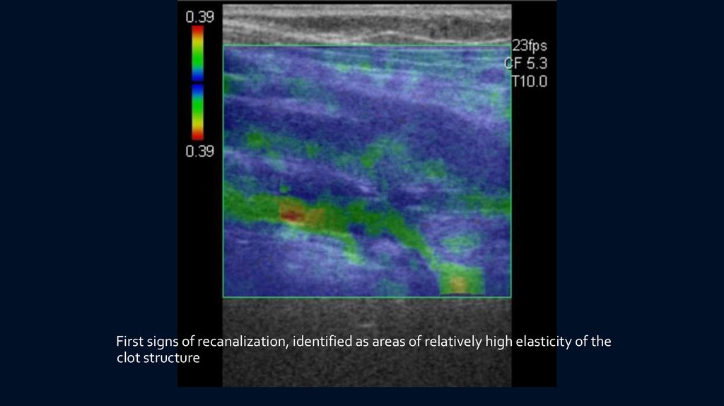
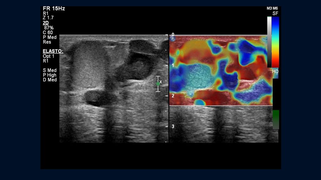
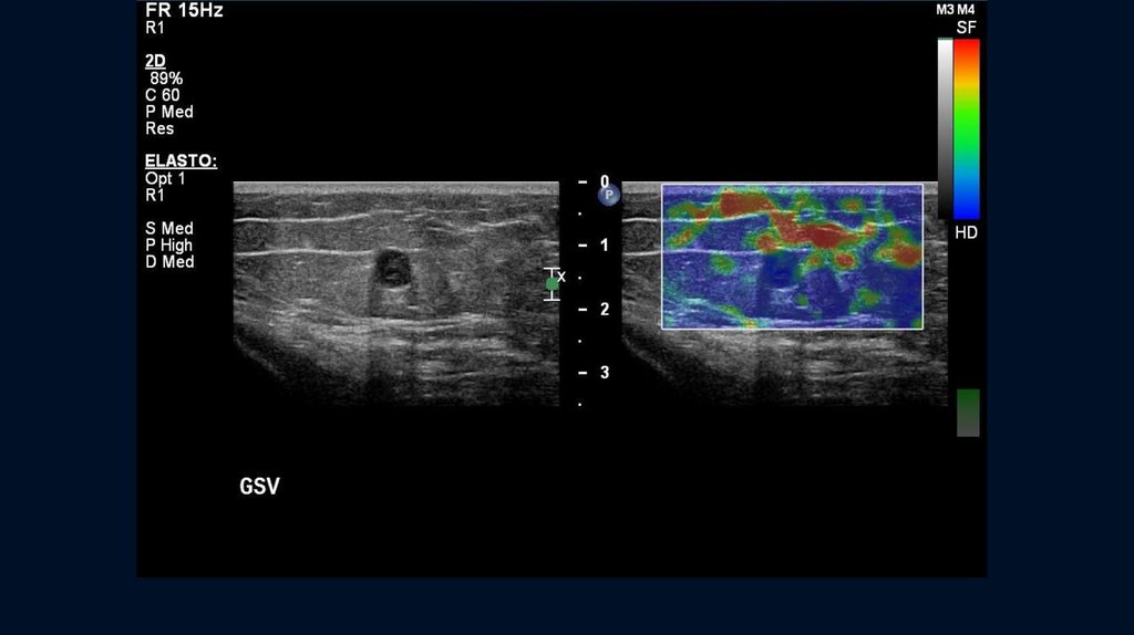
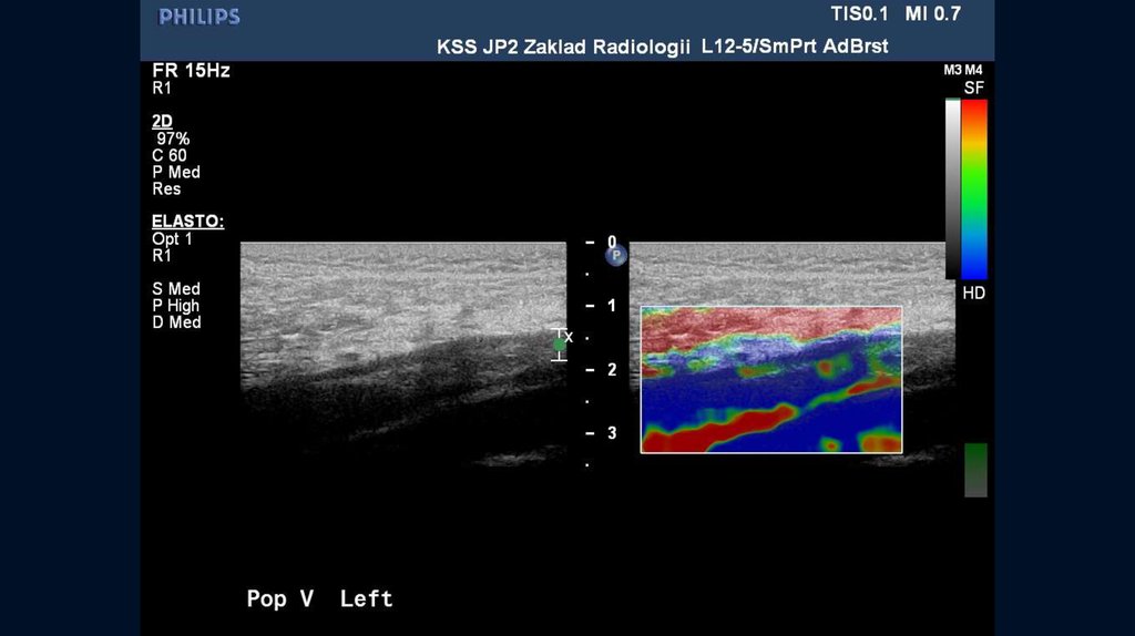
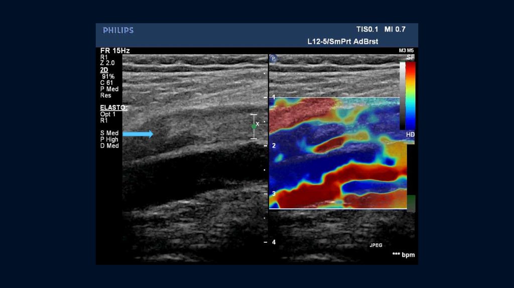
First signs of recanalization, identified as areas of relatively high elasticity of theclot structure
11.
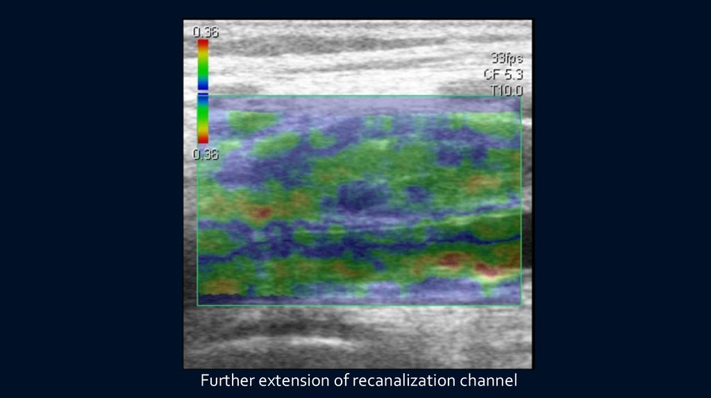
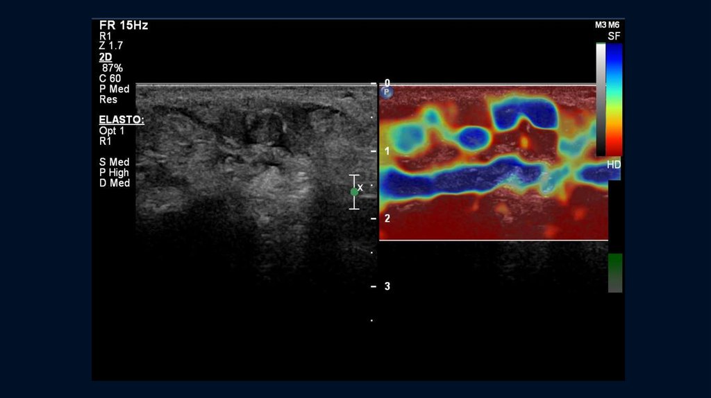
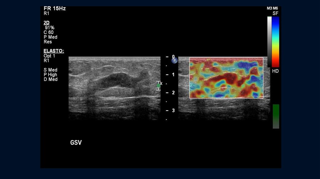
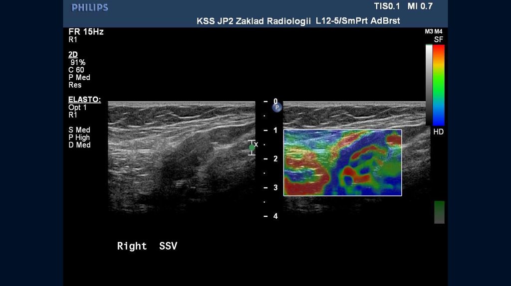
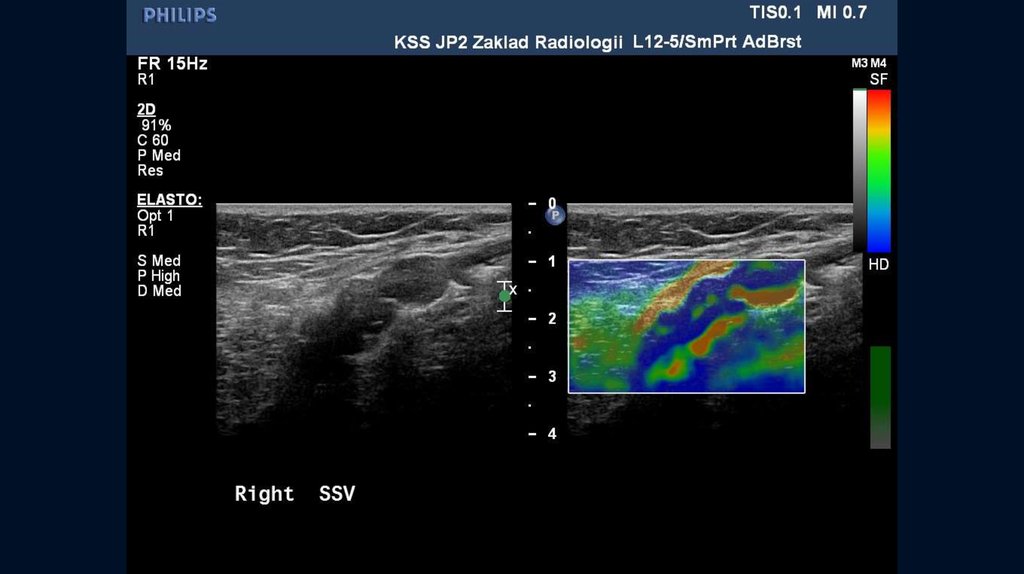
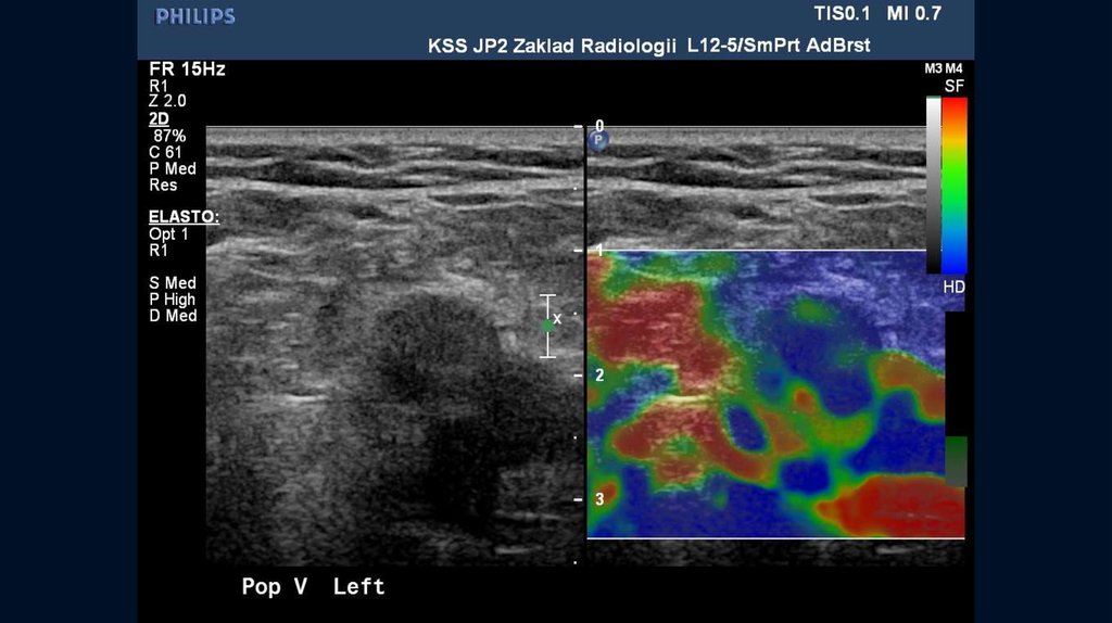
Further extension of recanalization channel12.
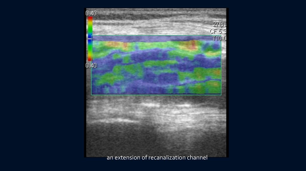
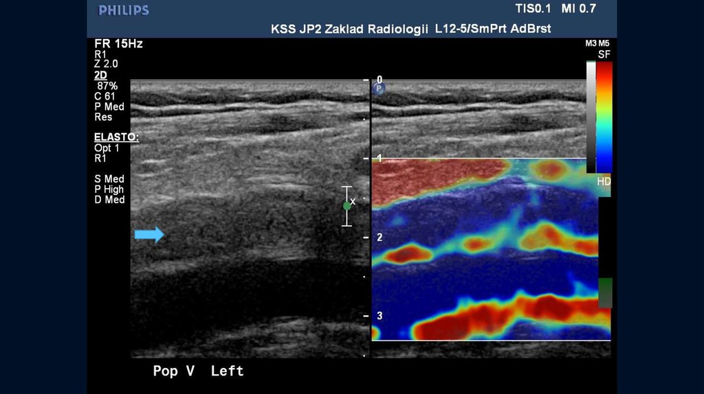
an extension of recanalization channel13.
14.
15.
16.
17.
18.
19.
20.
21.
22.
23.
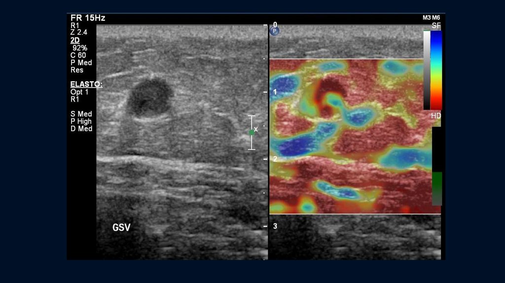
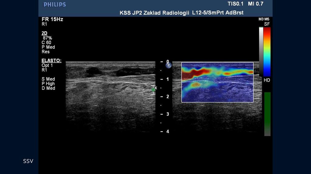
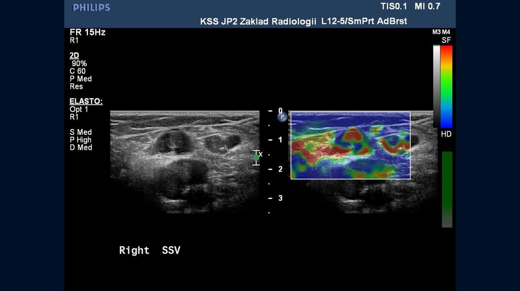
SSV24.
SSV25.
SSV26.
SSV27.
28.
29.
30.
31.
32.
33.
34.
35.
Forehead the thrombus36.
37. POPLITEAL VEIN
38.
39.
ConclusionsUSEG usage for dynamic monitoring of acute venous thrombosis can help in identifying
of initial signs of thrombus recanalization
Changes in clot structure can define probable prognosis of recanalization.
Elastography would be useful for evaluating the tissue characteristics of venous thrombi,
enabling selection of the treatment method and forecast of complications
Further review is desired
40.
REFERENCES1.USEFULNESS OF ULTRASONOGRAPHIC ELASTOGRAPHY FOR EVALUATION OF VENOUS THROMBOEMBOLISM
K. Shimizu,T. Tomaru, Cardiology Center,Toho University Sakura Hospital, Sakura
Circulation Journal Vol 73, Suppl 1, 2009
2.AGING OF VENOUS THROMBOSIS - COMPARISON BETWEEN HISTOLOGICAL AND TISSUE
ELASTICITY IMAGING K.Uno, A. Tonomura, M. Yamakawa, T. Ishizu, Y. Seo, S. Honma.
T. Shiina, K. Aonuma, Circulation Journal Vol 72, Suppl 1, 2008
3.ULTRASOUND ELASTICITY IMAGING CAN DETECT HIGH RISK PATIENTS WITH DEEP VEIN THROMBOSIS WHO
RESULTS IN PULMONARY EMBOLISM
H.Kato, M.Nishino, M. Hara, T. Yoshimura, D.Nakamura, Y. Lee, S. Nakatani, A. Hashimoto, K. Yamagami, Y. Egami, R. Shutta,
H. Yamaguchi, J.Tanouchi, Y. Yamada
Division of Cardiology, Osaka Rosai Hospital, Sakai, Circulation Journal Vol 73, Suppl 1, 2009
4.AGING OF VENOUS THROMBOSIS - COMPARISON BETWEEN HISTOLOGICAL AND TISSUE
ELASTICITY IMAGING
K. Uno, A. Tonomura, M. Yamakawa, T. Ishizu, Y. Seo, S. Honma, T. Shiina, K. Aonuma, Circulation Journal Vol 72, Suppl 1, 2008
5.ULTRASONIC ELASTOGRAPHY IMAGING OF DEEP VENOUS THROMBOSIS
K. Shimizu, T. Tomaru, K. Nakamura, K. Hirano, H. Noike, M. Takahashi, T. Sakurai. Department of Cardiovascularcenter, Sakura
Hospital,Toho University School of Medicine, Chiba, Circulation Journal Vol 71 Suppl 1 PJ-709, 2007
6.APPLICATION OF ULTRASONIC ELASTOGRAPHY FOR EVALUATION OF DEEP VENOUS THROMBOSIS
K. Shimizu, K. Nakamura, T. Hirohashi, et al.,Circulation Journal Vol 70 Suppl 1 PE-623, 2006
7.EXPERIENCE OF USING ULTRASOUND ELASTOGRAPHY FOR DYNAMIC MONITORING OF RECANALISATION OF
VENOUS THROMBOSIS OF LOWER EXTREMITIES IN ADULTS AND CHILDREN A. R. Zubarev, N. Krivosheeva, A.
Demidova, I. Rychkova, E. A. Zubareva; ECR 2012
8.SONOGRAPHISCHE KLASSIFIKATION DER TIEFEN VENOSEN THROMBOSE S.Siebers, B.Geier, M.Vogt, U.Scheipers,
A.Mumme, H.Ermert, Ultraschall in der Medizin, No. 25. Sept.2004, p.S35