 Медицина
Похожие презентации:


")


")

Takayasu’s arteritis
1.
TAKAYASU’S ARTERITISDr Katya Dolnikov
2017
D_katya@rambam.health.gov.il
2.
3.
4.
5.
6.
7.
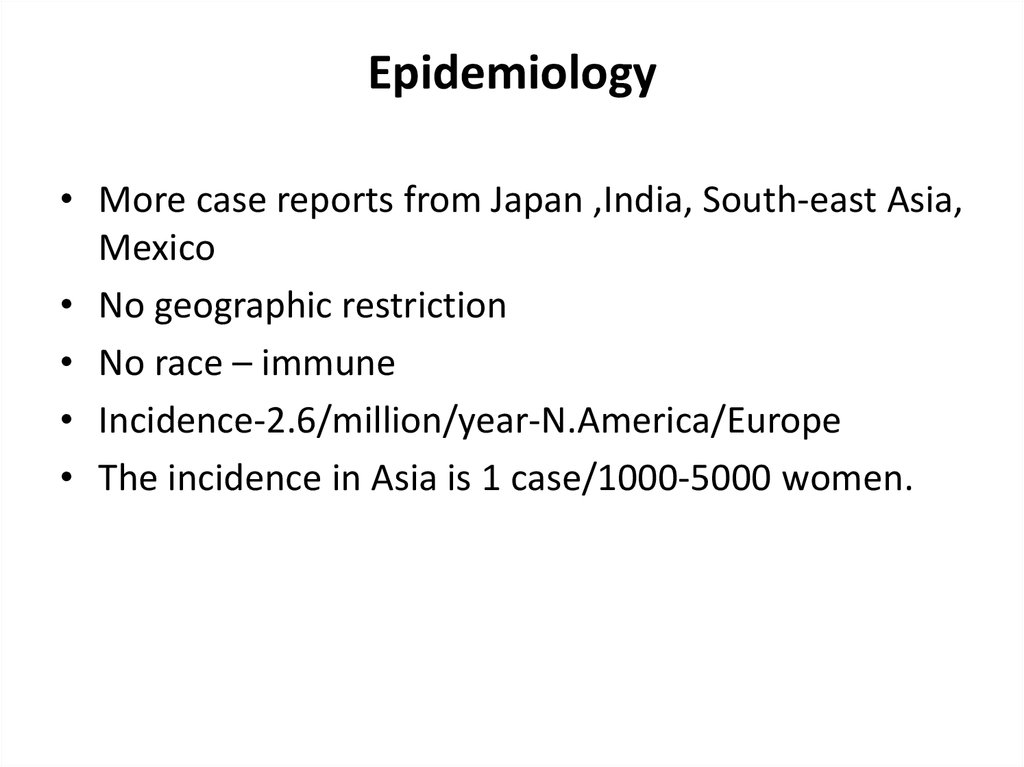
Epidemiology• More case reports from Japan ,India, South-east Asia,
Mexico
• No geographic restriction
• No race – immune
• Incidence-2.6/million/year-N.America/Europe
• The incidence in Asia is 1 case/1000-5000 women.
8.
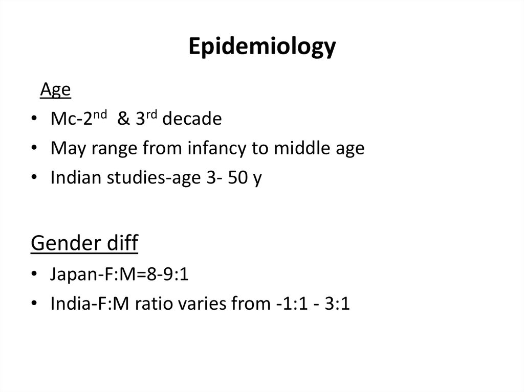
EpidemiologyAge
• Mc-2nd & 3rd decade
• May range from infancy to middle age
• Indian studies-age 3- 50 y
Gender diff
• Japan-F:M=8-9:1
• India-F:M ratio varies from -1:1 - 3:1
9.
Genetics• Japan - HLA-B52 and B39
• Mexican and Colombian patients - HLA-DRB1*1301
and HLA-DRB1*1602
• India- HLA- B 5, -B 21
10.
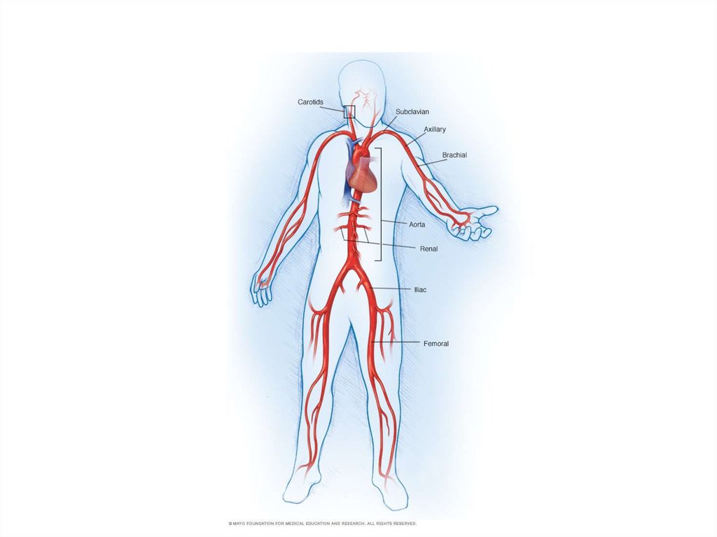
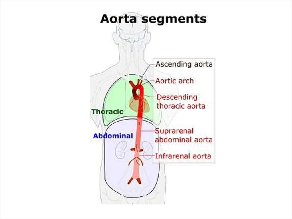
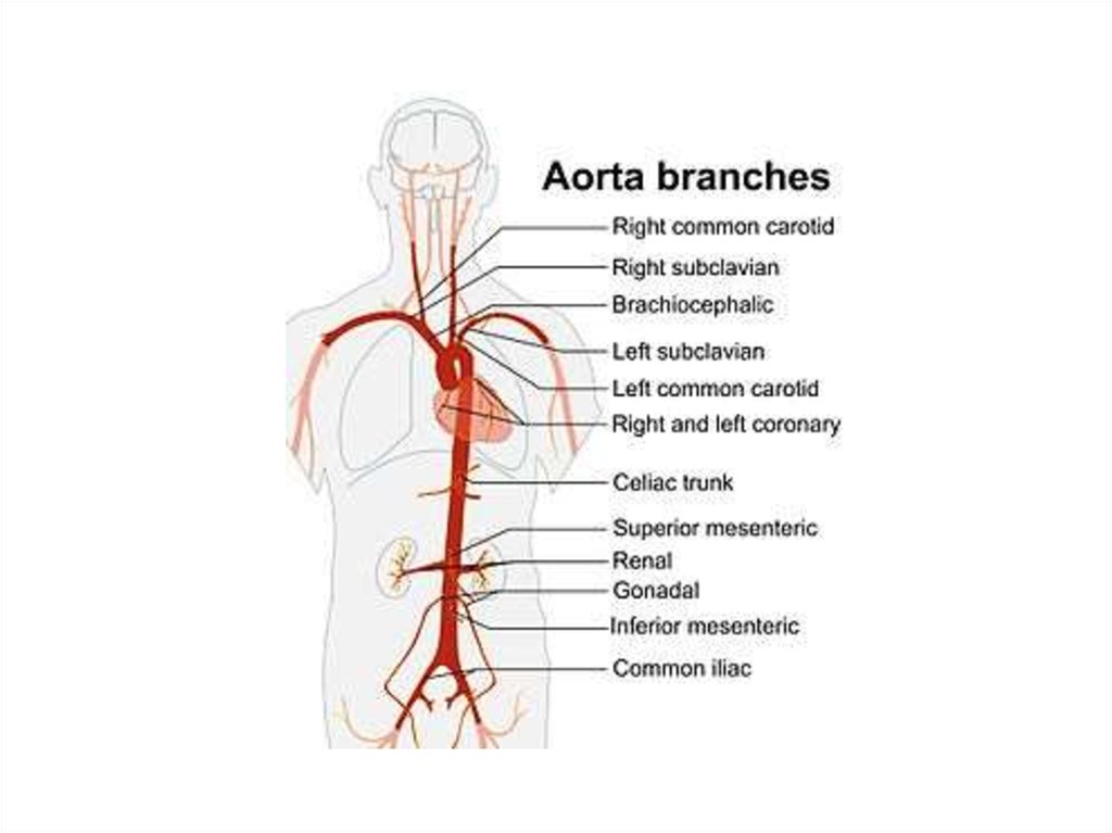
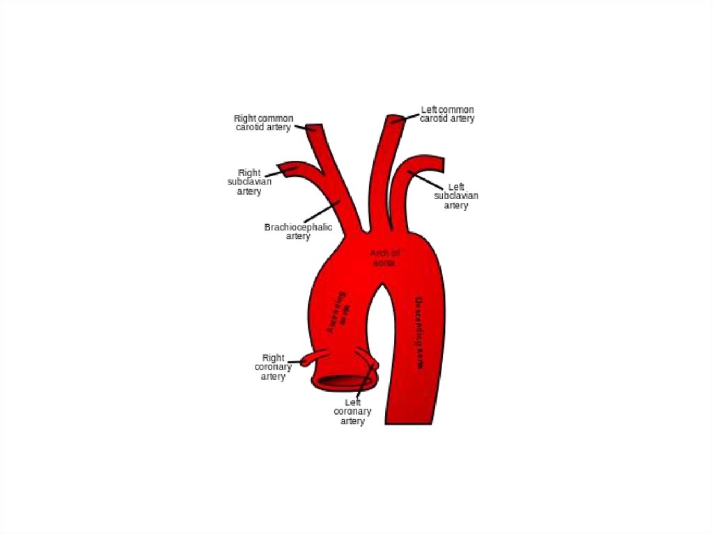
Histopathology• Idiopathic inflammatory arteritis of elastic arteries
resulting in occlusive/ ectatic changes
• Large vessels – Aorta and its main branches
(brachiocephalic, carotid, SCL, vertebral, RA)
• Coronary and PA involvement
• Aorta - usually not beyond IMA
• Multiple segments with skipped areas
or diffuse involvement
11.
Pathogenesis• Antigen-driven disease, with the site
of immunologic recognition events
being the adventitia.
• DC in adventitia activated by AG
release IL-18 and chemokines that
“recruit” T cells from vasa vasorum
to the vessel wall
• CD4+ T cells secrete interferon-γ→
stimulate macrophages and
multinucleated giant cells
• The results of this inflammatory
cascade are :
– granulomatous inflammation
– destruction of the internal elastic
lamina
– arterial wall hyperplasia, smooth
muscle cell proliferation, intimal
thickening, vascular occlusion
12.
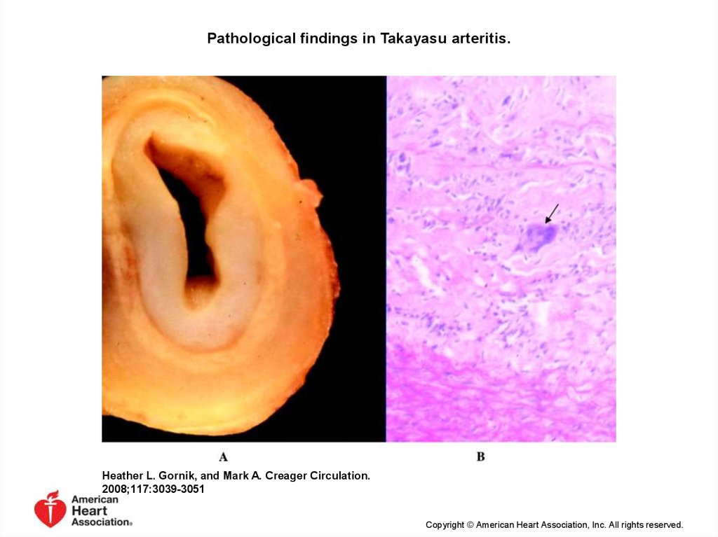
Pathological findings in Takayasu arteritis.Heather L. Gornik, and Mark A. Creager Circulation.
2008;117:3039-3051
Copyright © American Heart Association, Inc. All rights reserved.
13.
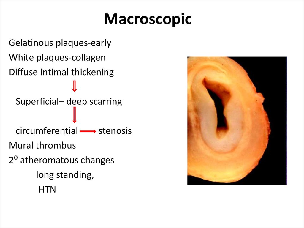
MacroscopicGelatinous plaques-early
White plaques-collagen
Diffuse intimal thickening
Superficial– deep scarring
circumferential
stenosis
Mural thrombus
2⁰ atheromatous changes
long standing,
HTN
14.
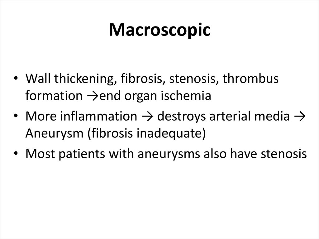
Macroscopic• Wall thickening, fibrosis, stenosis, thrombus
formation →end organ ischemia
• More inflammation → destroys arterial media →
Aneurysm (fibrosis inadequate)
• Most patients with aneurysms also have stenosis
15.
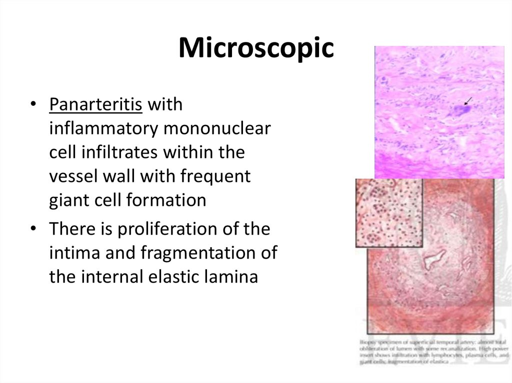
Microscopic• Panarteritis with
inflammatory mononuclear
cell infiltrates within the
vessel wall with frequent
giant cell formation
• There is proliferation of the
intima and fragmentation of
the internal elastic lamina
16.
17.
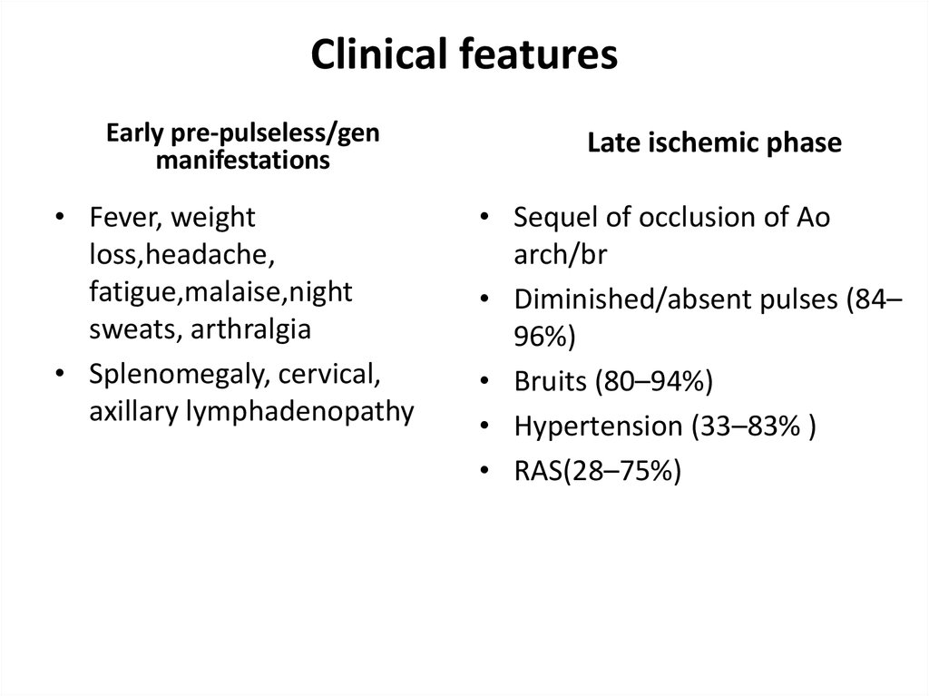
Clinical featuresEarly pre-pulseless/gen
manifestations
• Fever, weight
loss,headache,
fatigue,malaise,night
sweats, arthralgia
• Splenomegaly, cervical,
axillary lymphadenopathy
Late ischemic phase
• Sequel of occlusion of Ao
arch/br
• Diminished/absent pulses (84–
96%)
• Bruits (80–94%)
• Hypertension (33–83% )
• RAS(28–75%)
18.
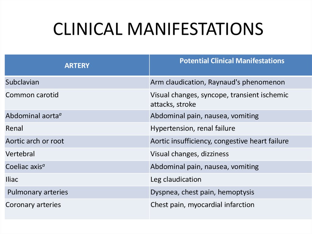
CLINICAL MANIFESTATIONSARTERY
Potential Clinical Manifestations
Subclavian
Arm claudication, Raynaud's phenomenon
Common carotid
Abdominal aortaa
Visual changes, syncope, transient ischemic
attacks, stroke
Abdominal pain, nausea, vomiting
Renal
Hypertension, renal failure
Aortic arch or root
Aortic insufficiency, congestive heart failure
Vertebral
Visual changes, dizziness
Coeliac axisa
Abdominal pain, nausea, vomiting
Iliac
Leg claudication
Pulmonary arteries
Dyspnea, chest pain, hemoptysis
Coronary arteries
Chest pain, myocardial infarction
19.
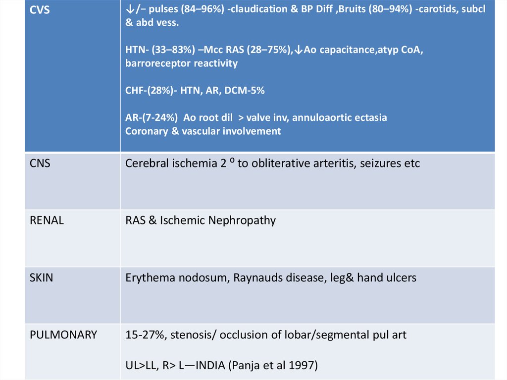
CVS↓/− pulses (84–96%) -claudication & BP Diff ,Bruits (80–94%) -carotids, subcl
& abd vess.
HTN- (33–83%) –Mcc RAS (28–75%),↓Ao capacitance,atyp CoA,
barroreceptor reactivity
CHF-(28%)- HTN, AR, DCM-5%
AR-(7-24%) Ao root dil > valve inv, annuloaortic ectasia
Coronary & vascular involvement
CNS
Cerebral ischemia 2 ⁰ to obliterative arteritis, seizures etc
RENAL
RAS & Ischemic Nephropathy
SKIN
Erythema nodosum, Raynauds disease, leg& hand ulcers
PULMONARY
15-27%, stenosis/ occlusion of lobar/segmental pul art
UL>LL, R> L—INDIA (Panja et al 1997)
20.
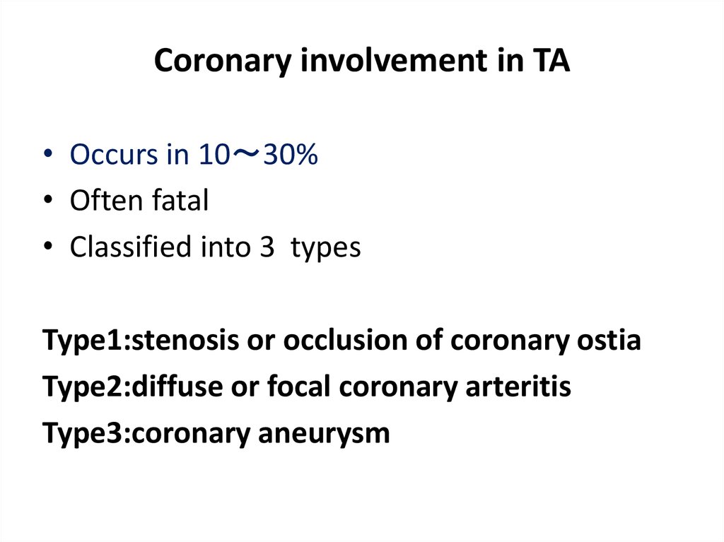
Coronary involvement in TA• Occurs in 10 30%
• Often fatal
• Classified into 3 types
Type1:stenosis or occlusion of coronary ostia
Type2:diffuse or focal coronary arteritis
Type3:coronary aneurysm
21.
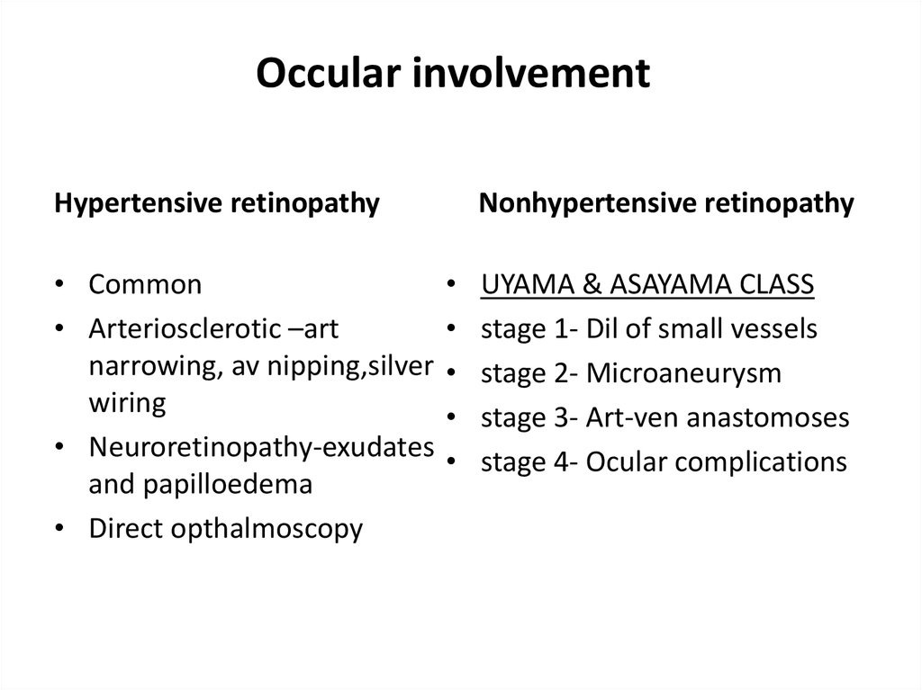
Occular involvementHypertensive retinopathy
• Common
• Arteriosclerotic –art
narrowing, av nipping,silver
wiring
• Neuroretinopathy-exudates
and papilloedema
• Direct opthalmoscopy
Nonhypertensive retinopathy
UYAMA & ASAYAMA CLASS
stage 1- Dil of small vessels
stage 2- Microaneurysm
stage 3- Art-ven anastomoses
stage 4- Ocular complications
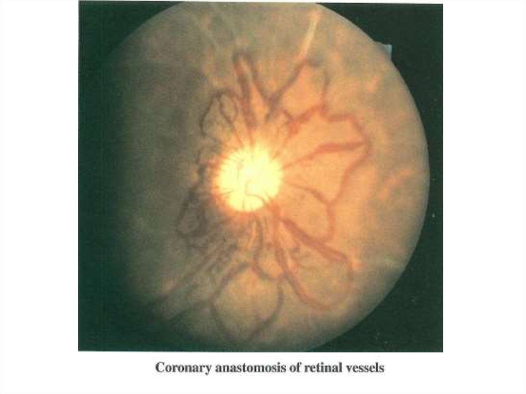
22.
23.
nee24.
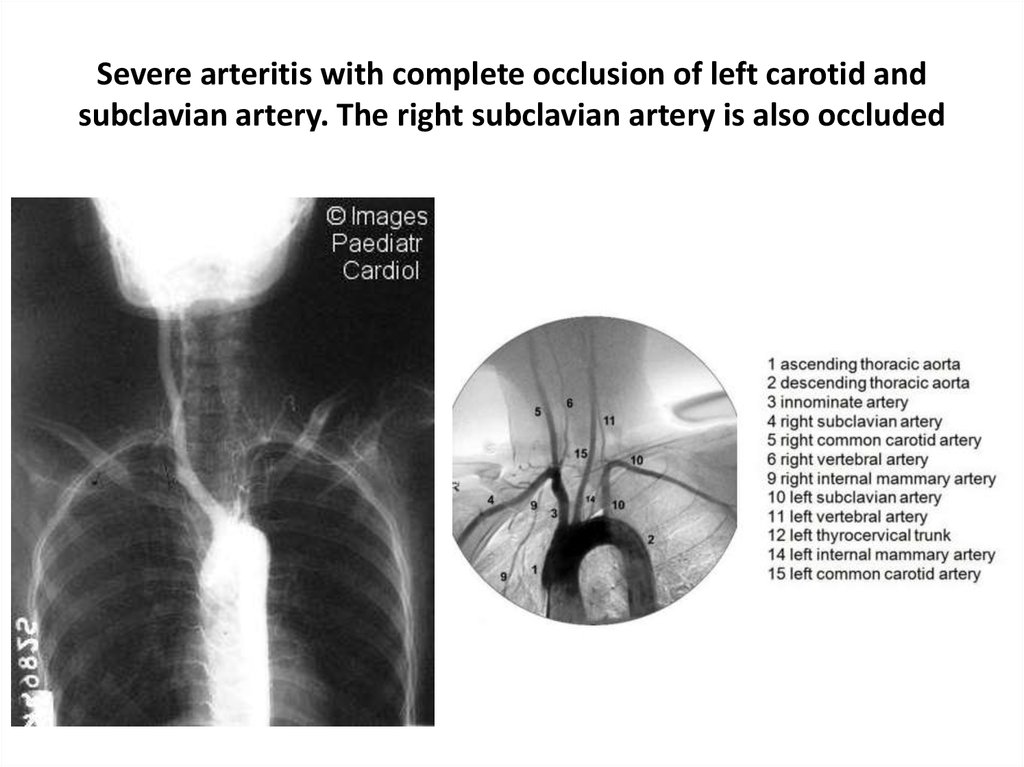
Severe arteritis with complete occlusion of left carotid andsubclavian artery. The right subclavian artery is also occluded
25.
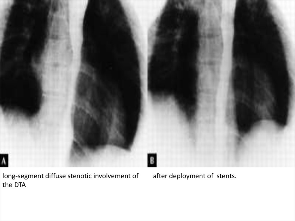
long-segment diffuse stenotic involvement ofthe DTA
after deployment of stents.
26.
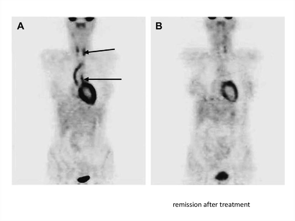
remission after treatment27.
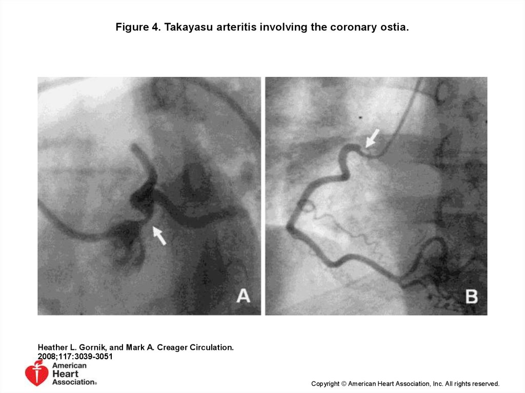
Figure 4. Takayasu arteritis involving the coronary ostia.Heather L. Gornik, and Mark A. Creager Circulation.
2008;117:3039-3051
Copyright © American Heart Association, Inc. All rights reserved.
28.
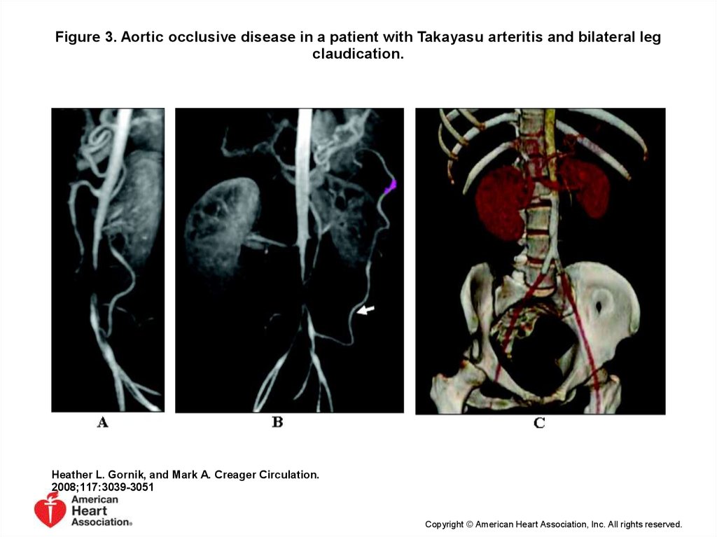
Figure 3. Aortic occlusive disease in a patient with Takayasu arteritis and bilateral legclaudication.
Heather L. Gornik, and Mark A. Creager Circulation.
2008;117:3039-3051
Copyright © American Heart Association, Inc. All rights reserved.
29.
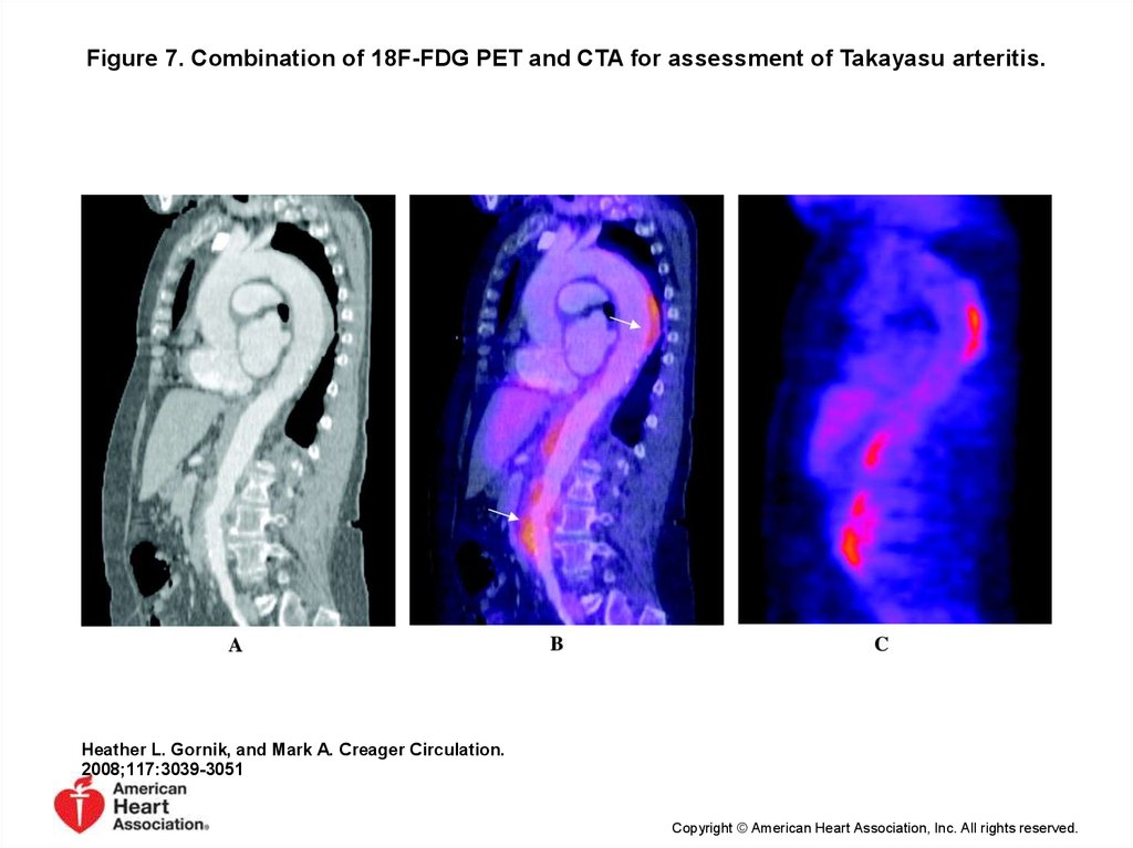
Figure 7. Combination of 18F-FDG PET and CTA for assessment of Takayasu arteritis.Heather L. Gornik, and Mark A. Creager Circulation.
2008;117:3039-3051
Copyright © American Heart Association, Inc. All rights reserved.
30.
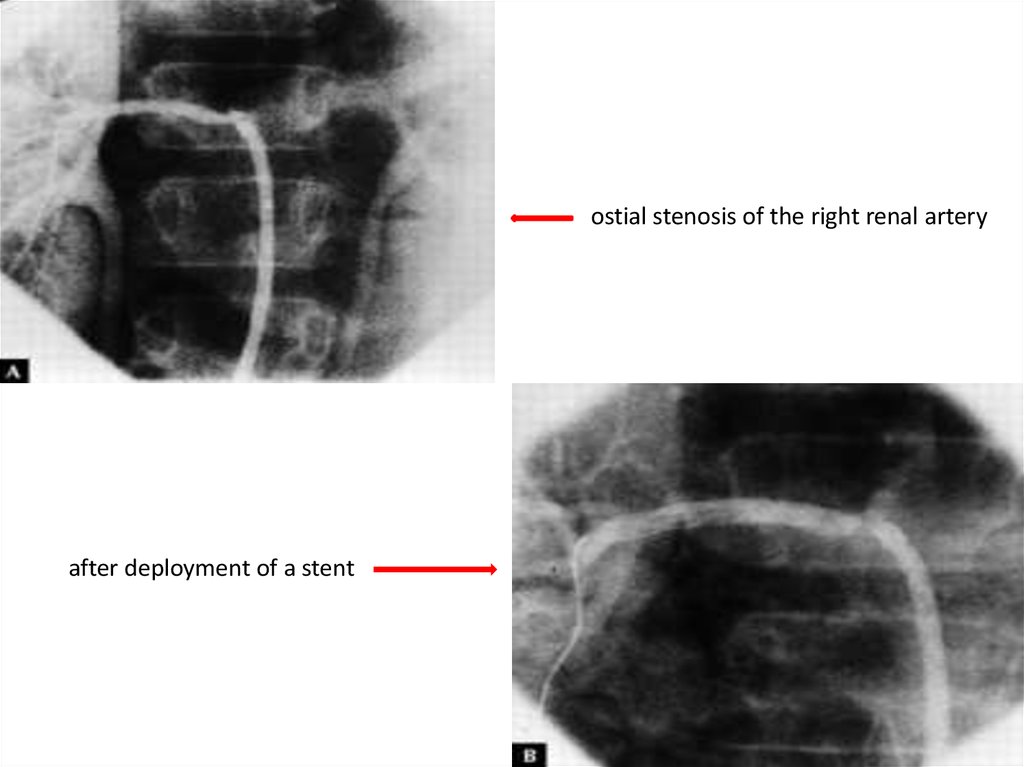
ostial stenosis of the right renal arteryafter deployment of a stent
31.
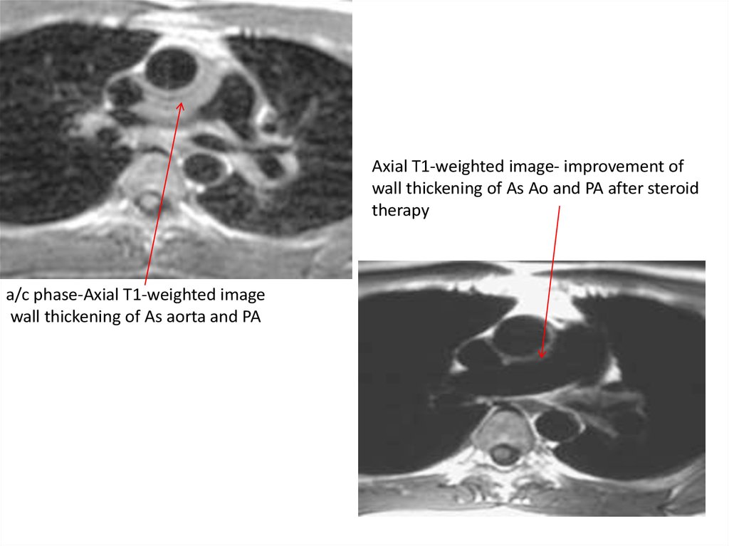
Axial T1-weighted image- improvement ofwall thickening of As Ao and PA after steroid
therapy
a/c phase-Axial T1-weighted image
wall thickening of As aorta and PA
32.
33.
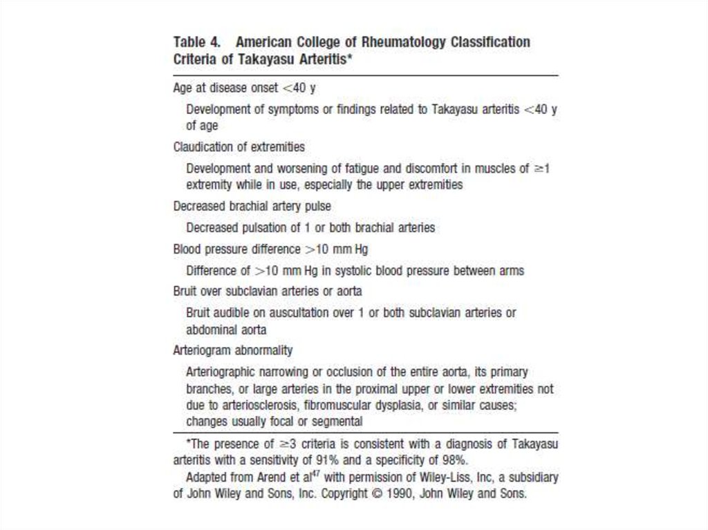
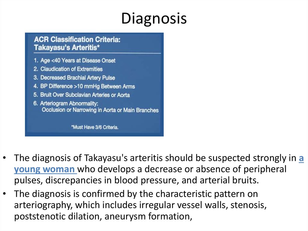
Diagnosis• The diagnosis of Takayasu's arteritis should be suspected strongly in a
young woman who develops a decrease or absence of peripheral
pulses, discrepancies in blood pressure, and arterial bruits.
• The diagnosis is confirmed by the characteristic pattern on
arteriography, which includes irregular vessel walls, stenosis,
poststenotic dilation, aneurysm formation,
34.
Treatment• Disease-related mortality most often occurs from congestive heart
failure, cerebrovascular events, myocardial infarction, aneurysm
rupture, or renal failure.
• The course of the disease is variable, and although spontaneous
remissions may occur, Takayasu's arteritis is most often chronic and
relapsing.
• Glucocorticoid therapy for acute signs and symptoms.
• An aggressive surgical and/or arterioplastic approach to stenosed
vessels. Unless it is urgently required, surgical correction of
stenosed arteries should be undertaken only when the vascular
inflammatory process is well controlled with medical therapy.
• In individuals who are refractory to or unable to taper
glucocorticoids, methotrexate in doses up to 25 mg per week has
yielded encouraging results.
• Anti-TNF therapies have encouraging results
35.
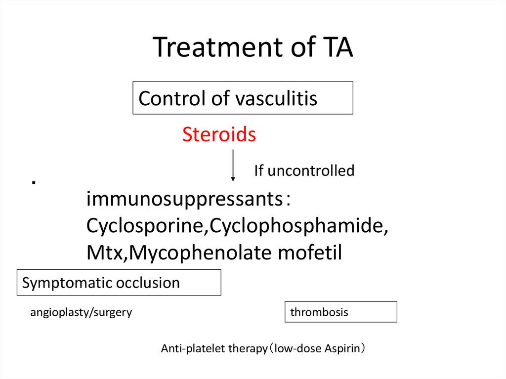
Treatment of TAControl of vasculitis
Steroids
・
If uncontrolled
immunosuppressants
Cyclosporine,Cyclophosphamide,
Mtx,Mycophenolate mofetil
Symptomatic occlusion
angioplasty/surgery
thrombosis
Anti-platelet therapy low-dose Aspirin
36.
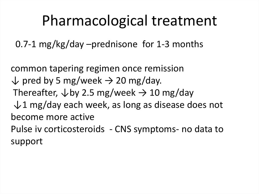
Pharmacological treatment0.7-1 mg/kg/day –prednisone for 1-3 months
common tapering regimen once remission
↓ pred by 5 mg/week → 20 mg/day.
Thereafter, ↓by 2.5 mg/week → 10 mg/day
↓1 mg/day each week, as long as disease does not
become more active
Pulse iv corticosteroids - CNS symptoms- no data to
support
37.
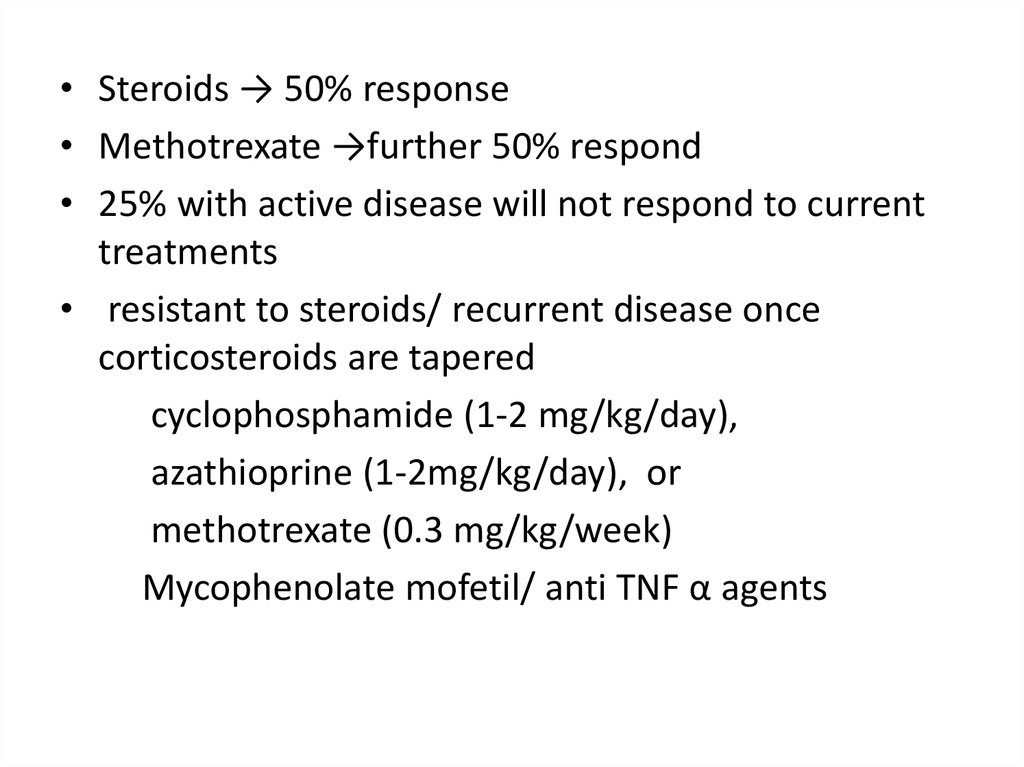
• Steroids → 50% response• Methotrexate →further 50% respond
• 25% with active disease will not respond to current
treatments
• resistant to steroids/ recurrent disease once
corticosteroids are tapered
cyclophosphamide (1-2 mg/kg/day),
azathioprine (1-2mg/kg/day), or
methotrexate (0.3 mg/kg/week)
Mycophenolate mofetil/ anti TNF α agents
38.
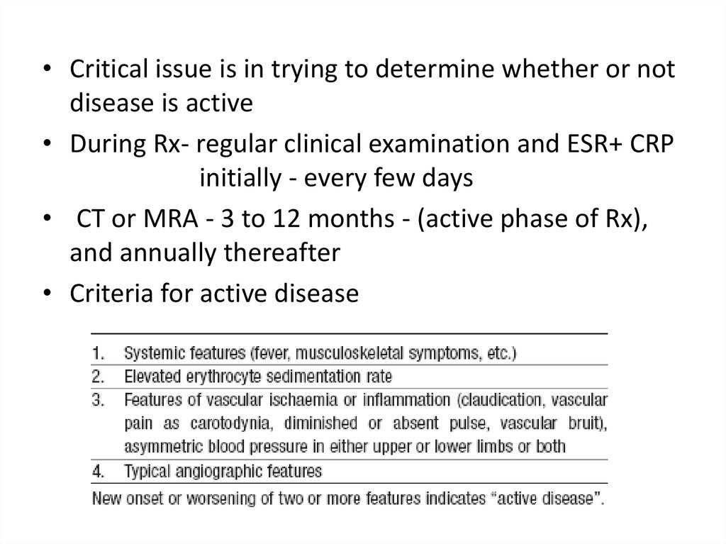
• Critical issue is in trying to determine whether or notdisease is active
• During Rx- regular clinical examination and ESR+ CRP
initially - every few days
• CT or MRA - 3 to 12 months - (active phase of Rx),
and annually thereafter
• Criteria for active disease
39.
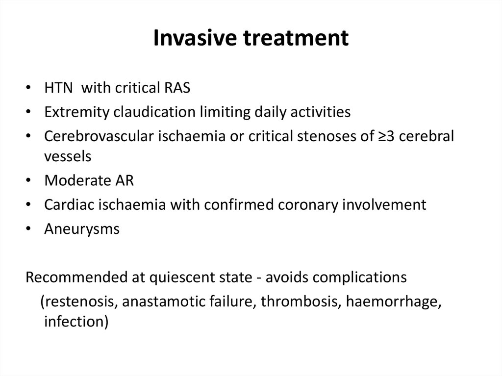
Invasive treatment• HTN with critical RAS
• Extremity claudication limiting daily activities
• Cerebrovascular ischaemia or critical stenoses of ≥3 cerebral
vessels
• Moderate AR
• Cardiac ischaemia with confirmed coronary involvement
• Aneurysms
Recommended at quiescent state - avoids complications
(restenosis, anastamotic failure, thrombosis, haemorrhage,
infection)