


 Медицина
Похожие презентации:


Takayasu disease (Nonspecific aortoarteritis)
1.
Takayasu disease(Nonspecific
aortoarteritis)
2. Content:
Historyof the discovery of the disease
definition
Epidemiology
Classification
Etiology
Pathogenesis
Clinical picture
case
Diagnostics
Treatment
3. History of the discovery of the disease by Dr. Takayasu Takayasu Mikita
4.
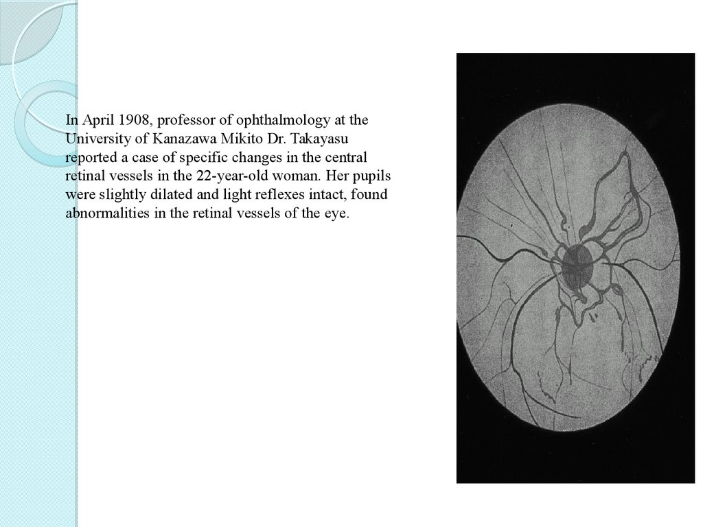
In April 1908, professor of ophthalmology at theUniversity of Kanazawa Mikito Dr. Takayasu
reported a case of specific changes in the central
retinal vessels in the 22-year-old woman. Her pupils
were slightly dilated and light reflexes intact, found
abnormalities in the retinal vessels of the eye.
5.
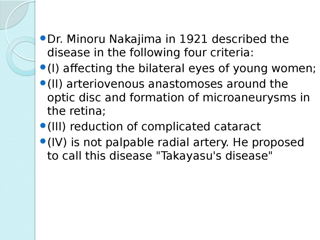
Dr.Minoru Nakajima in 1921 described the
disease in the following four criteria:
(I) affecting the bilateral eyes of young women;
(II) arteriovenous anastomoses around the
optic disc and formation of microaneurysms in
the retina;
(III) reduction of complicated cataract
(IV) is not palpable radial artery. He proposed
to call this disease "Takayasu's disease"
6. Monument Mikita Takayasu in Kanazawa University
7. definition
Takayasu'sdisease - rarely
diagnosed idiopathic disease, one
of the manifestations of chronic
granulomatous arteritis with a
primary lesion of the aorta and its
main branches characterized by a
tendency to recur.
8.
Inthe pathological process involved carotid,
subclavian artery and anonymous, at least pulmonary, coronary and renal arteries.
9.
ЭпидемиологияПо частоте встречаемости
географическеского расположения
Япония
Юговосточная
Азия
Мексика
Африка
Южная
Америка
10.
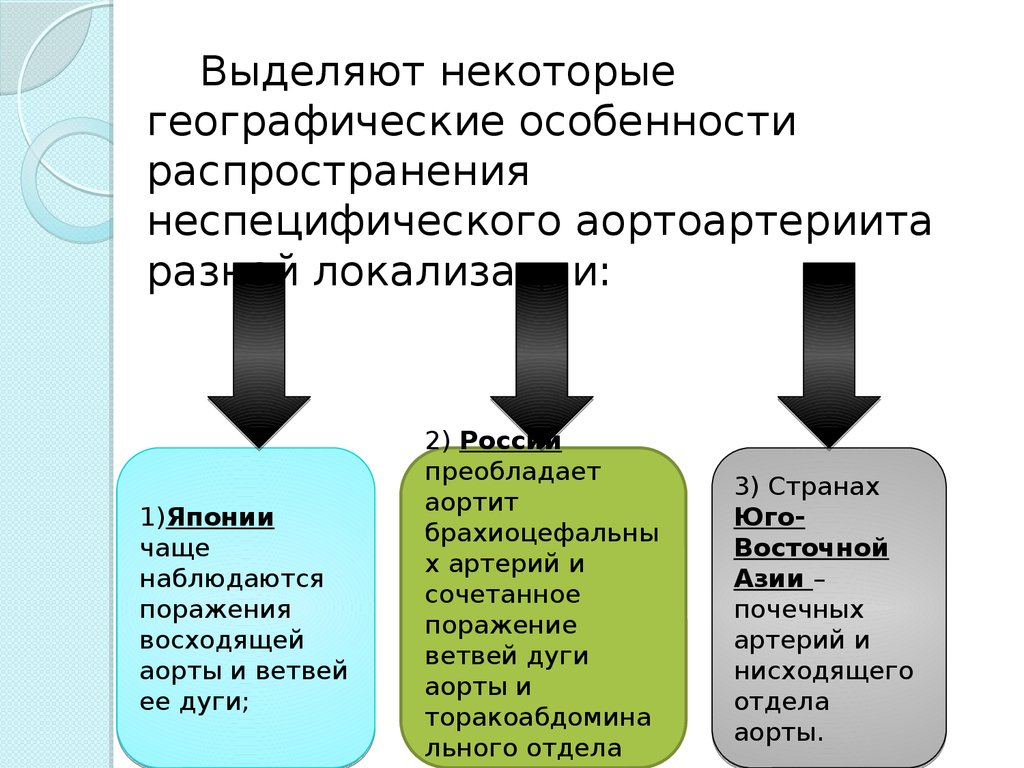
Выделяют некоторыегеографические особенности
распространения
неспецифического аортоартериита
разной локализации:
1)Японии
чаще
наблюдаются
поражения
восходящей
аорты и ветвей
ее дуги;
2) России
преобладает
аортит
брахиоцефальны
х артерий и
сочетанное
поражение
ветвей дуги
аорты и
торакоабдомина
льного отдела
3) Странах
ЮгоВосточной
Азии –
почечных
артерий и
нисходящего
отдела
аорты.
11.
По частоте ,принадлежности
полу
и
возрасту
2,6: 1 000000 населения в год.
15:1 преобладают женщины
12.
мужчины; 20%женщины; 80%
13.
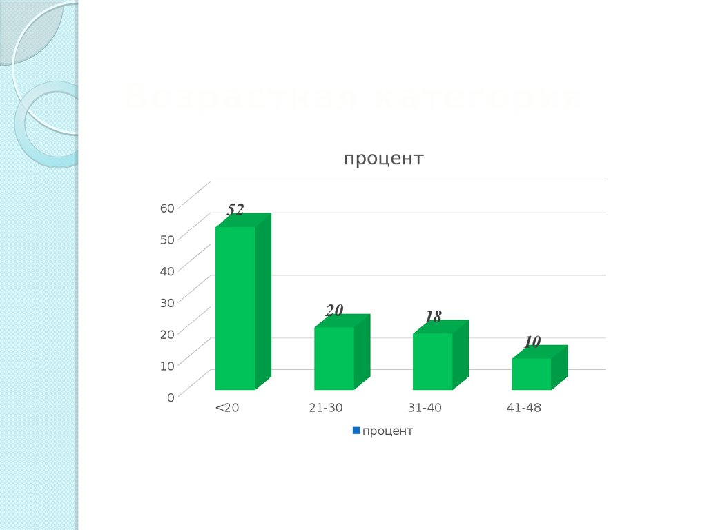
Возрастная категорияпроцент
60
52
50
40
30
20
18
20
10
10
0
<20
21-30
31-40
процент
41-48
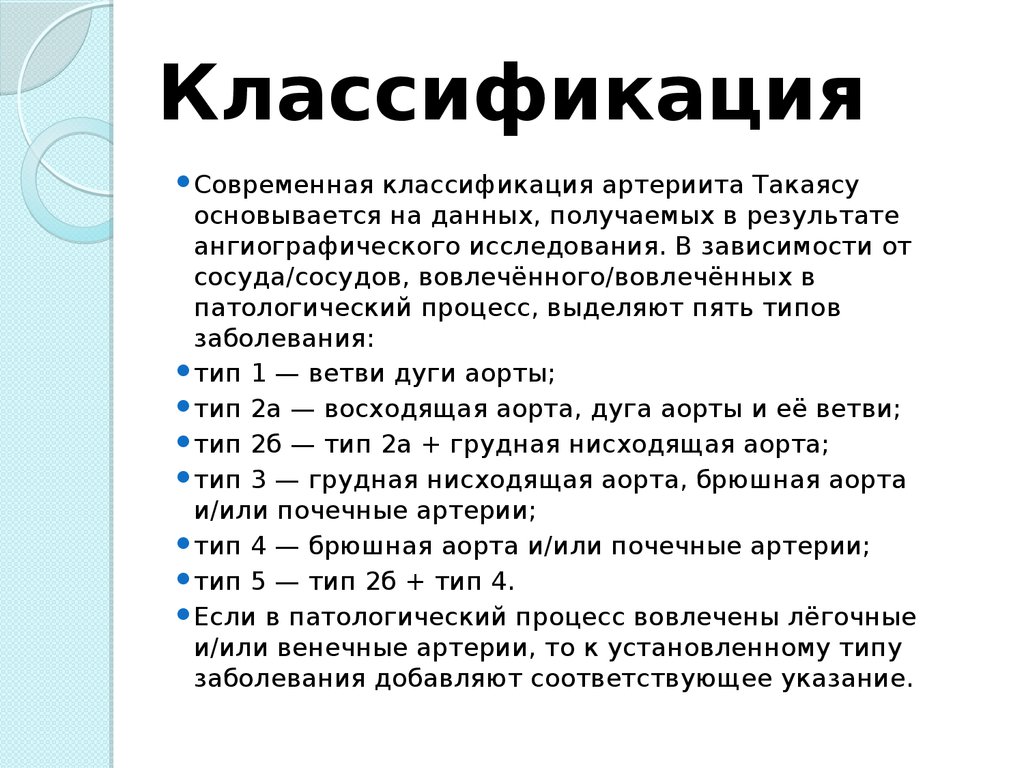
14. Классификация болезни Такаясу в зависимости от анатомии поражения:
Первый тип — поражена дугааорты и ветви, отходящие от нее
(артерии);
Второй тип — затрагивает
грудной и брюшной отдел аорты
15.
Третий тип — поражена дугааорты вместе с грудным и
брюшным отделами.
Четвертый тип – в болезнь
включается легочная артерия.
16.
КлассификацияСовременная
классификация артериита Такаясу
основывается на данных, получаемых в результате
ангиографического исследования. В зависимости от
сосуда/сосудов, вовлечённого/вовлечённых в
патологический процесс, выделяют пять типов
заболевания:
тип 1 — ветви дуги аорты;
тип 2а — восходящая аорта, дуга аорты и её ветви;
тип 2б — тип 2а + грудная нисходящая аорта;
тип 3 — грудная нисходящая аорта, брюшная аорта
и/или почечные артерии;
тип 4 — брюшная аорта и/или почечные артерии;
тип 5 — тип 2б + тип 4.
Если в патологический процесс вовлечены лёгочные
и/или венечные артерии, то к установленному типу
заболевания добавляют соответствующее указание.
17. Etiology
The etiology of systemic vasculitis has notbeen studied, but traced its connection with
infectious-allergic factors and autoimmune
aggression. Immunological reaction, which is
caused by bacteria antigens, is autoimmune
in nature. The result is appear an antibody to
its own tissues, in particular to tissues of the
arterial walls. So arise of inflammation.
18.
Suggest that the damage to the smoothmuscle cells mediated cytotoxic action of
CD8 T-cells, infiltrating the tissue, due to
spore-forming enzyme perforin and
granzyme B.
19.
Role of cytotoxlic damage of tissue withCD8 T cells was confirmed by the fact that
some molecules HLA I class, in particular
HLA-B52, expressed at patients in excess.
CD8 + T cells recognize antigens when
bound to HLA molecules of class.
20.
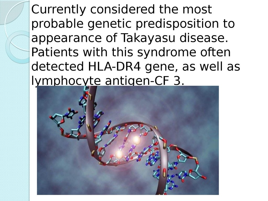
Currently considered the mostprobable genetic predisposition to
appearance of Takayasu disease.
Patients with this syndrome often
detected HLA-DR4 gene, as well as
lymphocyte antigen-CF 3.
21. Pathogenesis
The disease begins with the developmentof inflammation in the wall of the aorta
and its major branches.
As the progression of the inflammatory
process in the vessel walls accumulate
immune complexes;
Numerous micro anguishes occur inside
of the vascular membrane, leading to
increased sclerosis wall and thrombosis.
22.
Inthe later stages of the disease,
inflammatory elements are transformed
into common atherosclerotic lesions of
vessels involved in the process.
23.
Important feature of Takayasu syndromic issimultaneous presence in the vascular wall
various forms and phases of the
inflammatory process: the acute stage of
inflammation in one area of the vessel wall
is often combined with sclerotic changes in
the other.
24.
Clinic:* Ischemia of the
upper extremities weakness and pain in
the hands
* Numbness
* Low exercise
tolerance,
* The absence of
pulsation on one or
both subclavian,
brachial, radial artery,
* Cooling brushes.
25.
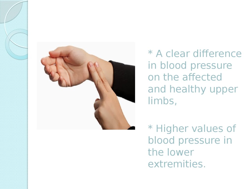
* A clear differencein blood pressure
on the affected
and healthy upper
limbs,
* Higher values of
blood pressure in
the lower
extremities.
26.
* Pain in the leftshoulder, the neck, the
left side of the chest
* Changed over the
arteries:
palpation – painfulness
auscultation - noise.
27.
Inflammatorylesion of the vertebral and
carotid arteries Takayasu's disease causes
neurological symptoms:
dizziness, confusion attention and memory,
decreased working ability
fainting.
28.
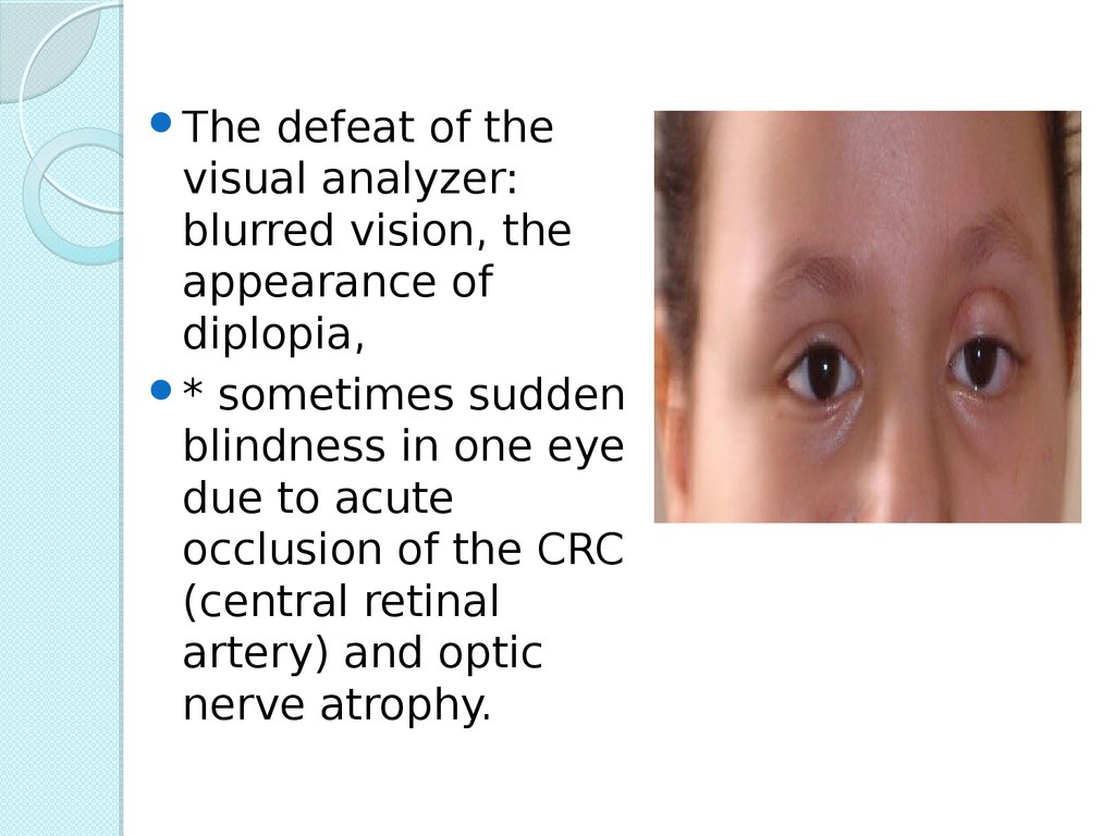
Thedefeat of the
visual analyzer:
blurred vision, the
appearance of
diplopia,
* sometimes sudden
blindness in one eye
due to acute
occlusion of the CRC
(central retinal
artery) and optic
nerve atrophy.
29.
Visiondue to
diplopia
30.
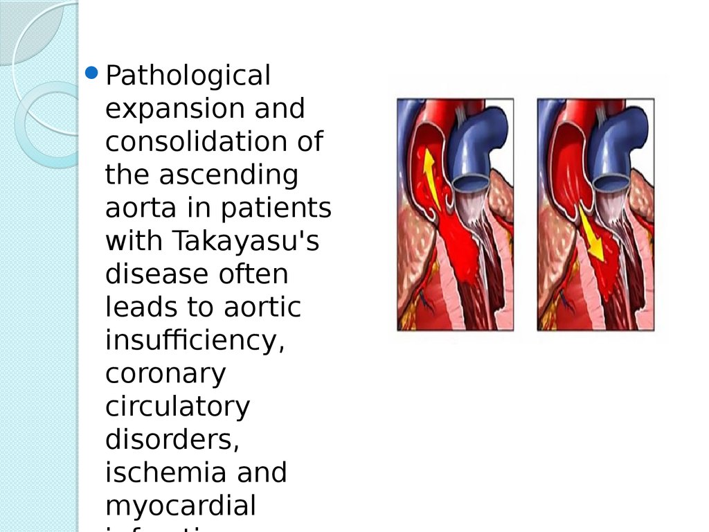
Pathologicalexpansion and
consolidation of
the ascending
aorta in patients
with Takayasu's
disease often
leads to aortic
insufficiency,
coronary
circulatory
disorders,
ischemia and
myocardial
31. Echograms with aortic insufficiency and aortic stenosis
32.
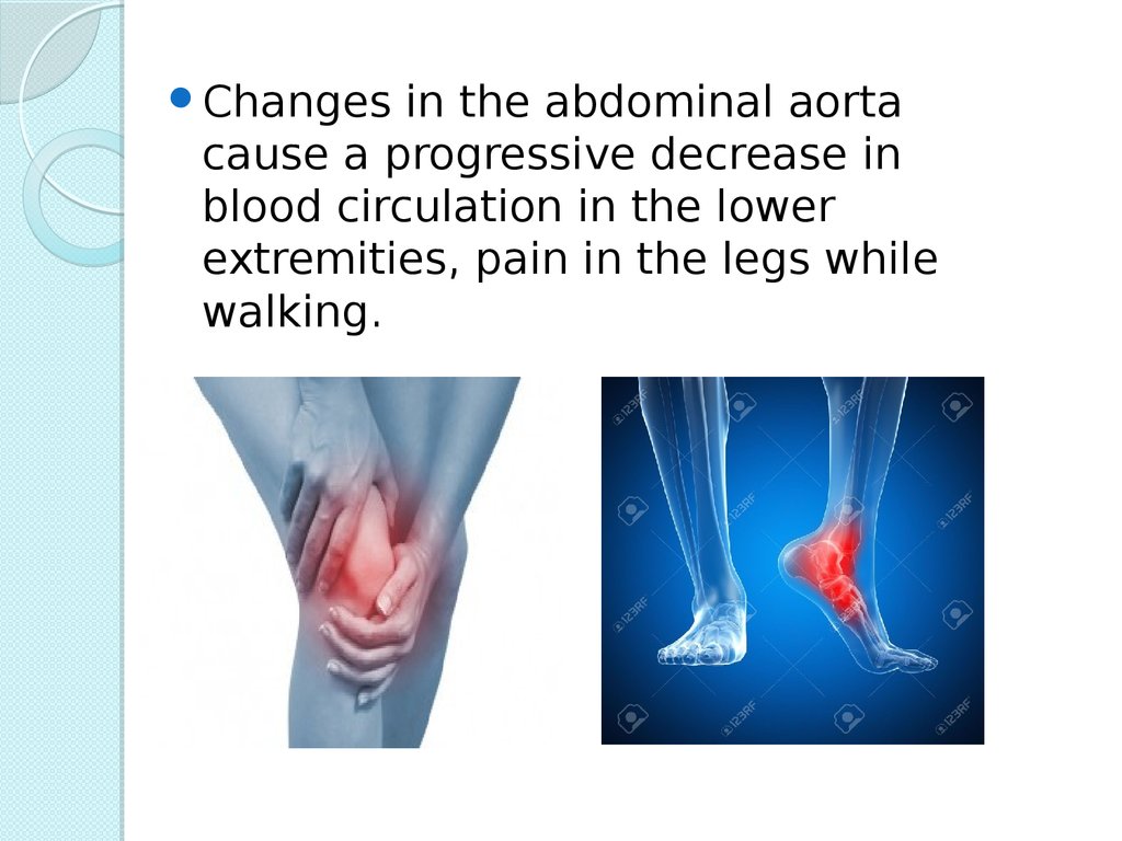
Changesin the abdominal aorta
cause a progressive decrease in
blood circulation in the lower
extremities, pain in the legs while
walking.
33.
Takayasu'sdisease is
marked articular
syndrome arthralgia
34.
Migratoryarthritis
with predominant
involvement of the
hand joints.
35.
Whenkidney disease
of the arteries:
proteinuria, hematuria,
rarely thrombosis. The
involvement of the
pulmonary artery
manifests itself with
pain in the chest,
shortness of breath,
development of
pulmonary
hypertension.
36.
Ospan Gulbanu 2002(14 years)В 24.02.2016 Came to NCP
and CS with complains:
weight loss
arthralgia,
general weakness.
37.
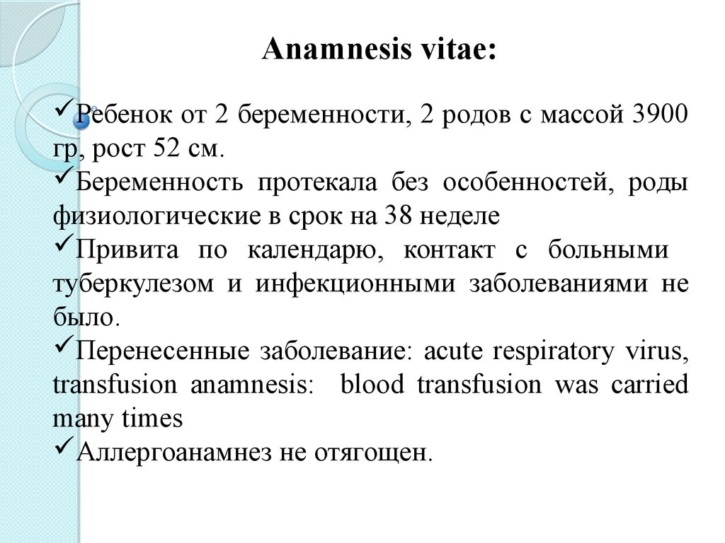
Anamnesis vitae:Ребенок от 2 беременности, 2 родов с массой 3900
гр, рост 52 см.
Беременность протекала без особенностей, роды
физиологические в срок на 38 неделе
Привита по календарю, контакт с больными
туберкулезом и инфекционными заболеваниями не
было.
Перенесенные заболевание: acute respiratory virus,
transfusion anamnesis: blood transfusion was carried
many times
Аллергоанамнез не отягощен.
38.
Status praesens:
Состояние средней степени тяжести, сознание
ясное.
Ребенок повышенного питания, правильного
телосложения.
Самочувствие не страдает. Тургор кожи
сохранен.
Лицо одутловатое, имеется легкая гиперемия на
щеках.
Кожные покровы: обычной окраски, чистые.
КСС: суставы визуально не изменены,
движение в них в полном объеме.
Периферические л/узлы не увеличены.
39.
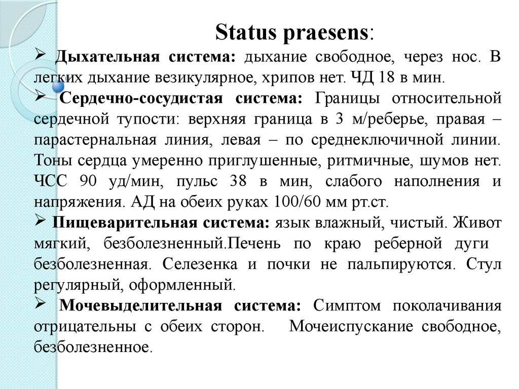
Status praesens:Дыхательная система: дыхание свободное, через нос. В
легких дыхание везикулярное, хрипов нет. ЧД 18 в мин.
Сердечно-сосудистая система: Границы относительной
сердечной тупости: верхняя граница в 3 м/реберье, правая –
парастернальная линия, левая – по среднеключичной линии.
Тоны сердца умеренно приглушенные, ритмичные, шумов нет.
ЧСС 90 уд/мин, пульс 38 в мин, слабого наполнения и
напряжения. АД на обеих руках 100/60 мм рт.ст.
Пищеварительная система: язык влажный, чистый. Живот
мягкий, безболезненный.Печень по краю реберной дуги
безболезненная. Селезенка и почки не пальпируются. Стул
регулярный, оформленный.
Мочевыделительная система: Симптом поколачивания
отрицательны с обеих сторон. Мочеиспускание свободное,
безболезненное.
40.
Лабораторные данные:ОАК: эр – 3,2 *1012/л;
Нв- 117г/л; тромб- 303т,
Л.- 6.5 *109/л; СОЭ – 5
мм/час.
ОАМ : уд. вес –1020,
реакция -кислая, белок –
отр., пл. эп. – 2 в п/зр.
БАК : АЛТ- 0.58
мккат/л, АСТ- 0.66
мюсат/л,билирубин
общий- 21.4 мколь/л,
билирубин прямой- 5.7
мколь/л
Инструментальные данные:
ЭхоКГ: АО-2.4 см, ЛД-2.64 см,
Тмжп-0,5см, Тзслж-0.5 см, ПЖ1.79 см, Толщ.Ст ПЖ-0,34 см,
ДЕ-1.55см, Ef-0.12 мм/ cек,
КДР – 3.75 см, КСР – 2.19 см,
КДО – 58 мл, КСО – 14.4 мл,
УО-43,6 мл, ФИ – 75%, DS
-43%.
В сечении по короткой оси:
Дао(см)-2.19
Дсла(см)-2.19 ветви 0.90 см
41.
Еchocardiography andDoppler
Echocardiographic indicators within
the age norm. Data for heart disease
have not been identified.
-the heart cavity is not
expanded
-the wall thickness of the
myocardium in the normal
range
- myocardial contractility
42.
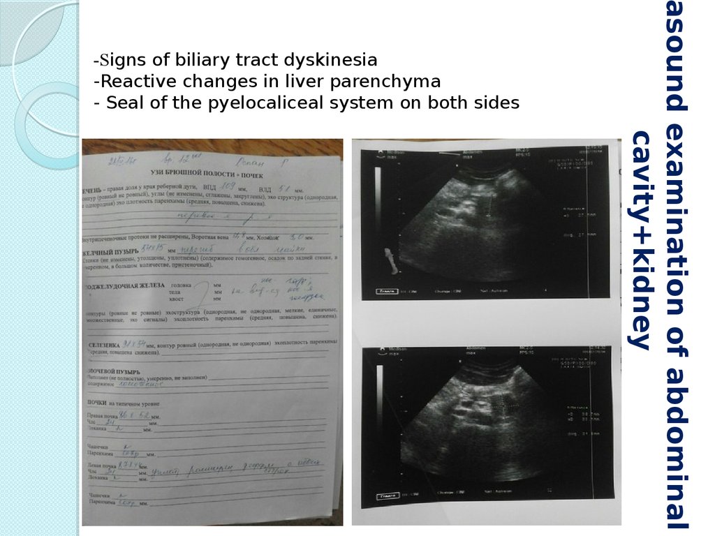
asound examination of abdominalcavity+kidney
-Signs of biliary tract dyskinesia
-Reactive changes in liver parenchyma
- Seal of the pyelocaliceal system on both sides
43.
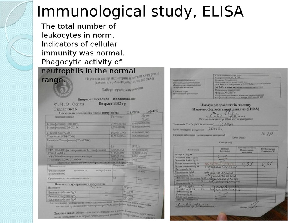
Immunological study, ELISAThe total number of
leukocytes in norm.
Indicators of cellular
immunity was normal.
Phagocytic activity of
neutrophils in the normal
range.
44. ЭЭГ
Невыраженный дисфункциональныеизменения, снижение порога
возбудимости синхронизирующих
срединных структур.
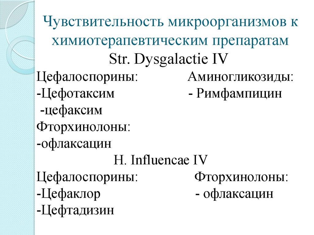
45. Результат микробиологического исследования
При исследовании маска из зеваВыделены Str. Dysgalactie IVH. Influencae IV
46.
Чувствительность микроорганизмов кхимиотерапевтическим препаратам
Str. Dysgalactie IV
Цефалоспорины:
Аминогликозиды:
-Цефотаксим
- Римфампицин
-цефаксим
Фторхинолоны:
-офлаксацин
H. Influencae IV
Цефалоспорины:
Фторхинолоны:
-Цефаклор
- офлаксацин
-Цефтадизин
47.
ЛечениеРост 143 см, Вес 38 кг, Пол:
женский
Режим щадящий
Диета № 15
Метилпреднизалон 16 мг 7:00- 1 тб
Аспаркам- 175 мг по 1 тб х 3 раза в
день
Методжет- 15 мг
Фолиевая кислота-1 тб х 3 раза в
день
Орашение зева раствором
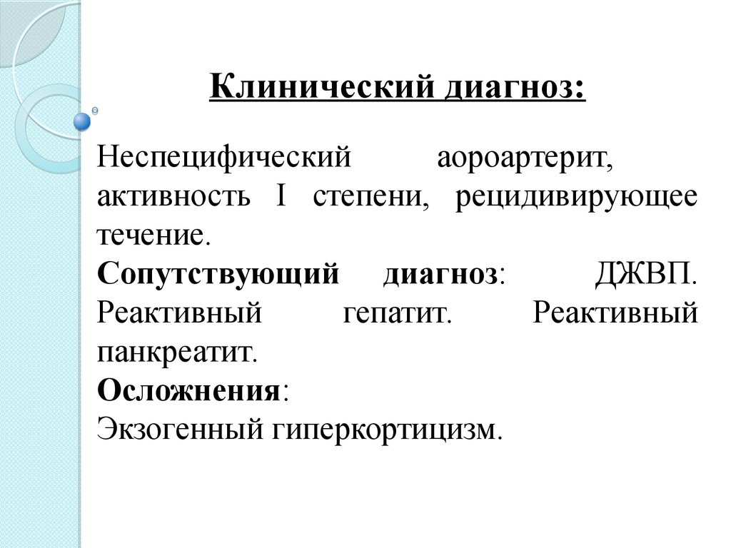
48.
Клинический диагноз:Неспецифический
аороартерит,
активность I степени, рецидивирующее
течение.
Сопутствующий диагноз:
ДЖВП.
Реактивный
гепатит.
Реактивный
панкреатит.
Осложнения:
Экзогенный гиперкортицизм.
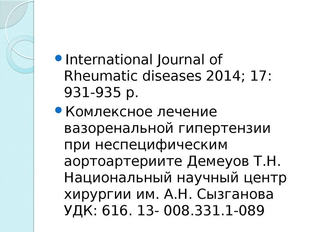
49.
InternationalJournal of
Rheumatic diseases 2014; 17:
931-935 p.
Комлексное лечение
вазоренальной гипертензии
при неспецифическим
аортоартериите Демеуов Т.Н.
Национальный научный центр
хирургии им. А.Н. Сызганова
УДК: 616. 13- 008.331.1-089