


 Медицина
Похожие презентации:


Giant cell arteritis
1. Giant cell arteritis
Dr Katya DolnikovD_katya@rambam.health.gov.il
2017
2.
3. Introduction
• Giant cell arteritis (GCA) is categorized as avasculitis of large- and medium-sized vessels
• Systemic symptoms are common in GCA and
vascular involvement can be widespread
• Targeting of the muscular arteries from cranial
branches of the aortic arch gives rise to the most
characteristic symptoms of GCA
• The most feared complication of GCA, visual loss,
is one potential consequence of such cranial
arteritis
4.
5. Epidemiology
• GCA is the most common systemic vasculitis• The lifetime risk of developing GCA is ~1% in women
and 0.5% in men
• The greatest risk factor for developing GCA is aging
• The disease almost never occurs before age 50
• Over 80 percent of patients are older than 70 years
• Ethnicity is a major risk factor for GCA. The highest
incidence figures are found in Scandinavian countries
• F>M
6. Clinical findings
• The onset of symptoms tends to be subacute• Abrupt presentations occurs less frequently
• Systemic symptoms are frequent and include
fever, fatigue, and weight loss
• Fever occurs in up to one-half of patients with
GCA and is usually low-grade
• In ~ 10% of patients constitutional
symptoms and/or laboratory evidence of
inflammation dominate the clinical presentation
and can be the only clues to the diagnosis
7.
8. Clinical findings - Headache
• Located over the temples, but can also befrontal or occipital or generalized
• The headaches can progressively worsen, or
wax and wane, sometimes subsiding
temporarily before treatment is started
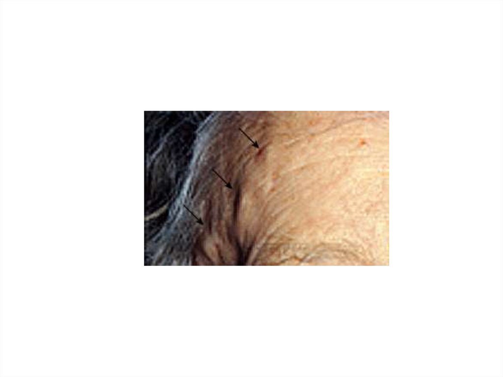
• Tenderness of the scalp to touch
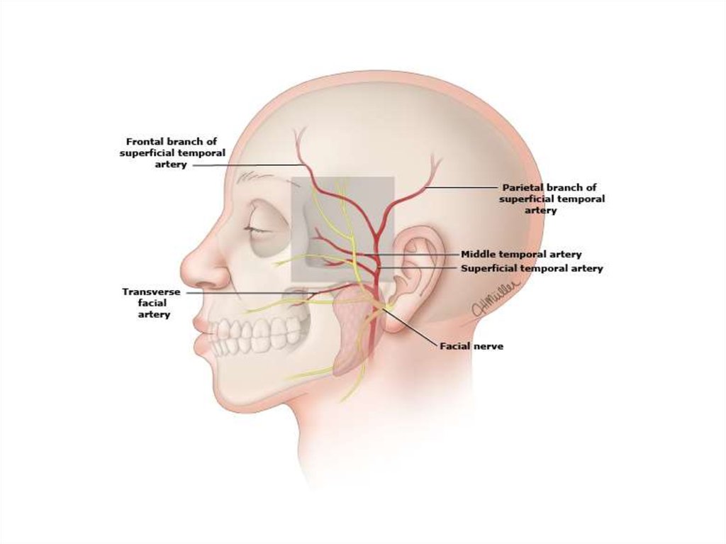
9. Temporal artery
10. Jaw Claudication
• Trismus-like symptoms• Fatigue of the muscles of mastication
• Rapid onset after the start of chewing and the ensuing
severity of pain
• Patients seldom recognize the significance of
symptoms of jaw claudication and must be questioned
directly about this symptom
• Claudication-like symptoms occasionally occur with
repeated swallowing and in the tongue during eating
• Jaw claudication is the symptom most highly
associated with a positive temporal artery biopsy
11. Vision
• Transient visual loss (amaurosis fugax) — Transientmonocular (and, rarely, binocular) impairment of vision can
be an early manifestation of GCA.
• Permanent vision loss — The most feared complication of
GCA. Commonly is painless and sudden, may be partial or
complete, and may be unilateral or bilateral. Even in the era
of effective therapy, permanent partial or complete loss of
vision in one or both eyes is reported 20% of patients
• Risk factors — prior transient visual loss as the strongest
predictor for subsequent permanent visual loss
• Diplopia
12. Large vessel GCA
• Involvement of the aorta and its major proximal branches especially in the upper extremities• The clinical consequences comprise aneurysms and
dissections of the aorta, particularly the thoracic aorta, as
well as stenosis, occlusion and ectasia of large arteries
• Axillary arteries, proximal brachial arteries - arterial bruits,
diminished or absent blood pressures, and arm claudication
may ensue. Cold intolerance is common, but explicit digital
ulcerations and gangrene are rare because of the adequacy
of collateral arterial supply
• Upper-extremity disease is bilateral, though not symmetric,
13.
14. External carotid artery- branches
Maxillary and dental pain
Facial swelling
Throat pain
Tongue pain
15.
16. Physical examination
• Pulses – carotid, brachial, radial, femoral,pedal
• Blood pressure
• Bruits – carotid or supraclavicular areas; over
the axillary, brachial, or femoral arteries; over
the abdominal aorta
• Cardiac auscultation
• Temporal a. examination
17.
18. AION
19. Laboratory findings
• Normochromic anemia is often present prior to therapyand improves promptly after the institution of
glucocorticoids
• Thrombocytosis
• The leukocyte count is usually normal, even in the setting
of widespread systemic inflammation.
• Serum albumin — moderately decreased at diagnosis but
responds quickly to the institution of glucocorticoids
• Hepatic enzymes — Elevated serum concentrations of
hepatic enzymes, especially the alkaline phosphatase,
occur in 25 to 35 percent of patients
• ESR and C-reactive protein — elevated
20. Diagnosis
• The diagnosis of giant cell arteritis (GCA)should be considered in a patient over the age
of 50 who complains of:
– New headaches
– Abrupt onset of visual disturbances
– Symptoms of polymyalgia rheumatica
– Jaw claudication
– Unexplained fever or anemia
– High ESR/CRP
21. Diagnosis
• Patient suspected of having GCA shouldundergo temporal artery biopsy
• ~85% sensitivity
• Other arteries can also be sampled
• Scheduling of the biopsy should NOT interfere
with the start of glucocorticoid therapy when
there is a significant concern about the
possibility of GCA
22. Biopsy-negative GCA
• The patient may not have GCA. If the clinical story isequivocal, then alternative diagnoses should be given
more weight
• The patient may have GCA involving only the great
vessels. Among patients with suggestive symptoms
(most often arm claudication), an imaging study should
be performed
• An empiric trial of glucocorticoid therapy may be
helpful. Failure of the patient’s symptoms to resolve
within one week of high-dose glucocorticoids argues
strongly against the diagnosis of GCA
23. Imaging
MRI/MRA
USD
Angiography
PET-CT
24. Treatment
• Uncomplicated GCA - 40 to 60 mgof prednisone in a single dose
• After achieving a daily dose of 10 mg, the
prednisone taper should be slow, such that
patients remain on some prednisone for 9 to
12 months. Tapering in 1 mg decrements per
month once the daily dose is less than 10 mg
is appropriate
25. Treatment
Add aspirin (80 to 100 mg/day) to reduce the risk of visual loss, transient ischemic
attacks, or stroke
PPI to prevent GI damage
If there is a strong suspicion of GCA as the cause of visual loss - intravenous
pulse methylprednisolone – 1gr for 3 days.
This is then followed by oral therapy with 1 mg/kg per day (maximum of
60 mg/day), as recommended above for uncomplicated GCA. For patients without
contraindications to anticoagulation, warfarin therapy in addition to lowdose aspirin may also be considered in this setting.
Annual chest radiographs for up to 10 years to identify asymptomatic thoracic
aortic
Self-limited course over several months to several years. The glucocorticoid dose
can eventually be reduced and discontinued in the majority of patients. GCA may
not adversely affect overall survival
Permanent partial or complete loss of vision in one or both eyes has been
observed in 15 to 20 percent of patients