


")
")


")


 Медицина
Похожие презентации:


Pulmonary Hypertension-Pathways, Diagnostic
1. Pulmonary Hypertension-Pathways, Diagnostic.
Made by,RISHABH GURU,
3rd year, PSMU
2. PH - History
History of smoking
ETOH/recreational drug use
Systemic hypertension
Cyanosis/murmur as a child
Joint/musculoskeletal pain
Raynaud’s Syndrome
FH of unexplained early cardiopulmonary disease
Use of appetite suppressant drugs
3. Pulmonary circulation
• Low resistance, high compliance vascularbed
• Only organ to receive entire cardiac output
(CO)
• Changes in CO as well as pleural/alveolar
pressure affect pulmonary blood flow
• Different reactions compared to the systemic
circulation
• Normally in a state of mild vasodilation
4. Outline
• Review classification of pulmonary hypertension(PH)
• Pulmonary arterial hypertension (PAH)
• Evaluation of PH and how to differentiate PAH
from other forms of PH
• PH and cardiac, renal and hepatic transplantation
• Review PAH-approved therapy and treatment of
non-Group 1 PH
5. Classification of Pulmonary Hypertension (PH)
• 1) Pulmonary arterial hypertension• 2) Pulmonary hypertension due to left heart disease
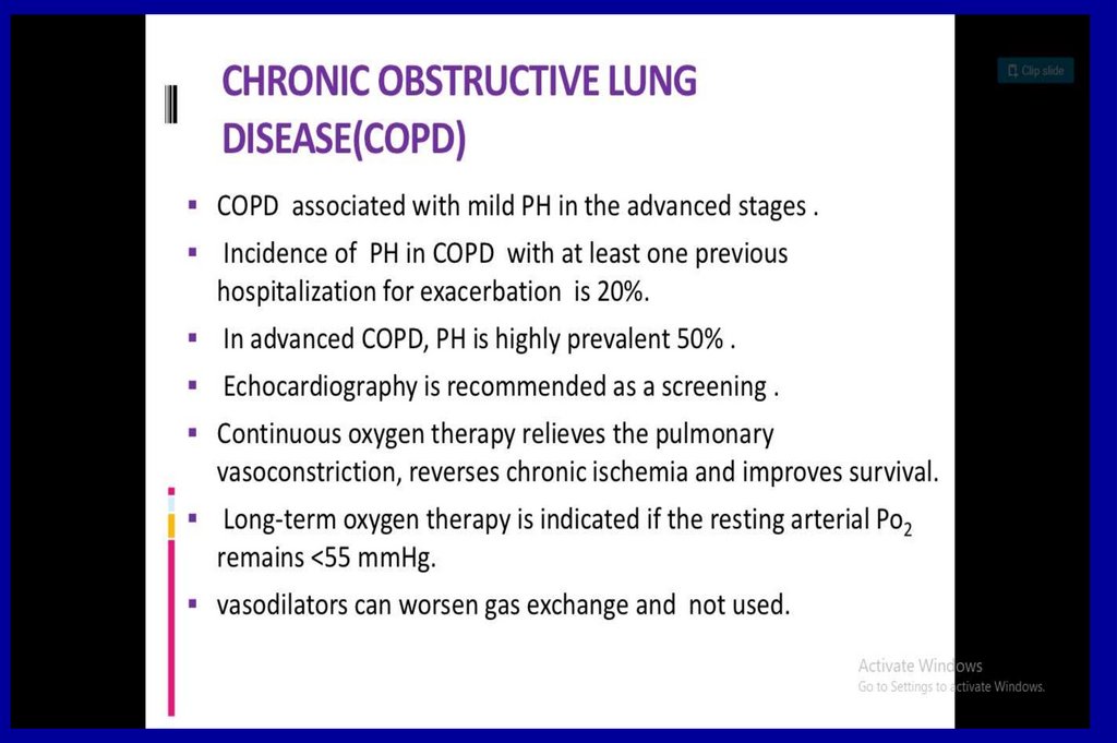
• 3) Pulmonary hypertension due to lung diseases and/or
hypoxia
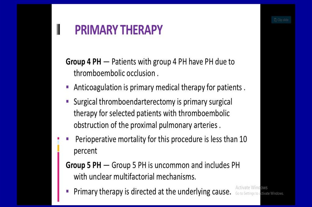
• 4) Chronic Thromboembolic pulmonary hypertension
(CTEPH)
• 5) Pulmonary Hypertension with unclear and/or
multifactorial mechanism
6. Vascular Pressure in Systemic and Pulmonary Circulations (mm Hg)
120/80, mean 93Systemic
Circulation
25/8, mean 14
Arteries
Arteries
Right
Atrium
Mean >6
Left
Atrium
Mean 5
Pulmonary
Circulation
Lung
Body
SVR= 17.6
PVR= 1.8
Veins
Right
Ventricle
25/2-5
Left
Ventricle
120/5-10
Veins
7. PH- Symptoms
DOE
Fatigue, weakness
Chest pain
LE or abdominal swelling
Syncope
Not typical of PAH: orthopnea
8.
9.
10.
11.
12.
13.
14.
15.
16.
17.
18.
19.
20.
21.
22.
23.
24.
25.
26.
27.
28.
29.
30.
31.
32.
33.
34.
35.
36.
37.
38.
39.
40.
41.
42.
43. Algorithm illustrating general diagnostic workup for pediatric pulmonary arterial hypertension
44. Classification of PAH, Group 1
• Idiopathic PAH (formerly primary pulmonaryhypertension, PPH)
• Heritable
Drug/toxin induced
• Associated with:
–
–
–
–
–
–
Connective tissue diseases
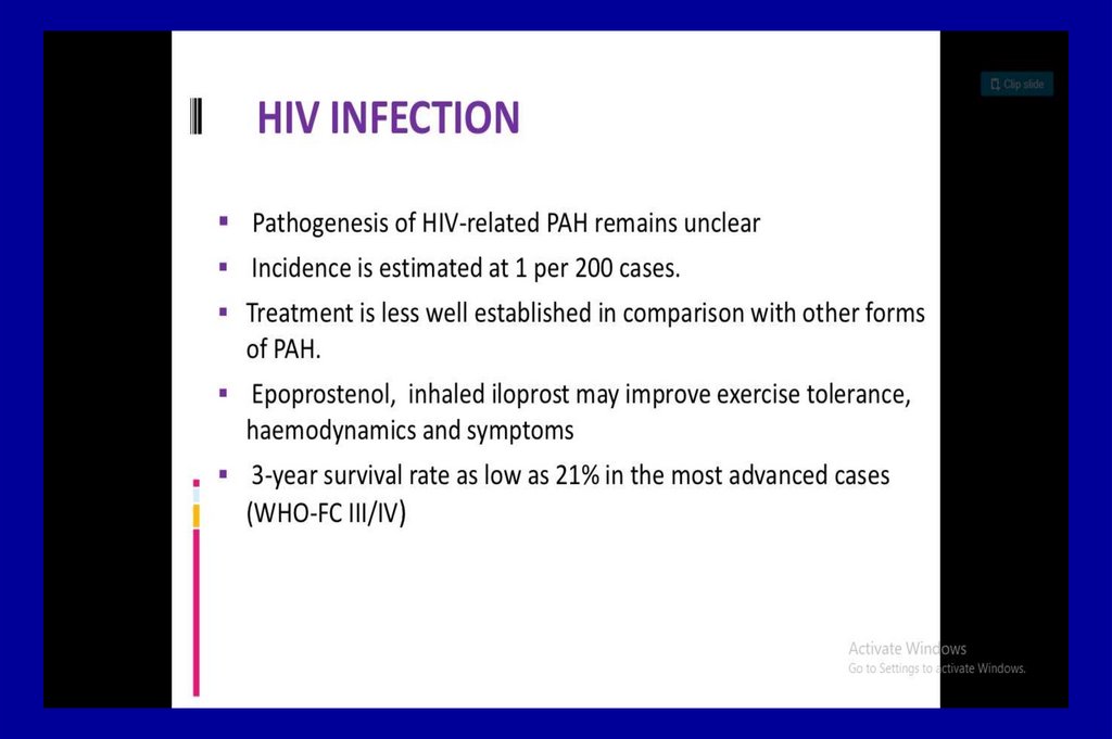
HIV infection
Portal hypertension
Systemic to pulmonary shunts
Schistosomiasis
Chronic hemolytic anemia
45. Group 2 PH
• Pulmonary hypertension owing to left heartdisease
– Systolic dysfunction
– Diastolic dysfunction
– Valvular disease
– Pulmonary venous obstruction
46. PH with unclear or multifactorial mechanisms: Group 5
1.Hematologic disorders2.Systemic disorders: vasculitis
3.Metabolic disorders
4.Others: chronic renal failure on dialysis
47. Pathogenesis : An Integrated View
GeneticPredisposition
Proliferation
Other Risk
Factors
Thrombosis
Altered Pathways
and Mediators
Vasoconstriction
Vascular
Remodeling
Inflammation
48. Evaluation for PH
ECG
Chest x-ray
V/Q scan or contrasted spiral CT (+/- angiogram)
PFTs
Exercise oximetry
Echocardiogram
Right heart catheterization w/vasodilator testing
Labs: CBC, CMP, INR, ANA, HIV, TFTs
49.
50. PH - Radiographic studies
• CXR:-large proximal PA with peripheral
tapering (pruning)
-cardiomegaly due to enlarged RA, RV
-pleural effusion is uncommon
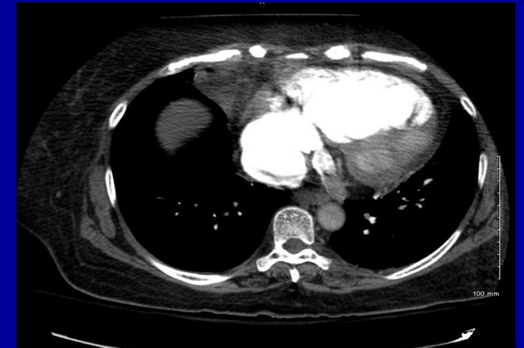
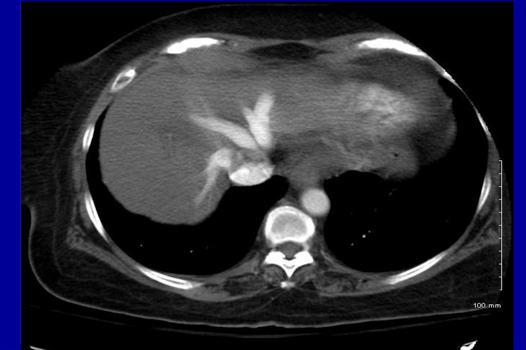
• CT:
-PA >aorta
-cardiomegaly, enlarged RV
-pericardial effusion
51. CXR in PAH
52. CXR in Eisenmenger Syndrome
53. Mitral Stenosis
54. Enlarged main PA on CT Standard view Coronal view
Enlarged main PA on CTStandard view
A
PA
Coronal view
55.
56.
57. Ventilation Perfusion Lung Scan
PAHPerf
Vent
CTEPH
Perf
Vent
58. CTEPH: Pulmonary Angiography
• Confirms diagnosis of CTEPH inpatients with PH
• Assess thrombus accessibility
• Distinct angiographic patterns
– “Web” narrowing
– Poststenotic dilatation
– Proximal occlusion
– “Pouch” defects
59. Organized Clot Removed at Surgery
60. Pulmonary Function tests
• No characteristic changes• Mandatory to screen for significant restrictive
or obstructive lung disease
• Diffusing capacity often significantly reduced
in patients with scleroderma (<50%)
61. RV, RA Enlargement on Echocardiogram
RVLV
RA
Normal
LA
PH
62. Other Helpful Diagnostic Tests (Determined by patient’s history)
High resolution chest CT
Cardiopulmonary exercise study
Polysomnography
Arterial blood Gas
Hepatitis serologies
Left heart catheterization, evaluation of
coronary arteries
63. Echocardiographic findings in ESRD patients undergoing transplant
Patients WithPHT
(n = 85)
Patients Without PHT
(n = 415)
LV, diastole (cm)
4.9 ± 0.5
4.7 ± 2.0
.3
LV, systole (cm)
3.2 ± 0.5
3.1 ± 0.5
.8
Right ventricle (cm)
3.3 ± 0.5
3.2 ± 0.4
.8
Left atrium (cm)
4.0 ± 0.7
3.5 ± 0.6
<.0001
Right atrium (cm)
3.7 ± 0.5
3.3 ± 0.4
<.0001
Diastolic dysfunction (%)
18.8
21.4
.6
Systolic dysfunction (%)
22.4
13.5
.04
49.7 ± 7.9
52.3 ± 6.9
.002
78.8
59.8
.001
Echocardiographic Data
LV ejection fraction (%)
LV hypertrophy (%)
P Value
64. Treatment of non PAH-pulmonary hypertension
• Pulmonary Venous Hypertension:• Treat heart failure with afterload reduction
– Systolic or diastolic
• MV or AV disease
– Replace the valve
• Pulmonary vein stenosis
– Pulmonary vein stenting
65. Treatment of non PAH-pulmonary hypertension
• PH associated with disorders of the respiratorysystem and/or hypoxemia:
– Rx of hypoxemia is often the main therapy
• PH due to chronic thromboembolic disease:
– Thromboendarterectomy for proximal disease
– Can consider PAH therapy for distal disease
66. Adjunctive treatments of PAH
Anticoagulation
Diuretics
Digoxin
Oxygen
Calcium channel blockers
Exercise
Salt restriction
67. Specific PAH Treatment
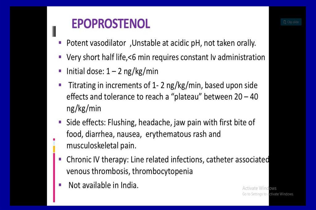
Epoprostenol (generic and Flolan®)
Treprostinil (Remodulin®)
Prostaglandins
Iloprost (Ventavis®)
Bosentan (Tracleer®)
Endothelin receptor
antagonists (ERAs)
Ambrisentan (Letairis®)
Tadalifil (Adcirca®)
Phosphodiesterase 5
inhibitors (PDE5 inhibitors)
Sildenafil (Revatio®)
68. PAH Determinants of Risk
.Lower Risk
Determinants of Risk
Higher Risk
No
Clinical evidence of
RV failure
Yes
Gradual
Progression
Rapid
II, III
WHO class
IV
Longer (>400 m)
6MWD
Shorter (<300 m)
Minimally elevated
BNP
Very elevated
Minimal RV dysfunction
Echocardiographic findings
Pericardial effusion,
significant RV dysfunction
Normal/near normal
RAP and CI
Hemodynamics
High RAP, low CI
69. Take Home Points
• PH can not be diagnosed by Echo alone, need athorough evaluation for all patients
• Right heart catheterization is necessary in ALL patients
to accurately diagnose PH
• PAH is a progressive disease, even with Rx
• Make sure the patient has PAH before treating
• Despite multiple therapies, lung transplantation is the
only curative treatment for PAH
• PH negatively impacts outcome of all solid organ
transplants