


 Медицина
Похожие презентации:

")

")


Fat Embolism Syndrome
1.
Fat Embolism SyndromeDr. S. Parthasarathy
MD., DA., DNB, MD (Acu),
Dip. Diab. DCA, Dip. Software statistics
PhD (physio)
Mahatma gandhi medical college and research
institute, puducherry, India
2.
History• In 1861, Zenker described fat droplets in the
lung capillaries of a railroad worker who
sustained a fatal thoracoabdominal crush
injury.
• In 1873, Bergmann was first to establish the
clinical diagnosis of fat embolism syndrome.
3.
What is it ??• complex with potentially catastrophic
cardiopulmonary and cerebral dysfunction
• Three problems :
• dyspnoea, petechiae and mental confusion
4.
DefinitionsFat Emboli: Fat particles or droplets
travel through the circulation
Fat Embolism: fat emboli passes into
the bloodstream and lodges within a
blood vessel.
Fat Embolism Syndrome (FES):
serious manifestation of fat embolism
occasionally causes multi system
dysfunction, the lungs are always
involved and next is brain
5.
Fulminant fat embolism• sudden intravascular liberation of a large
amount of fat causing pulmonary vascular
obstruction, severe right heart failure, shock
and often death within the first 1-12 h of
injury
6.
Etiology7.
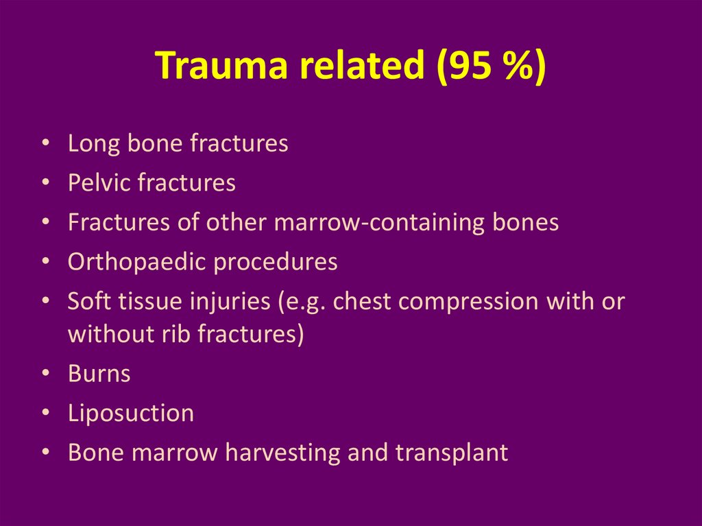
Trauma related (95 %)Long bone fractures
Pelvic fractures
Fractures of other marrow-containing bones
Orthopaedic procedures
Soft tissue injuries (e.g. chest compression with or
without rib fractures)
• Burns
• Liposuction
• Bone marrow harvesting and transplant
8.
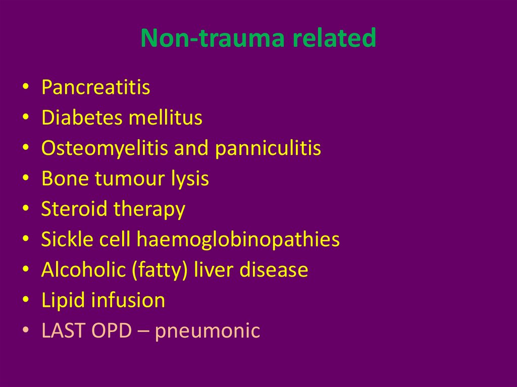
Non-trauma relatedPancreatitis
Diabetes mellitus
Osteomyelitis and panniculitis
Bone tumour lysis
Steroid therapy
Sickle cell haemoglobinopathies
Alcoholic (fatty) liver disease
Lipid infusion
LAST OPD – pneumonic
9.
fat emboli also can arisefrom circulating lipoproteins
10.
What is frequent ??• lower extremity and pelvic trauma,
• intramedullary nailing of long-bone fractures,
• hip arthroplasty, and knee arthroplasty
11.
Incidence ??• incidence of FES was 1 %
• But multiple fractures, adults, high velocity
injuries, cementing, hypovolumia
• It can be upto 33 %
12.
Lethal dose• The acute lethal dose of fat ranges from 20-50
ml.
• The volume of marrow fat from a femur is
approximately 70-100 ml.
• Mortality – 10 – 20 %
13.
Pathophysiology ??• The Mechanical theory (Gauss)
• Biochemical theory (Lehmann and Moore)
• Coagulation theory
14.
The Mechanical theory (Gauss)• Trauma to long bones releases fat droplets
• (10-40 μm in diameter)
• fat droplets enter the torn veins near long bone (
intramedullary pressure is higher than the venous
pressure)
• They enter lungs
• perivascular hemorhage and edema- picture of
ARDS
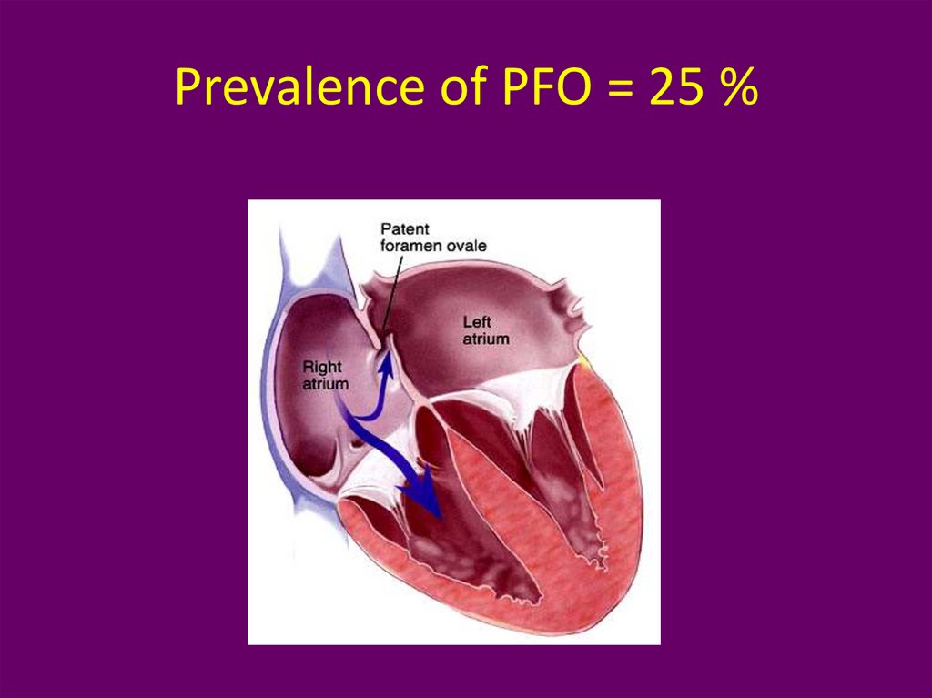
• but smaller ones ( 7- 10 mic.) travel to systemic
circulation via ? Patent foramen ovale -
15.
Prevalence of PFO = 25 %16.
Biochemical theory• Embolized fat is degraded in plasma to free fatty
acids.
• FFA can cause lung injury, cardiac contractile
dysfunction
• CRP appears to be responsible for lipid
agglutination and may also participate in the
mechanism of non-traumatic FES.
17.
Coagulation theory• Tissue thromboplastin is released with marrow
elements following long bone fractures.
• Activates intravascular coagulation
• fibrin and fibrin degradation products, leukocytes,
platelets and fat globules combine to increase
pulmonary vascular permeability
• Catecholamines are involved
18.
• Can it happen in sickle celldisease ??
19.
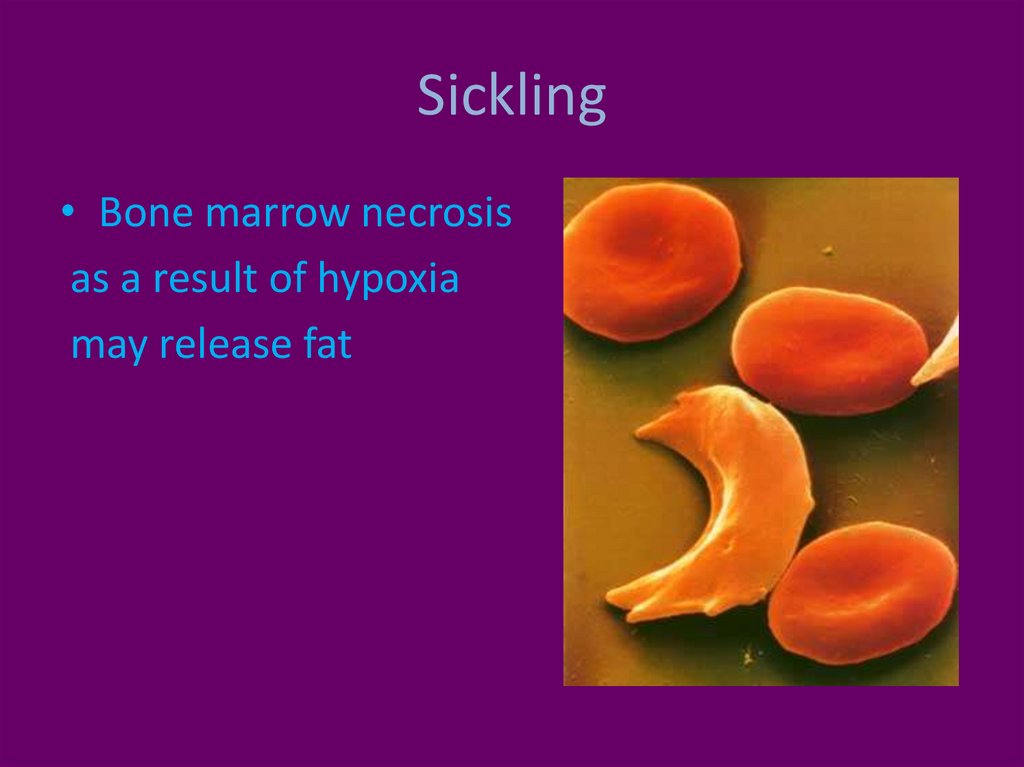
Sickling• Bone marrow necrosis
as a result of hypoxia
may release fat
20.
• Number of theories means• Poorly understood ??
21.
Clinical Features• 12-72 hrs after the initial injury
• Rarely two weeks
22.
Features• Respiratory changes – 95 %
• Cerebral changes – 60 %
• petechiae (33% - 60 %).
• Not necessary to follow one by one
23.
Respiratory changes• Dyspnoea, tachypnoea and hypoxaemia are
the most frequent early findings.
• Respiratory failure as ARDS
24.
Cerebral• The more common presentation is with an acute
confusional state
• but focal neurological signs including hemiplegia,
aphasia, apraxia, visual field disturbances have been
described.
• Seizures and decorticate posturing have also been
seen.
• Fortunately, almost all neurological deficits are
transient and fully reversible.
25.
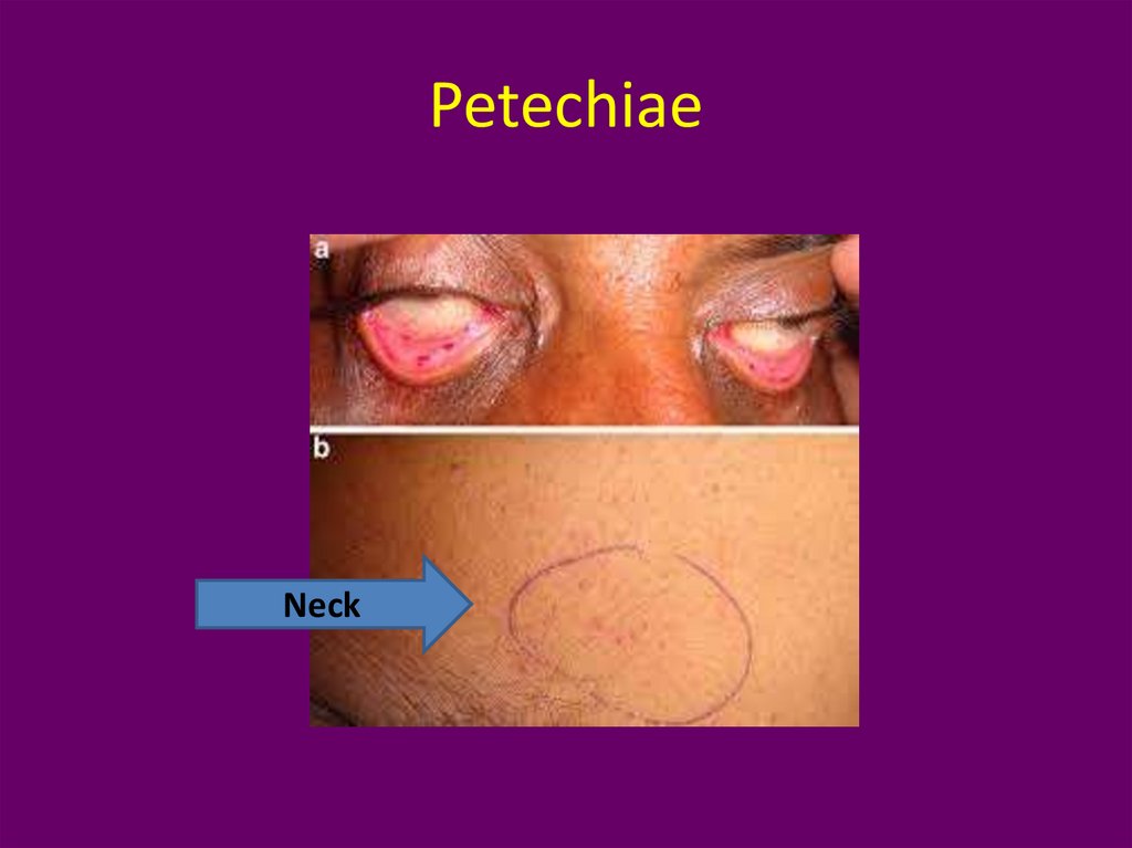
Petechiae• Embolization of small dermal capillaries leading
to extravasation of erythrocytes. This produces a
petechial rash in the conjunctiva, oral mucous
membrane and skin folds of the upper body
especially the neck and axilla
• No relation to platelets
• Self limiting (36 hours to seven days)
26.
PetechiaeNeck
27.
Petechiae• Petechiae only rarely appear on the legs and they are
never seen on the face or the posterior aspect of the
body. WHY ??
• May be –
• fat globules float and therefore distribute to
branches of the aorta that arise from the top
of the arch, and to the side of the body that is
uppermost
28.
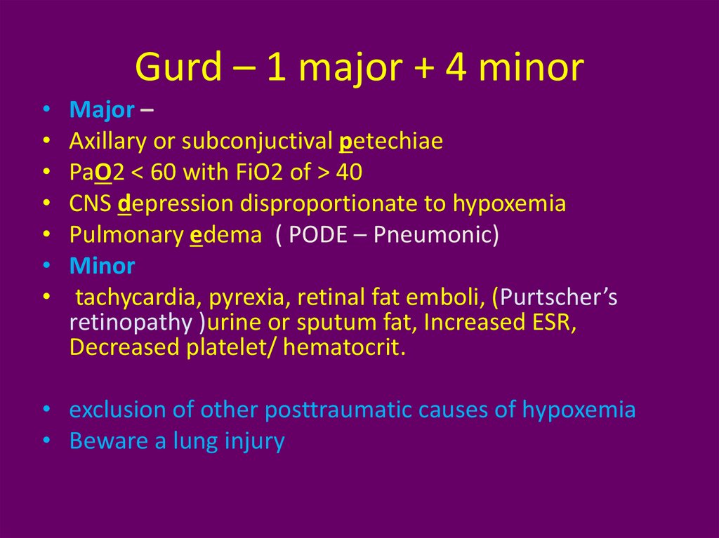
Gurd – 1 major + 4 minorMajor –
Axillary or subconjuctival petechiae
PaO2 < 60 with FiO2 of > 40
CNS depression disproportionate to hypoxemia
Pulmonary edema ( PODE – Pneumonic)
Minor
tachycardia, pyrexia, retinal fat emboli, (Purtscher’s
retinopathy )urine or sputum fat, Increased ESR,
Decreased platelet/ hematocrit.
• exclusion of other posttraumatic causes of hypoxemia
• Beware a lung injury
29.
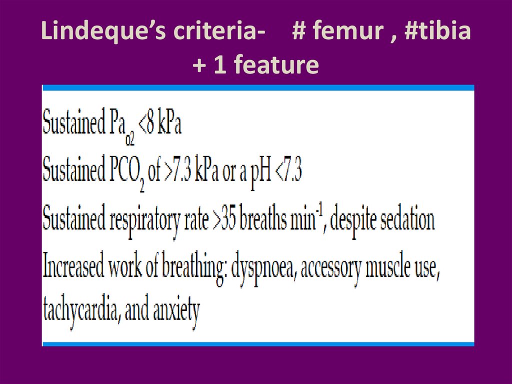
Lindeque’s criteria- # femur , #tibia+ 1 feature
30.
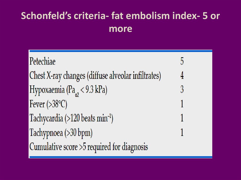
Schonfeld’s criteria- fat embolism index- 5 ormore
31.
The features are acute, but not abrupt32.
How to confirm ??• High index of suspicion and some
investigations
33.
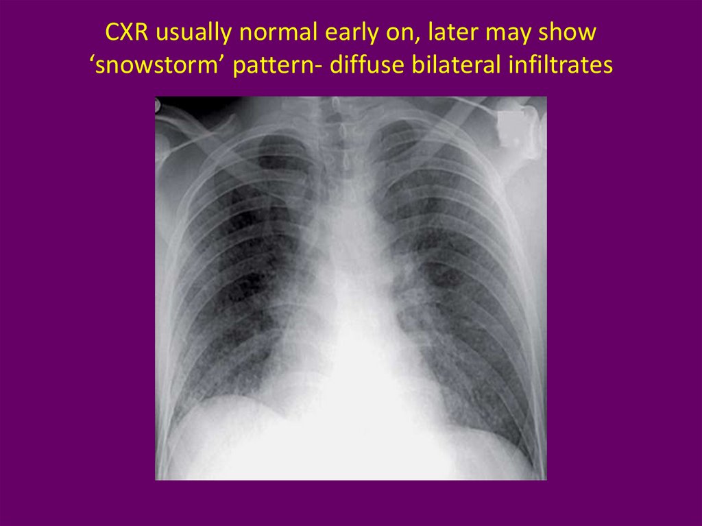
CXR usually normal early on, later may show‘snowstorm’ pattern- diffuse bilateral infiltrates
34.
Lab values• Arterial blood gases :
• This reveals a low partial pressure of oxygen and a
low partial pressure of CO2 with respiratory alkalosis.
• An unexplained anemia (70% of patients) and
thrombocytopenia (platelet count <1,50,000 mm-3 in
up to 50% of patients.
Hypocalcemia (due to binding of free fatty acids to
calcium) and elevated serum lipase have also been
Reported
Hypofibrinogenemia
35.
CVS• ECG : sinus tachycardia ; Non specific ST T
changes, RBBB,
• Lung scan : ? V/Q mismatch.
• Transesophageal echocardiography : Fat
droplets. PFO, Rt sided dilatation if present
36.
Broncho alveolar lavage• BAL : fat droplets.
• The staining of cells with oil red O after
recovery by a standard 150- to 200-mL lavage
can identify intracellular fat droplets.
• Can be there in minimal fat embolism – but!!
• quantitative count of lavage cells containing
fat of greater than 30% being significant of fat
embolism syndrome
37.
CT Brain• White matter petechiae
• Cerebral edema
• Rarely cerebral atrophy due to
• full embolisation
38.
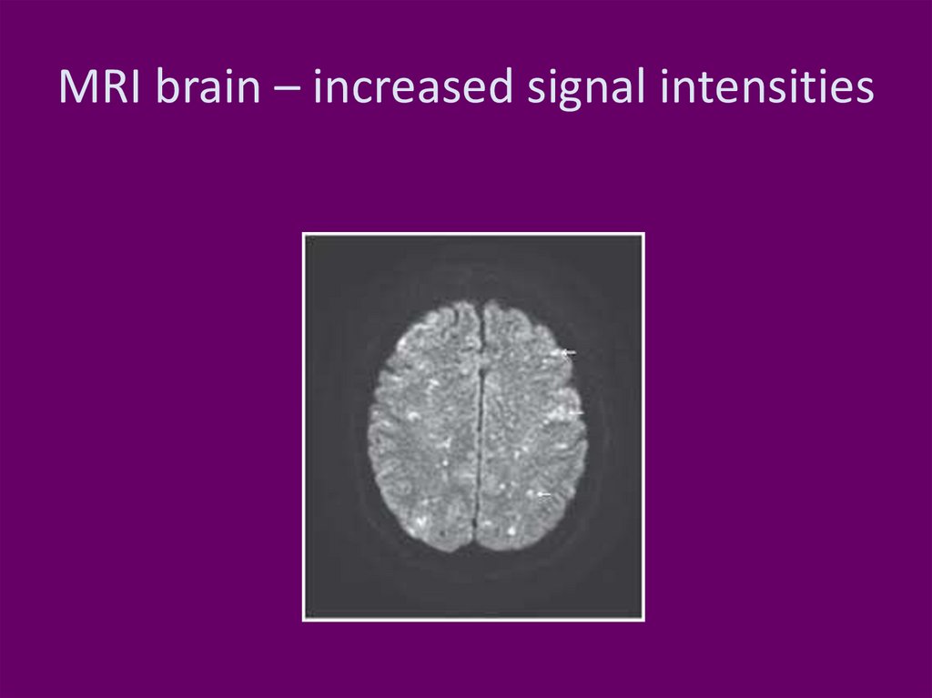
MRI brain – increased signal intensities39.
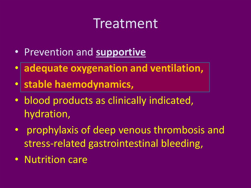
TreatmentPrevention and supportive
adequate oxygenation and ventilation,
stable haemodynamics,
blood products as clinically indicated,
hydration,
• prophylaxis of deep venous thrombosis and
stress-related gastrointestinal bleeding,
• Nutrition care
40.
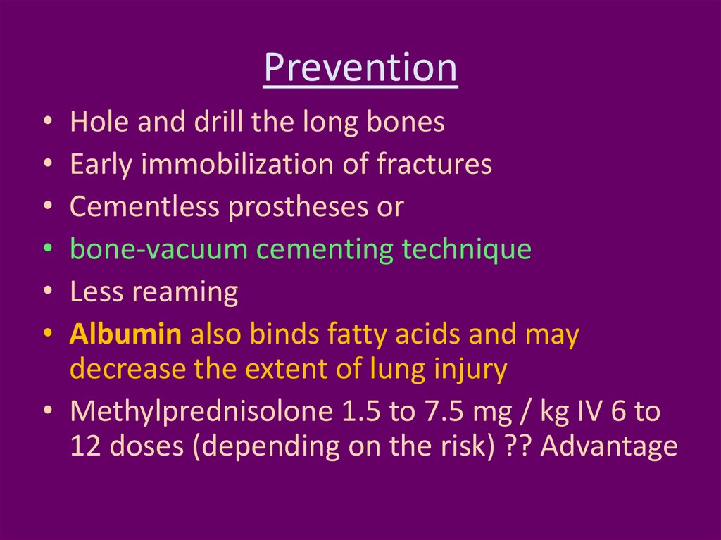
PreventionHole and drill the long bones
Early immobilization of fractures
Cementless prostheses or
bone-vacuum cementing technique
Less reaming
Albumin also binds fatty acids and may
decrease the extent of lung injury
• Methylprednisolone 1.5 to 7.5 mg / kg IV 6 to
12 doses (depending on the risk) ?? Advantage
41.
Prevention• during cementing
• Hydration
• Oxygenation
• No nitrous
42.
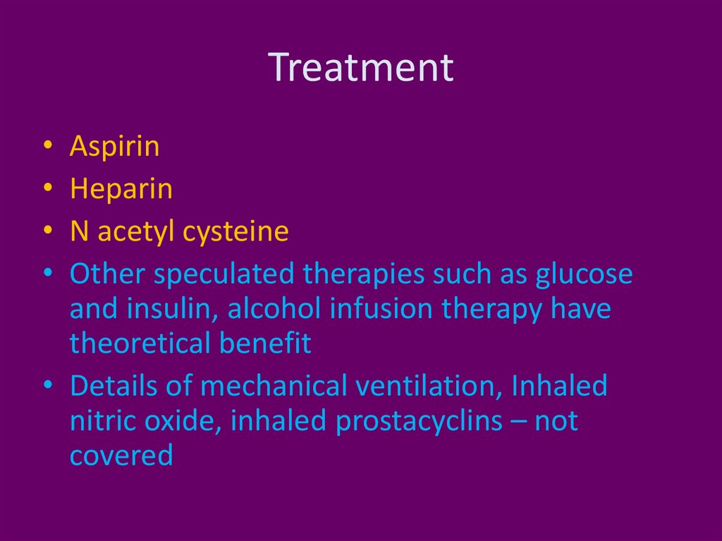
TreatmentAspirin
Heparin
N acetyl cysteine
Other speculated therapies such as glucose
and insulin, alcohol infusion therapy have
theoretical benefit
• Details of mechanical ventilation, Inhaled
nitric oxide, inhaled prostacyclins – not
covered
43.
Prognosis who survived• The prognosis for patients who survive fat
embolism is good, with recovery from the fat
embolism syndrome usually being complete
within 2-4 weeks.
• neurological signs may remain for up to 3
months
44.
SummaryDefinitions
Incidence
Etiology
lethal dose
Theories
Prevention
Treatment