


 Медицина
Похожие презентации:


Radiologic diagnostics of chest cavity
1.
Radiologic diagnostics of chestcavity
Жакенова Д.К.
Визуальная диагностика
КазНМУ
2.
Methods of respiratory organs examination:Radiography
Radioscopy
Bronchography
Angiopulmonography
Ultrasound diagnostics
CT scan
Magnetic resonance imaging
Radionuclide Diagnostics
PET
3.
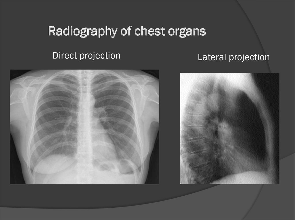
Radiography of chest organsDirect projection
Lateral projection
4.
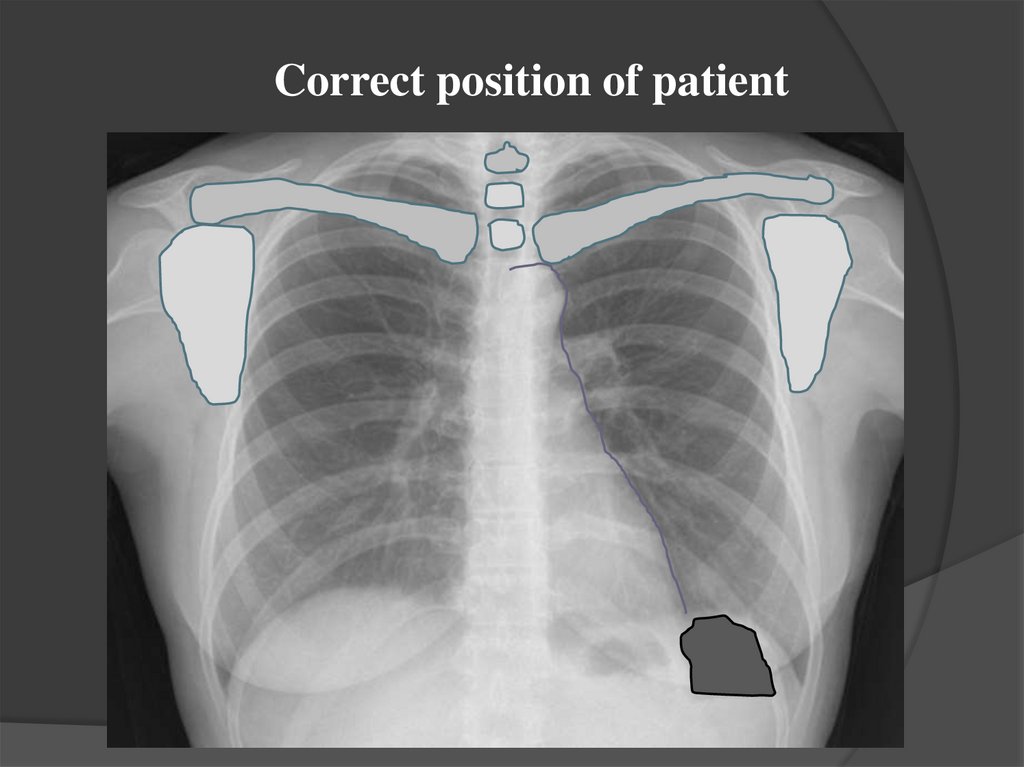
Correct position of patient5.
Normal X-ray anatomy of the lungs6
1
2
3
9
7
4
5
8
1 — anterior end of rib; 2 — trachea and major bronchi; 3 —ribs;
4 —right lower-lobe artery; 5 — diaphragm; 6 — posterior end of
rib;
7 —root of the left lung; 8 — left breast contour, 9- root of the
right lung
6.
Pulmonary fields7.
Roots of the lungsthe left root above the right is 1-1.5 cm
II-IV
ribs
II-IV
intercostal
space
Root width = 2.5 cm
8.
Pulmonary patternCardiac diaphragmatic sinuses
costodiaphragmatic sinuses
9.
SHADOW OF THE HEARTArc of the Heart
right
1- ascending aorta
2 -Arc of
the right
atrium
left
1-aortic arch
2-arc pulmonary
trunk
3-eye of the left
atrium
4-left
ventricular
arch
10.
Cardiothoracic index =transverse size of heart
transverse size of thorax
transverse size of heart
transverse size of thorax
= 0,4-0,5
11.
Atriovasal angleascending
aorta
Atriovasal angle
Right atrium
12.
Segments of the lungs in a straight projection1
1-2
3
2
3
4
4
8
5
5
8
13.
Lung segments in lateral projection1
2
3
6
4
5
10
9 7 8
8
14.
Segments of the lungs in a straight projection (front, back)15.
Median tomography1-trachea
2-right main bronchus
3-left main bronchus
4-right upper lobe
bronchus
5-right mid-lobe
bronchus
6-right lower lobe
bronchus
7-left upper lobe
bronchus
16.
Linear tomography17.
Lymph nodes of the mediastinumparatracheal lymph nodes
Tracheobronchial lymph nodes
Bronchopulmonary lymph no
Bifurcation lymph nodes
18.
Art. carotis comm. dext.Art. carotis comm. sin.
V. jugularis int. dext.
Art. subclavia dext.
V. jugularis int. sin.
V. subclavia dext
V. subclavia sin.
V. anonyma dext.
Art. anonyma dext.
V. cava sup.
Art. subclavia sin.
V. anonyma sin.
Arcus aortae
Art. pulm. sin.
19. X-ray examination methods
- Radiography- Radioscopy
- Linear tomography
- Fluorography
20.
RadioscopyX-ray examination method in which an X-ray
image of an object is obtained on the monitor
screen in real time.
21.
RadioscopyX-ray tube
X-ray radiation
fluorescent screen
22.
Indications:- polypositional study
- real-time evaluation of the function
- conducting the catheterization, angioplasty under the
control of radioscopy
Disadvantages:
- high radiation load
- subjectivity of data
- lack of documentation
23. Methods of radioscopy a - orthoscopy, b - trochoscopy, v-lateroscopy
24.
25.
RadiographyAn X-ray examination method in which a
fixed X-ray image of an object is obtained on
a film or in computer memory.
26.
Advantages of radiography:- better detectability of small parts
- less radiation load
- the possibility of an objective assessment for
follow-up and comparison
27.
28.
29.
30.
31.
32.
FluorographyThe method of X-ray examination consists in
photographing the image from a fluorescent screen,
screen of an electron-optical converter or systems
intended for the subsequent digitization of images, on a
film of a format 100х100, 110х110 mm.
33.
FluorographyX-ray tube
X-ray radiation
fluorescent
screen
camera
10 см
34.
Advantages of fluorography :- low cost of research
- the possibility of conducting mass
verification studies
Disadvantages of fluorography:
- high radiation load
- ban on conducting research for persons
under 15 years of age
35.
Main applications- Chest examination for early detection of
tuberculosis
36.
37.
Linear tomographyThe method of X-ray examination is to obtain an image of
an object at a specified depth.
38.
39.
40.
Main applicationsInvestigation of pulmonary parenchyma, trachea and major
bronchi, intrathoracic lymph nodes, paranasal sinuses,
larynx, separate structures of the spine.
41. Tomography of chest organs
42. Evaluation of the quality of the radiograph
Completeness of volume : the whole chest from the tops of the lungsto the costal-diaphragmatic sinuses is displayed
Position of the patient: the same distance between the medial contours
of the clavicles and the spinous process of the vertebra (Th 3), the
scapula is outward from the pulmonary fields, clavicles are arranged
horizontally.
Precision: clear contours of the diaphragm, front segments of the ribs,
visualization of all elements of the pulmonary pattern and the contours
of the heart (in adults, the sharpness is evaluated by the left contour, in
children up to 1 year on the right contour of the heart )
Contrast : equally expressed black, white and gray colors
Hardness: on the front radiographs, the outlines of the first 3-4
thoracic vertebrae, located above the middle shadow, on the lateral - a
clear image of the head of the humerus, a clear visualisation of the
elements of the pulmonary pattern
43. Прямая проекция
Критерии правильновыполненной
рентгенограммы
Видны
легочные поля на всем
протяжении и диафрагмальные
синусы
Изображение лопаток не
наслаивается на легочную ткань
Ключицы расположены
горизонтально
Расстояние от средней линии
(остистые отростки) до
грудинных краев ключиц
одинаково с обеих сторон
44. Прямая проекция
Ширина задних отрезковребер значительно
меньше передних
Контуры задних
отрезков ребер более
четкие, чем контуры
передних
45. Исследование на вдохе и выдохе
Рентгенография легких производится в фазу глубокого,но не форсированного вдоха.
Диафрагма справа в норме располагается на уровне
переднего отрезка 6-го ребра, слева на одно ребро
ниже.
На выдохе снижается прозрачность легочных полей,
сердце становится более широким.
46.
Фазы дыхания ВдохВыдох
Выполняется
В фазу глубокого, но не На выдохе
форсированного вдоха.
Диафрагма
6-7 ребро
Уплощена
4-5 ребро
Выпуклая
Легочный
рисунок
Легкое
Обычный
Усилен и сгущен
Прозрачность обычная
Прозрачность
понижена в средних и
нижних отделах
Ребра
Расположены косо
Расположены
горизонтально
Сердце
Узкое и расположено
вертикально
Широкое, расположено
горизонтально
47. Inspiration and expiration examination
46
48. Боковая проекция
Критерии правильновыполненной
рентгенограммы
Видны легочные поля на
всем протяжении (верхушки
и реберно-диафрагмальные
синусы).
Четкое изображение
грудины в боковой проекции
Отчетливо прослеживаются
рентгеноанатомические
детали