


 Медицина
Похожие презентации:

")


. Class Nematoda. Lesson 5")
")

Rework topic - 5 - schistosomes
1.
Medical Academy namedafter S.I. Georgievsky of
Vernadsky CFU.
REWORK TOPIC -5- SCHISTOSOMES
Prepared by – Mehak Mehla
Scientific Leader – Svetlana Smirnova
2.
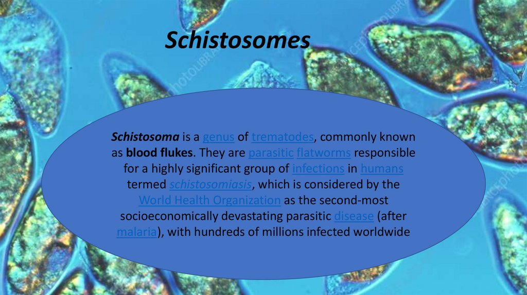
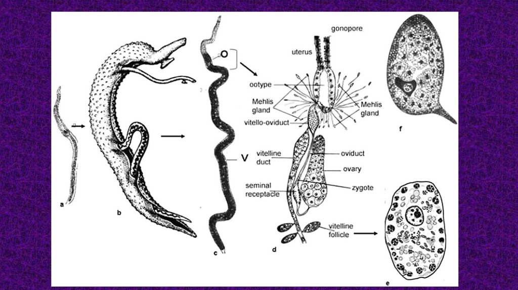
SchistosomesSchistosoma is a genus of trematodes, commonly known
as blood flukes. They are parasitic flatworms responsible
for a highly significant group of infections in humans
termed schistosomiasis, which is considered by the
World Health Organization as the second-most
socioeconomically devastating parasitic disease (after
malaria), with hundreds of millions infected worldwide
3.
Scientificclassification
Kingdom:
Animalia
Phylum:
Platyhelminthes
Class:
Trematoda
Order:
Diplostomida
Family:
Schistosomatidae
Genus:
Schistosoma
4.
GEOGRAPHICALGeographical distribution
DISTRIBUTION
Africa, Brazil, Cambodia, the Caribbean, China, Corsica, Indonesia, Laos,
the Middle East, the Philippines, Suriname, and Venezuela.[22] There had
been no cases in Europe since 1965, until an outbreak occurred on
Corsica
5.
6.
Haematobium groupMansoni group
Japonicum group
Mansoni group
TAXONOMY
7.
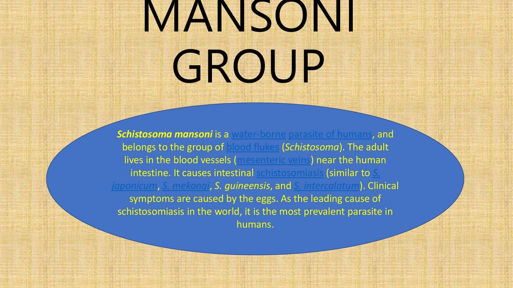
MANSONIGROUP
Schistosoma mansoni is a water-borne parasite of humans, and
belongs to the group of blood flukes (Schistosoma). The adult
Schistosomalives
mansoni
is a water-borne
parasite
of humans,
belongs to the
in the blood
vessels (mesenteric
veins)
near the and
human
group of blood
flukes
(Schistosoma).
The adult lives(similar
in theto
blood
vessels
intestine.
It
causes
intestinal
schistosomiasis
S.
(mesenteric veins) near the human intestine. It causes intestinal
japonicum,(similar
S. mekongi,
guineensis, and
S. intercalatum).
Clinical and S.
schistosomiasis
to S.S.japonicum,
S. mekongi,
S. guineensis,
intercalatum).
Clinical are
symptoms
are
caused
bythe
theleading
eggs. As
theofleading cause
symptoms
caused by
the
eggs. As
cause
of schistosomiasis
in the
it isitthe
most
prevalent
schistosomiasis
in world,
the world,
is the
most
prevalentparasite
parasite in
in humans.
humans.
8.
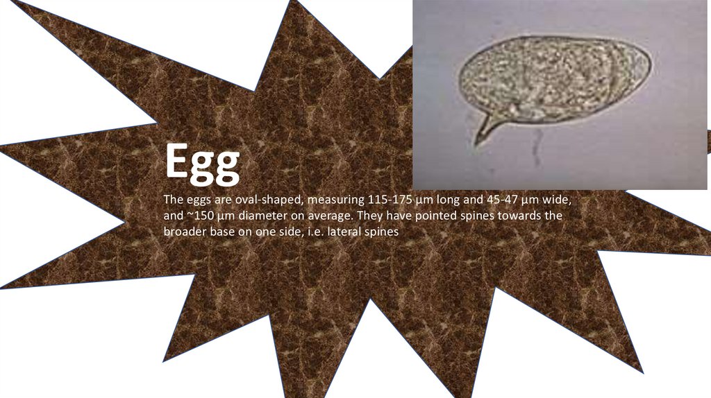
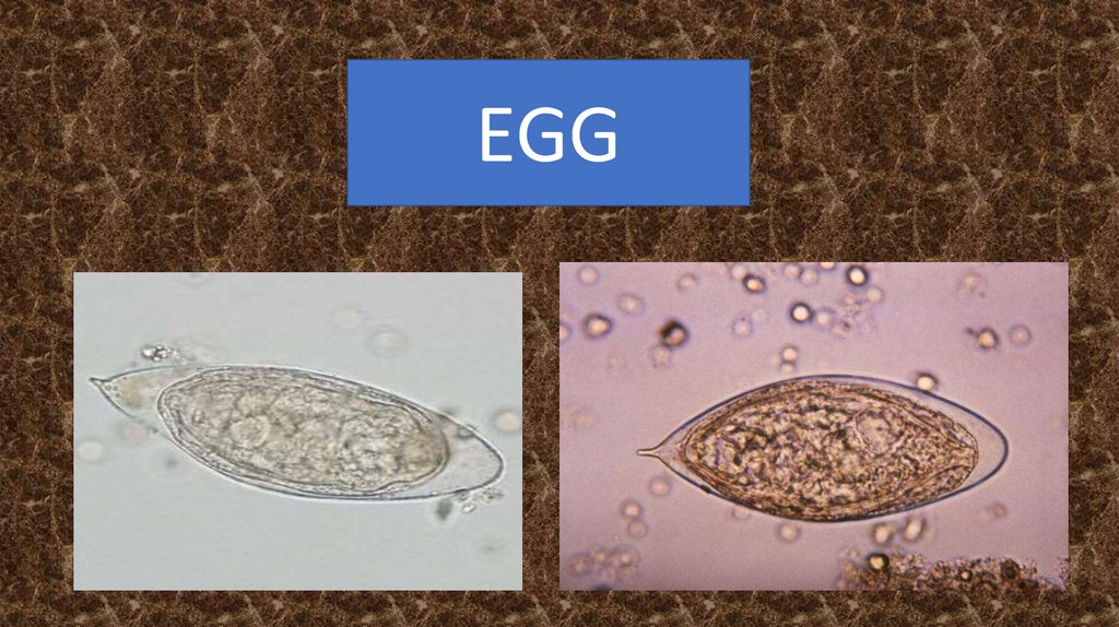
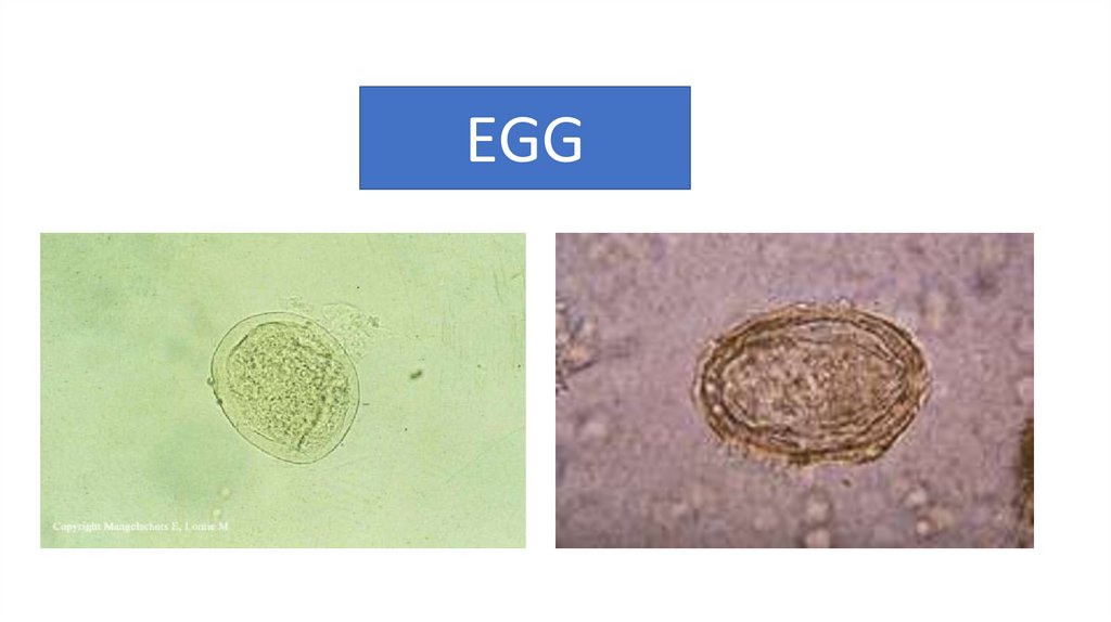
EggThe eggs are oval-shaped, measuring 115-175 µm long and 45-47 µm wide,
and ~150 µm diameter on average. They have pointed spines towards the
broader base on one side, i.e. lateral spines
9.
10.
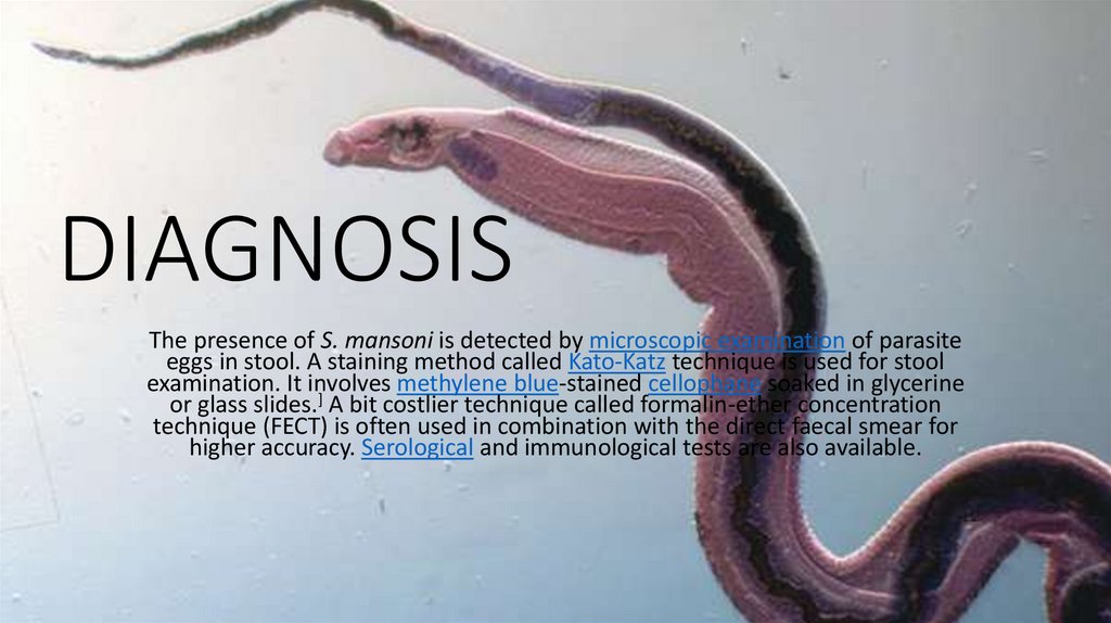
DIAGNOSISThe presence of S. mansoni is detected by microscopic examination of parasite
eggs in stool. A staining method called Kato-Katz technique is used for stool
examination. It involves methylene blue-stained cellophane soaked in glycerine
or glass slides.] A bit costlier technique called formalin-ether concentration
technique (FECT) is often used in combination with the direct faecal smear for
higher accuracy. Serological and immunological tests are also available.
11.
HAEMATOBIUM GROUPSchistosoma haematobium (urinary blood fluke)
is a species of digenetic HVS
trematode, belonging to
a group (genus) of blood flukes (Schistosoma). It
is found in Africa and the Middle East. It is the
major agent of schistosomiasis, the most
prevalent parasitic infection in humans
12.
EGG13.
14.
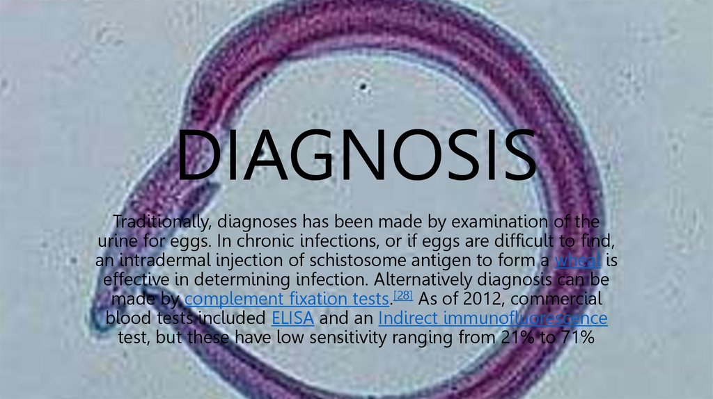
DIAGNOSISTraditionally, diagnoses has been made by examination of the
urine for eggs. In chronic infections, or if eggs are difficult to find,
an intradermal injection of schistosome antigen to form a wheal is
effective in determining infection. Alternatively diagnosis can be
made by complement fixation tests.[28] As of 2012, commercial
blood tests included ELISA and an Indirect immunofluorescence
test, but these have low sensitivity ranging from 21% to 71%
15.
JAPONICUM GROUPSchistosoma japonicum is an important parasite and one
infectious
agents
of schistosomiasis.This
ciesof
ofthe
wildmajor
mammals,
including
9 carnivores,
16 rodents, one primate
(Human),
twohas
insectivores
and three
and therefore
can be
parasite
a very wide
host artiodactyls
range, infecting
at leastit31
zoonosis.
species of wild considered
mammals,a true
including
9 carnivores, 16
rodents, one primate (Human), two insectivores and three
artiodactyls and therefore it can be considered a true
zoonosis.
16.
EGG17.
18.
DIAGNOSISMicroscopic identification of eggs in stool or urine is the most practical
method for diagnosis. Stool examination should be performed when
infection with S. mansoni or S. japonicum is suspected, and urine
examination should be performed if S. haematobium is suspected. Eggs
can be present in the stool in infections with all Schistosoma species.