


")


 Медицина
Похожие презентации:


Oral diagnosis
1.
Dr. Anas AlmisuratiBDS, MSc Periodontology and Oral Medicine (Cairo
University).
Assistant lecturer periodontology and oral medicine department
Zawia University
2.
• Oral Diagnosis• It is the art of using scientific knowledge to identify
oral disease processes and to distinguish one
disease from another.
3.
• Types of oral diagnosis :1) - Comprehensive oral diagnosis :• The diagnostic assessment for all dental problems as revealed
by : • Full history
• clinical examination
• Use of diagnostic aids (INVESTIGATION ,,,, BIOPSY,,,,,)
• It is done for the patients requiring total dental care.
4.
2) Emergency diagnosis :• It is the immediate diagnosis of the patient'scomplaint that requires immediate attention and
management by the dentist
(acute dental pain, accidental
fractures,…).
• The emergency interferes with obtaining
adequate history or full clinical examination
(only the area of chief complaint).
5.
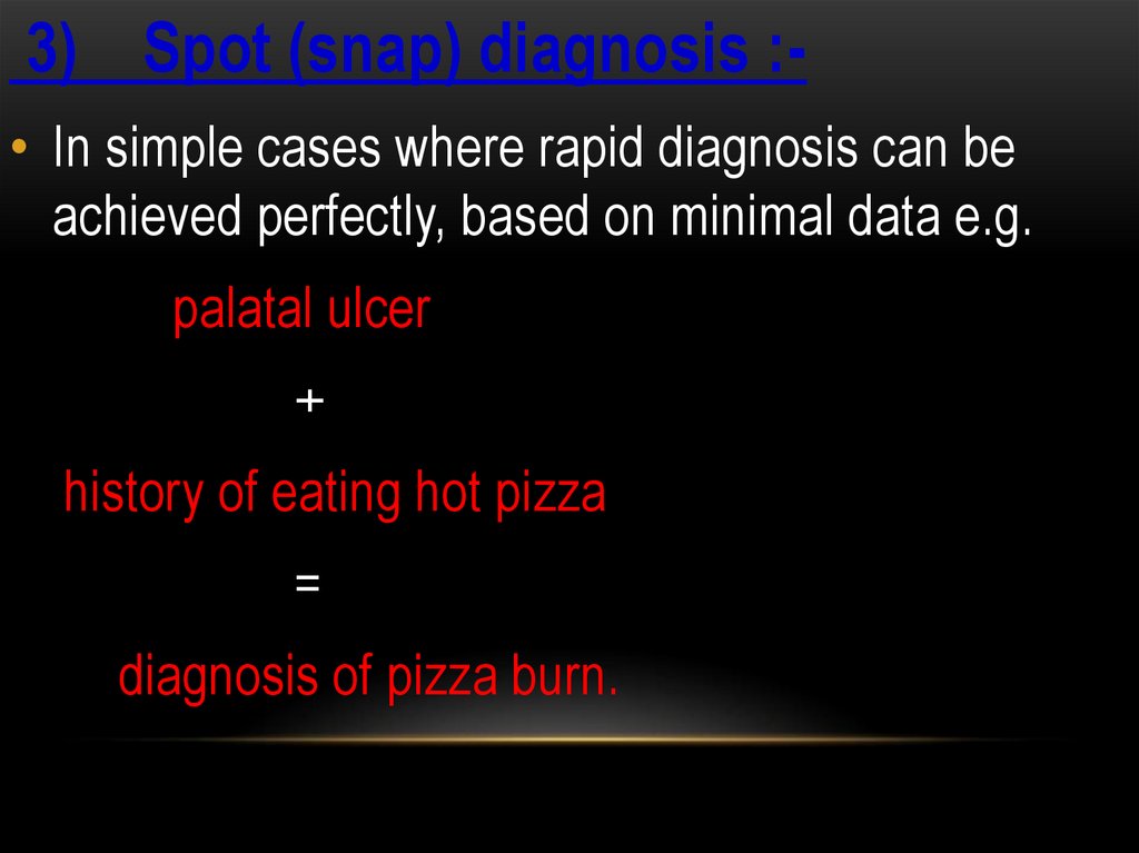
3) Spot (snap) diagnosis :• In simple cases where rapid diagnosis can beachieved perfectly, based on minimal data e.g.
palatal ulcer
+
history of eating hot pizza
=
diagnosis of pizza burn.
6.
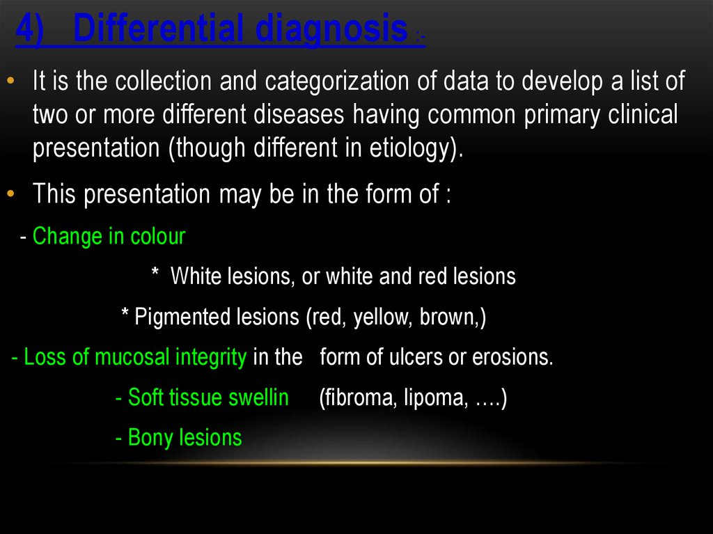
4) Differential diagnosis :• It is the collection and categorization of data to develop a list oftwo or more different diseases having common primary clinical
presentation (though different in etiology).
• This presentation may be in the form of :
- Change in colour
* White lesions, or white and red lesions
* Pigmented lesions (red, yellow, brown,)
- Loss of mucosal integrity in the form of ulcers or erosions.
- Soft tissue swellin
- Bony lesions
(fibroma, lipoma, ….)
7.
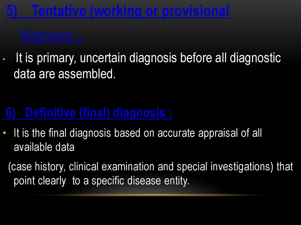
5) Tentative (working or provisionaldiagnosis :
It is primary, uncertain diagnosis before all diagnostic
data are assembled.
6) Definitive (final) diagnosis :
• It is the final diagnosis based on accurate appraisal of all
available data
(case history, clinical examination and special investigations) that
point clearly to a specific disease entity.
8.
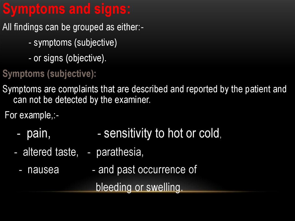
Symptoms and signs:All findings can be grouped as either:- symptoms (subjective)
- or signs (objective).
Symptoms (subjective):
Symptoms are complaints that are described and reported by the patient and
can not be detected by the examiner.
For example,:-
- pain,
- sensitivity to hot or cold ,
- altered taste, - parathesia,
- nausea
- and past occurrence of
bleeding or swelling.
9.
• Signs (objective findings):Objective findings are the changes or deviations
from normal that can be detected by the examiner.
• For example,:- discoloration of teeth or soft tissues,
- swelling,
- tenderness to palpation
10.
• Treatment plan:Treatment plan may take one of two forms:
• A. Emergency or immediate treatment
plan:-
• B. Comprehensive or long-range
treatment plan:-
11.
• The diagnostic methodIt is the application of a scientific method to reach a final diagnosis.
• Elements of the scientific diagnostic method include:
11- Collection of information.
2- Evaluation of the information.
3- Diagnostic decision.
4- Reassessment.
12.
• 1-Collection of information for reaching adiagnosis include:
1 – Patient history.
2 – Clinical examination.
3 – Diagnostic aids.
13.
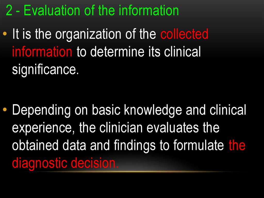
2 - Evaluation of the information• It is the organization of the collected
information to determine its clinical
significance.
• Depending on basic knowledge and clinical
experience, the clinician evaluates the
obtained data and findings to formulate the
diagnostic decision.
14.
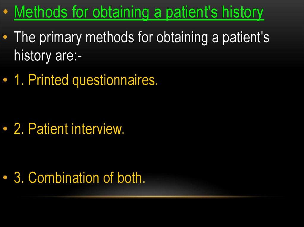
• Methods for obtaining a patient's history• The primary methods for obtaining a patient's
history are:• 1. Printed questionnaires.
• 2. Patient interview.
• 3. Combination of both.
15.
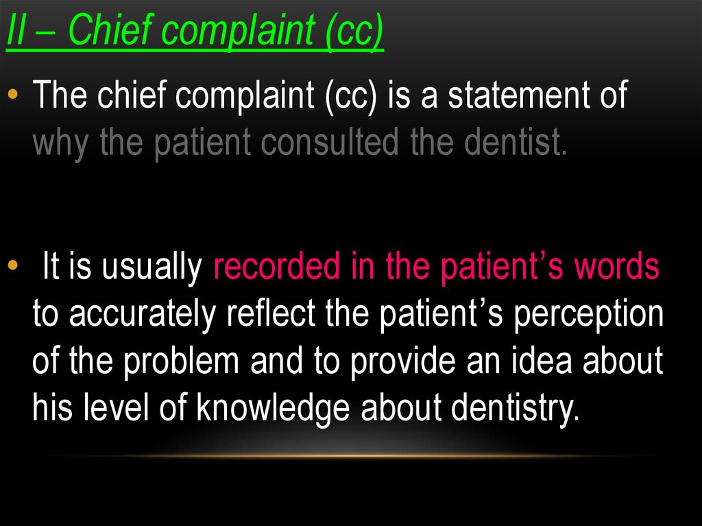
II – Chief complaint (cc)• The chief complaint (cc) is a statement of
why the patient consulted the dentist.
• It is usually recorded in the patient’s words
to accurately reflect the patient’s perception
of the problem and to provide an idea about
his level of knowledge about dentistry.
16.
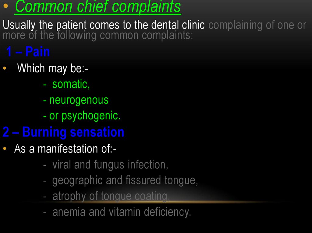
• Common chief complaintsUsually the patient comes to the dental clinic complaining of one or
more of the following common complaints:
1 – Pain
• Which may be:- somatic,
- neurogenous
- or psychogenic.
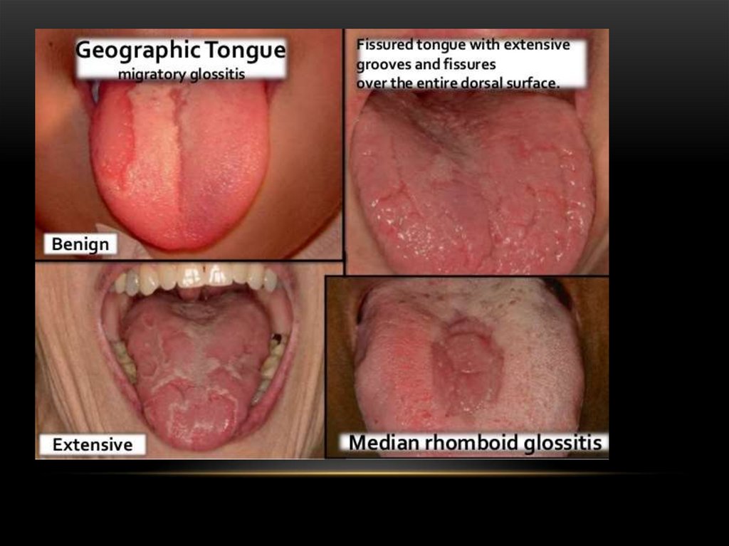
2 – Burning sensation
• As a manifestation of:- viral and fungus infection,
- geographic and fissured tongue,
- atrophy of tongue coating,
- anemia and vitamin deficiency.
17. pain
PAIN18.
3 – Paraesthesia and numbness• Caused by vitamin deficiency, pressure on the mandibular nerve
such as :- neurofibromatosis,
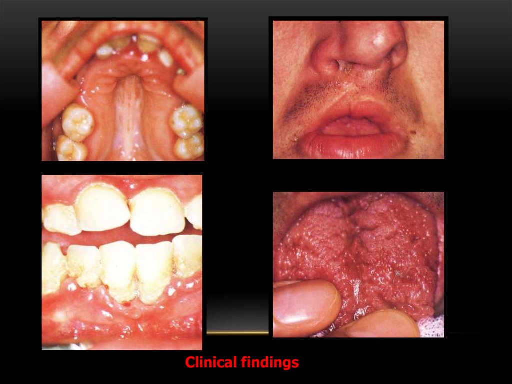
- injury to the trigeminal nerve,
- trauma from anaethetic needles
- and following surgical procedures.
• Also, it may be caused by:- diabetes,
- pernicious anemia,
- syphilis
- and prolonged use of some medications such as:- streptomycin,
- sedatives,
- tranquilizers
19.
4 - Sensitivity• Sensitivity to hot, cold and sweats may result from decayed
teeth, pulpitis or exposed roots.
5 – Bleeding
• Bleeding or hemorrhage may occur accidentally or following
surgery including extraction.
• It may result from different causes such as :- trauma,
- post-operative infection
- or even uncontrolled blood disorders.
• Gingival bleeding may be the early manifestation of periodontal
problems.
• The patient may complaint of bleeding gums spontaneously or
on slight provocation such as tooth brushing or eating hard food.
20.
6 – Swelling- Soft tissue swelling such as:-
- facial cellulitis
- and glandular swelling
- hard tissue swelling such as:- Paget’s disease
- ameloblastoma.
21.
7 – Oral ulceration• Ulceration of the oral mucous membrane are
multiple and caused by different etiologic
factors.
• The most common oral ulcerations in dental
practice are:- recurrent aphthous ulceration
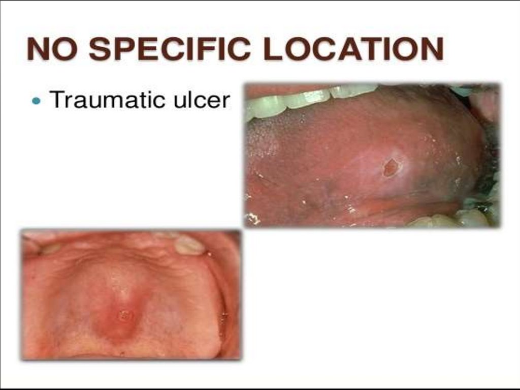
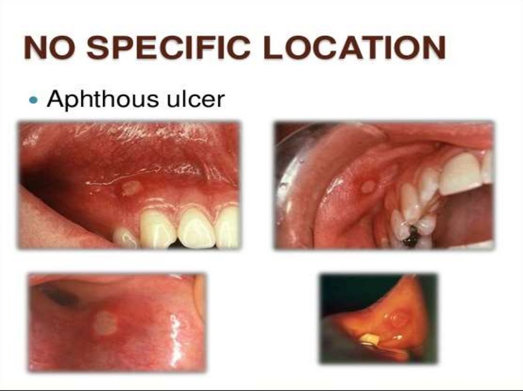
- and traumatic ulcers.
22.
8 – T.M.J. disorders• Patients with T.M.J. disorders may complaint of:- clicking in jaw joint
- and unilateral pain
felt in the ear and radiates to the angle of
the mandible with or without
limitation of jaw function.
23.
9 – Functional disorders• The patient complaint may result from functional
disorders such as:- dysphagia
- xerostomia,
which is a clinical
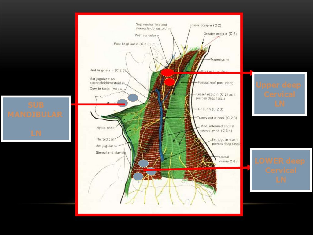
manifestation of salivary
gland dysfunction not
representing a disease
entity.
24.
10 – Bad breath (halitosis)• It results from either extra-oral or more
commonly oral causes especially poor oral
hygiene.
• Dental infection
• In some instances the cause may be
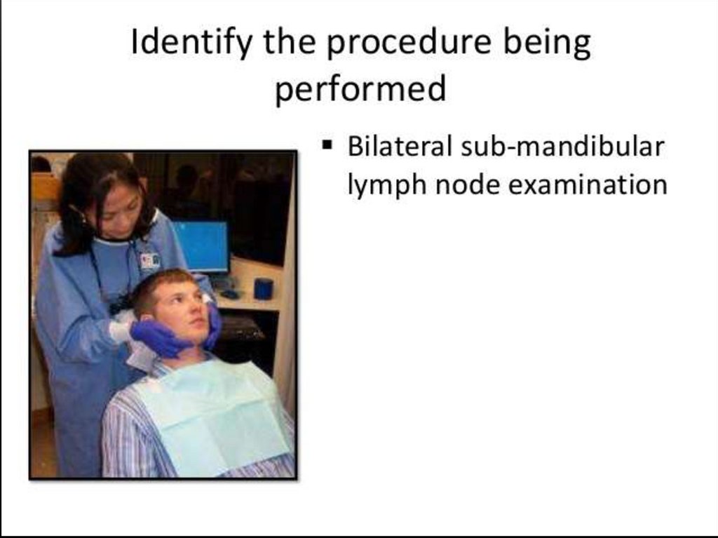
psychogenic.
25.
11- Esthetic problem• Orthodontic treatment or malposed teeth may be the only
complaint of certain age group of patients.
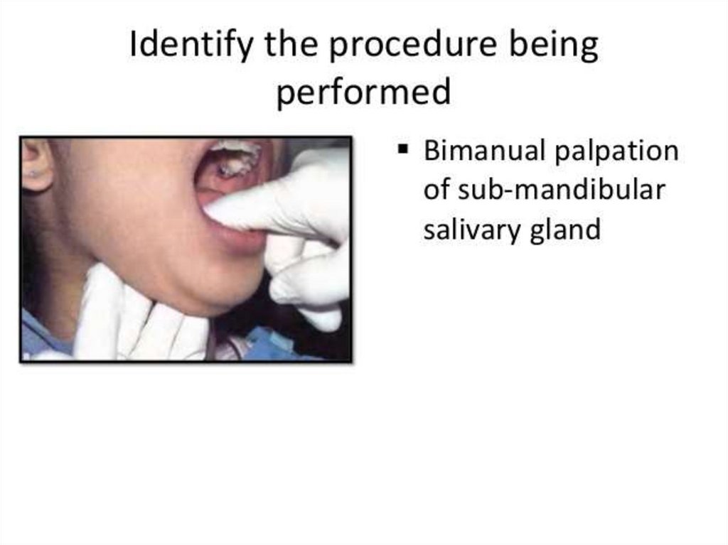
• Also, discolored or hypoplastic teeth may result in psychological
esthetic problem for many individuals.
• It should be noted that in many cases of gum recession and
exposure of the roots especially of the anterior teeth, the main
complaint of the patient is bad esthetic.
26.
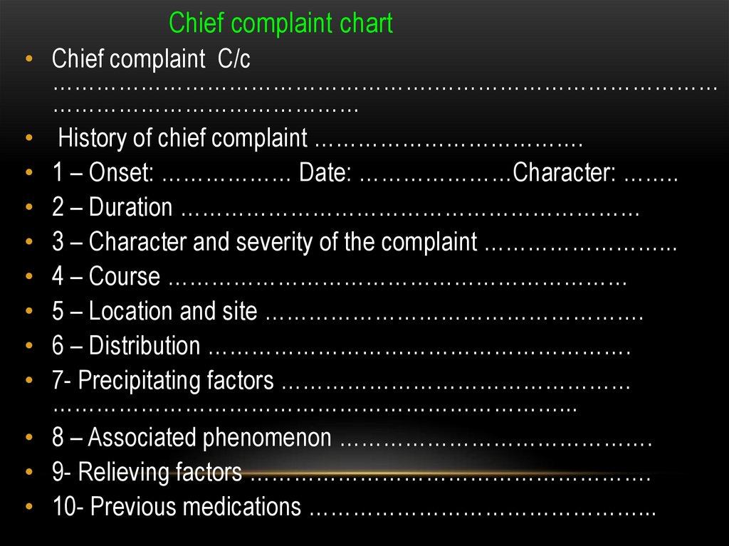
Chief complaint chart• Chief complaint C/c
…………………………………………….…………………………………
……………………………………
• History of chief complaint ……………………………….
• 1 – Onset: ……………… Date: …………………Character: ……..
• 2 – Duration ………………………………………………………
• 3 – Character and severity of the complaint ……………………...
• 4 – Course ………………………………………………………
• 5 – Location and site …………………………………………….
• 6 – Distribution ………………………………………………….
• 7- Precipitating factors …………………………………………
……………………………………………………………...
• 8 – Associated phenomenon …………………………………….
• 9- Relieving factors ……………………………………………….
• 10- Previous medications ………………………………………...
27.
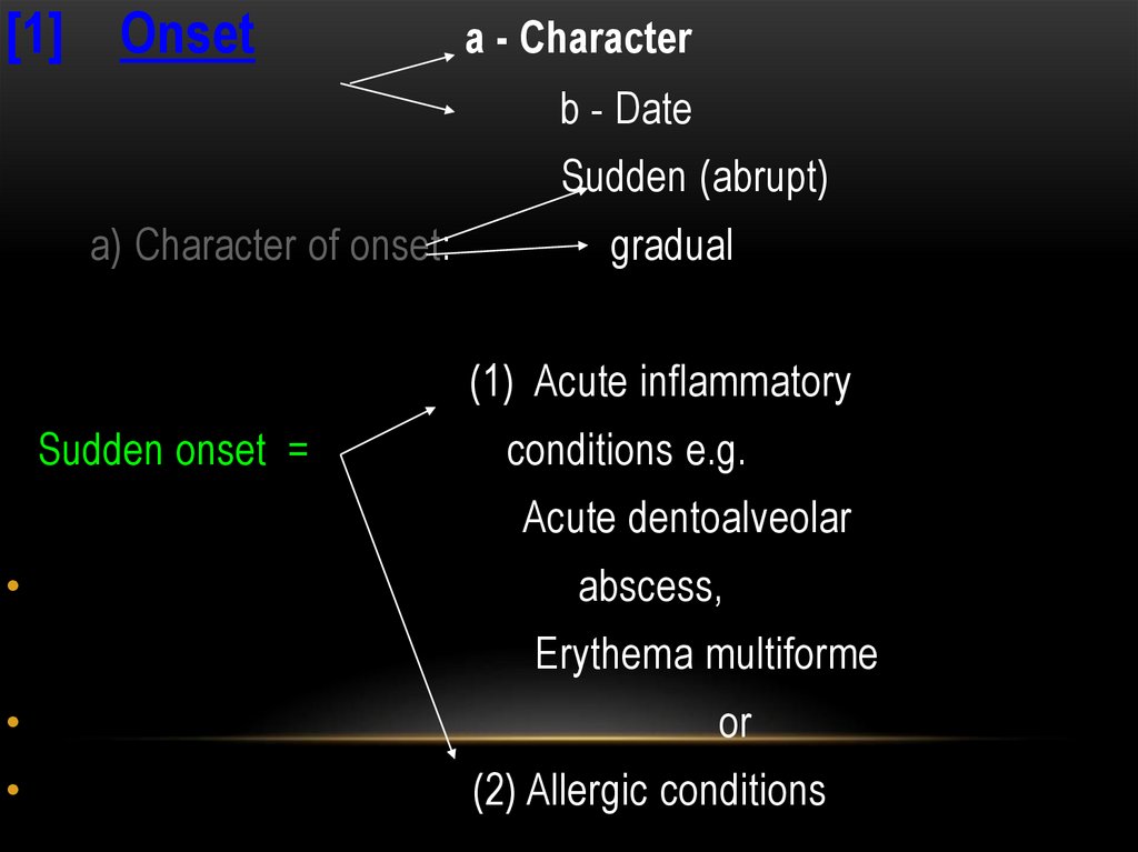
[1] Onseta - Character
b - Date
Sudden (abrupt)
a) Character of onset:
gradual
(1) Acute inflammatory
Sudden onset =
conditions e.g.
Acute dentoalveolar
abscess,
Erythema multiforme
or
(2) Allergic conditions
28.
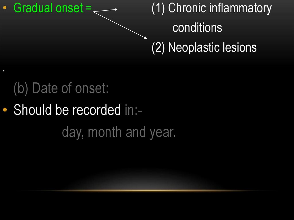
• Gradual onset =(1) Chronic inflammatory
conditions
(2) Neoplastic lesions
.
(b) Date of onset:
• Should be recorded in:day, month and year.
29.
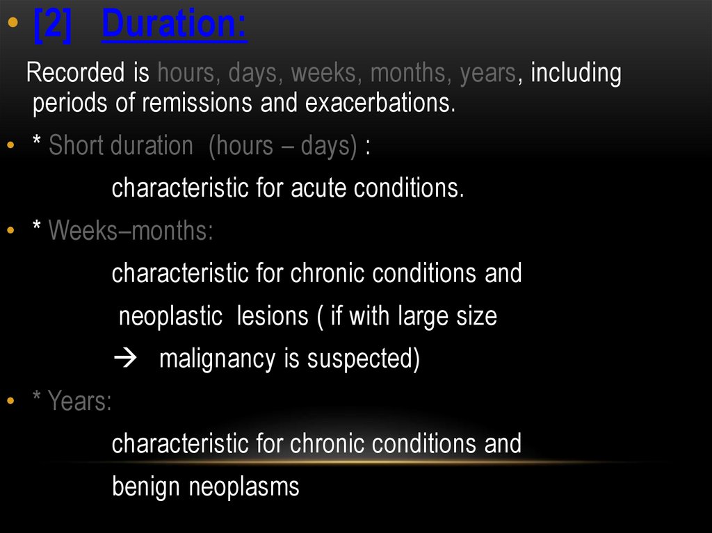
• [2] Duration:Recorded is hours, days, weeks, months, years, including
periods of remissions and exacerbations.
• * Short duration (hours – days) :
characteristic for acute conditions.
• * Weeks–months:
characteristic for chronic conditions and
neoplastic lesions ( if with large size
malignancy is suspected)
• * Years:
characteristic for chronic conditions and
benign neoplasms
30.
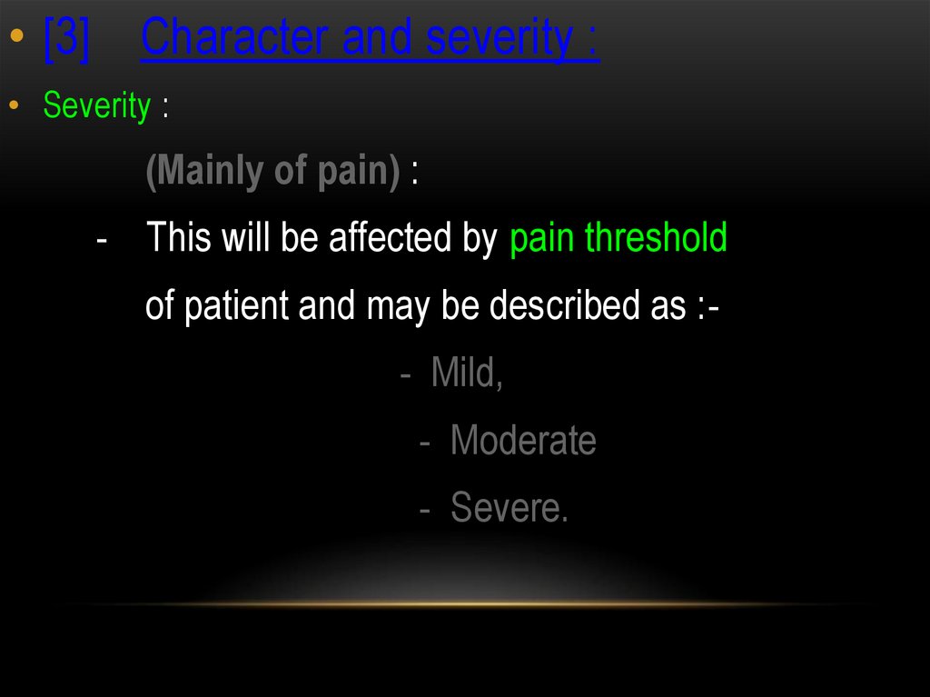
• [3] Character and severity :• Severity :
(Mainly of pain) :
- This will be affected by pain threshold
of patient and may be described as :- Mild,
- Moderate
- Severe.
31.
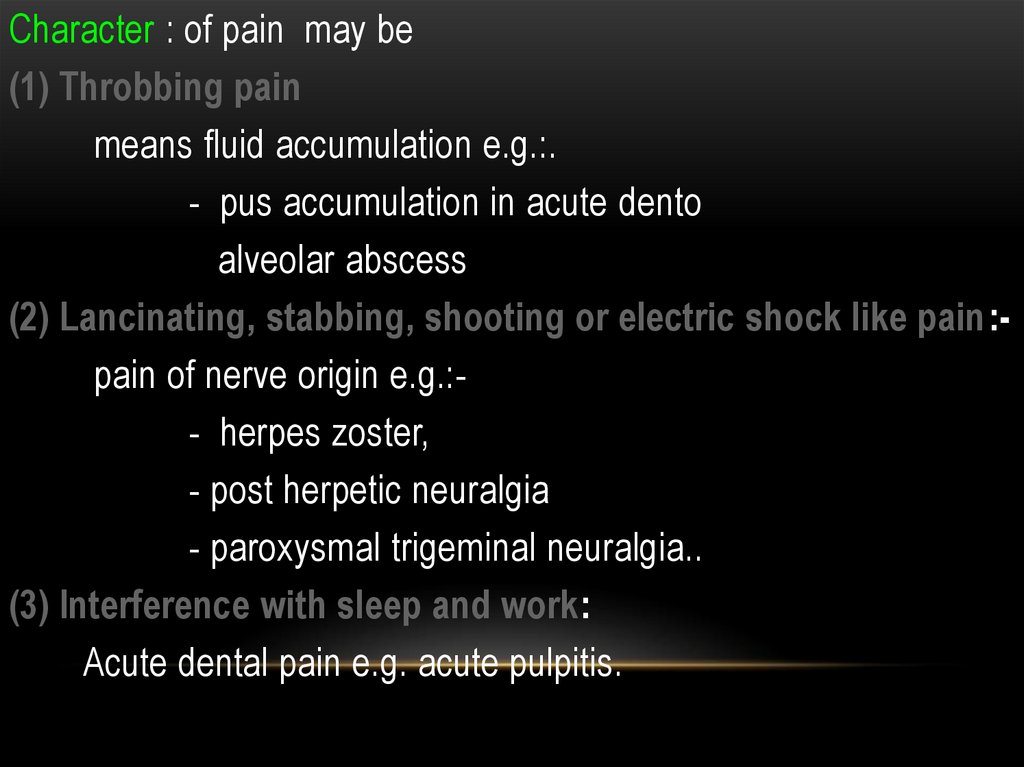
Character : of pain may be(1) Throbbing pain
means fluid accumulation e.g.:.
- pus accumulation in acute dento
alveolar abscess
(2) Lancinating, stabbing, shooting or electric shock like pain:pain of nerve origin e.g.:- herpes zoster,
- post herpetic neuralgia
- paroxysmal trigeminal neuralgia..
(3) Interference with sleep and work:
Acute dental pain e.g. acute pulpitis.
32.
[4] Location and site:* Location :
- The anatomical area : tongue,
cheek, gingiva, etc..
* Site:
- The specific area in an
anatomical location e.g. lateral
aspect of the tongue
N.B. Sometimes pain may be referred
from its origin to a remote area.
33.
[5] Course:Could be recorded as:
• Progressive:
(increasing in severity) e.g.
- tumours,
- acute inflammatory lesions.
• Regressive:
( decreasing in severity) e.g.
- self drained abscess.
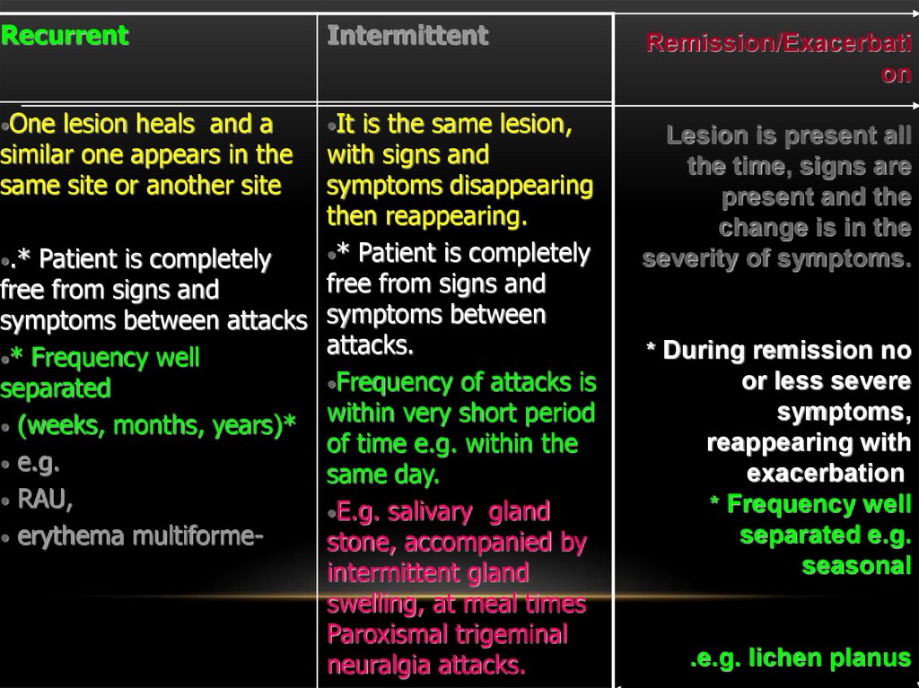
• Recurrent, intermittent, remission and exacerbation
34.
RecurrentIntermittent
•One
•It
lesion heals and a
similar one appears in the
same site or another site
is the same lesion,
with signs and
symptoms disappearing
then reappearing.
•* Patient is completely
•.* Patient is completely
free from signs and
free from signs and
symptoms between attacks symptoms between
attacks.
•* Frequency well
•Frequency of attacks is
separated
within very short period
• (weeks, months, years)*
of time e.g. within the
• e.g.
same day.
• RAU,
•E.g. salivary gland
• erythema multiformestone, accompanied by
intermittent gland
swelling, at meal times
Paroxismal trigeminal
neuralgia attacks.
Remission/Exacerbati
on
Lesion is present all
the time, signs are
present and the
change is in the
severity of symptoms.
* During remission no
or less severe
symptoms,
reappearing with
exacerbation
* Frequency well
separated e.g.
seasonal
.e.g. lichen planus
35.
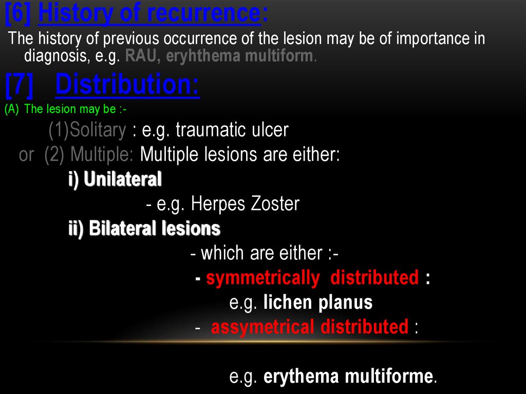
[6] History of recurrence:The history of previous occurrence of the lesion may be of importance in
diagnosis, e.g. RAU, eryhthema multiform.
[7] Distribution:
(A) The lesion may be :-
(1)Solitary : e.g. traumatic ulcer
or (2) Multiple: Multiple lesions are either:
i) Unilateral
- e.g. Herpes Zoster
ii) Bilateral lesions
- which are either :- symmetrically distributed :
e.g. lichen planus
- assymetrical distributed :
e.g. erythema multiforme.
36.
[8] Precipitating factors and relation toother activities:• *Pain may increase by eating, swallowing, sleeping, cold or hot
drinks:which are then called "precipitating factors" (ppt).
• According to ppt factors diagnosis could be guessed:
e.g. Any exposed dentin will lead to
sensitivity with thermal changes
specially cold,
e.g. carious lesions, exposed root dentin
37.
[9]Relieving factors:• Factors which relieve chief complaint e.g.:- Rest,
- Medications as simple
analgesics,
- Vasodilators
- Morphine should be noted.
38.
[10] Associated phenomena:• These are manifestations associated with the complaint:
● Fever
( acute abscess).
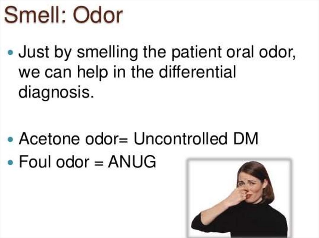
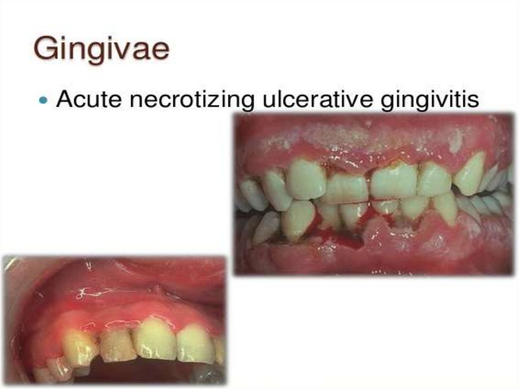
● Foetid odour + pain + bleeding gingiva +
mild fever + lymphadenopathy
(ANUG.)
• Others:
e.g. nausea, vomiting trismus, numbness,...etc.
all have value in diagnosis of cases.
39.
[11] Previous medication:Mouth washes, analgesics, antibiotics, previously used by
the patient, and their effect on c/c., as well as duration of
treatment should be noted. e.g. :-
• ● Mouth wash:
patient may use anti inflammatory mouth
wash as benzydamine hydrochloride, if
pain is relieved, therefore pain is of
gingival origin, if not, therefore it is of
dental origin
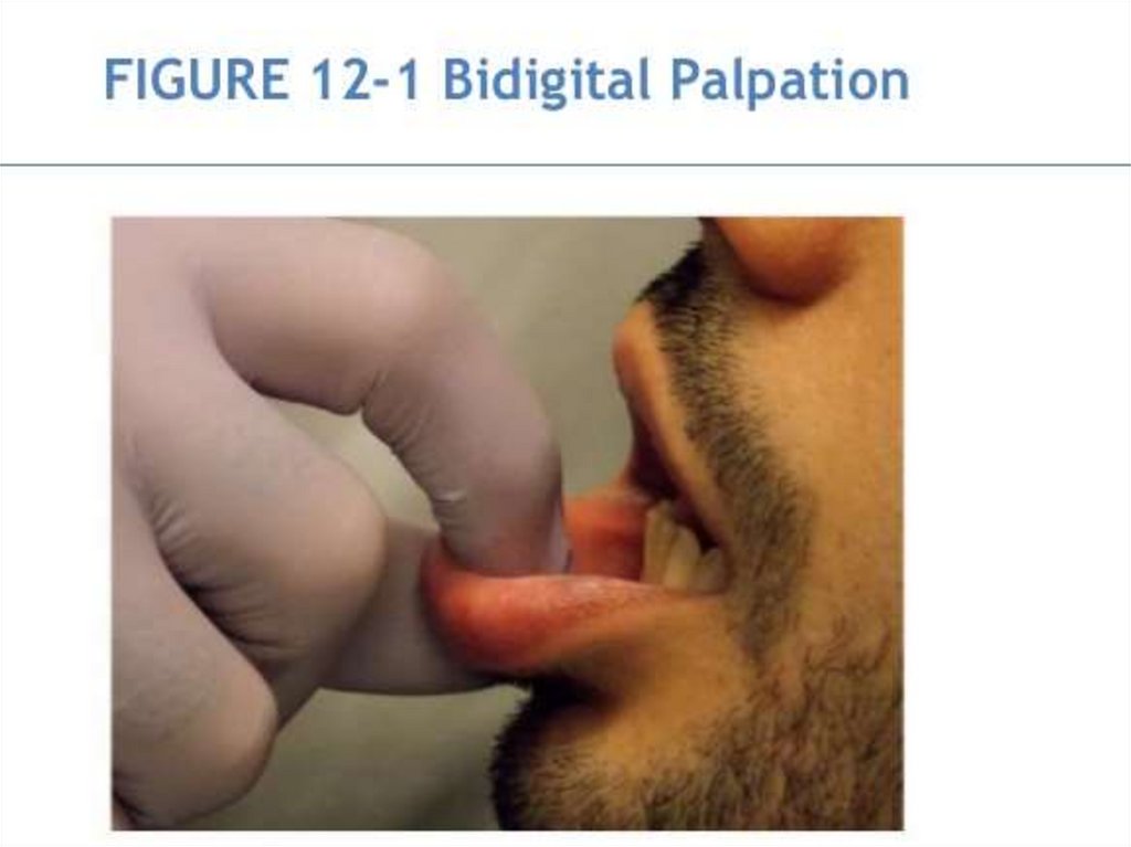
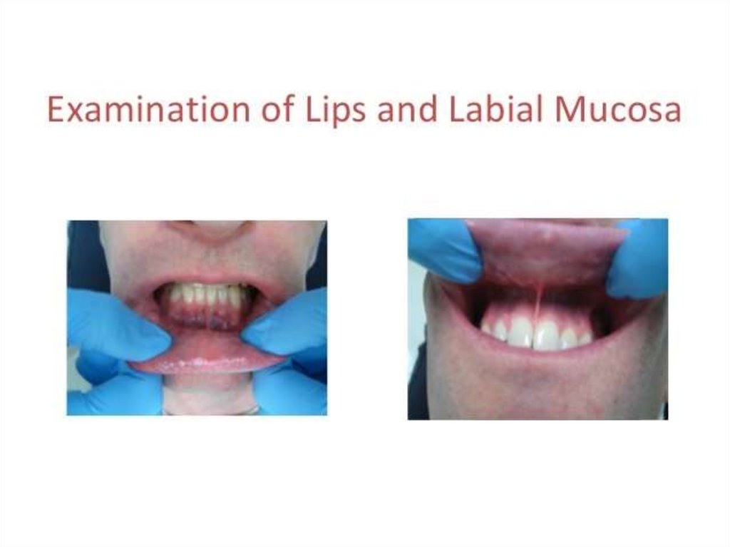
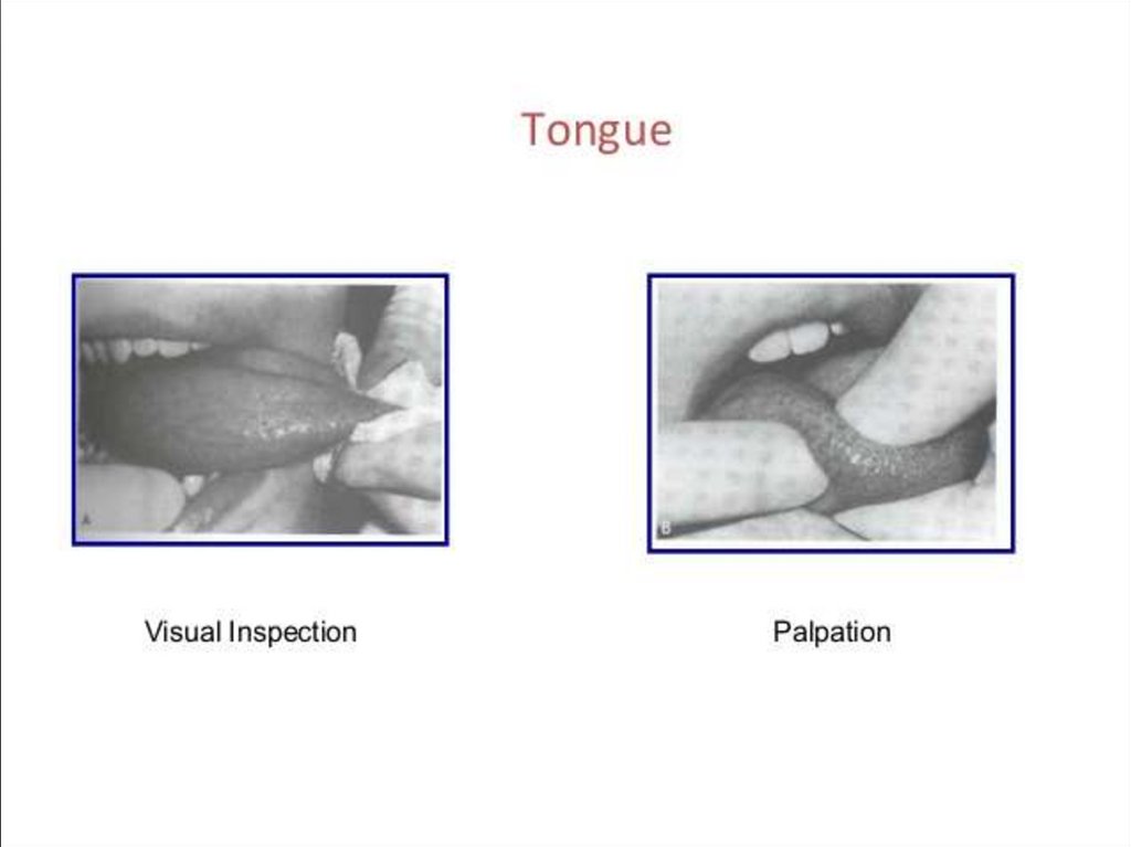
40. CLINICAL EXAMINATION
EXTRA ORALEXAMINATION
INTRA ORAL
EXAMINATION
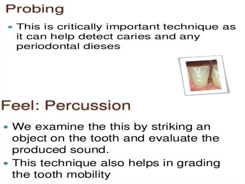
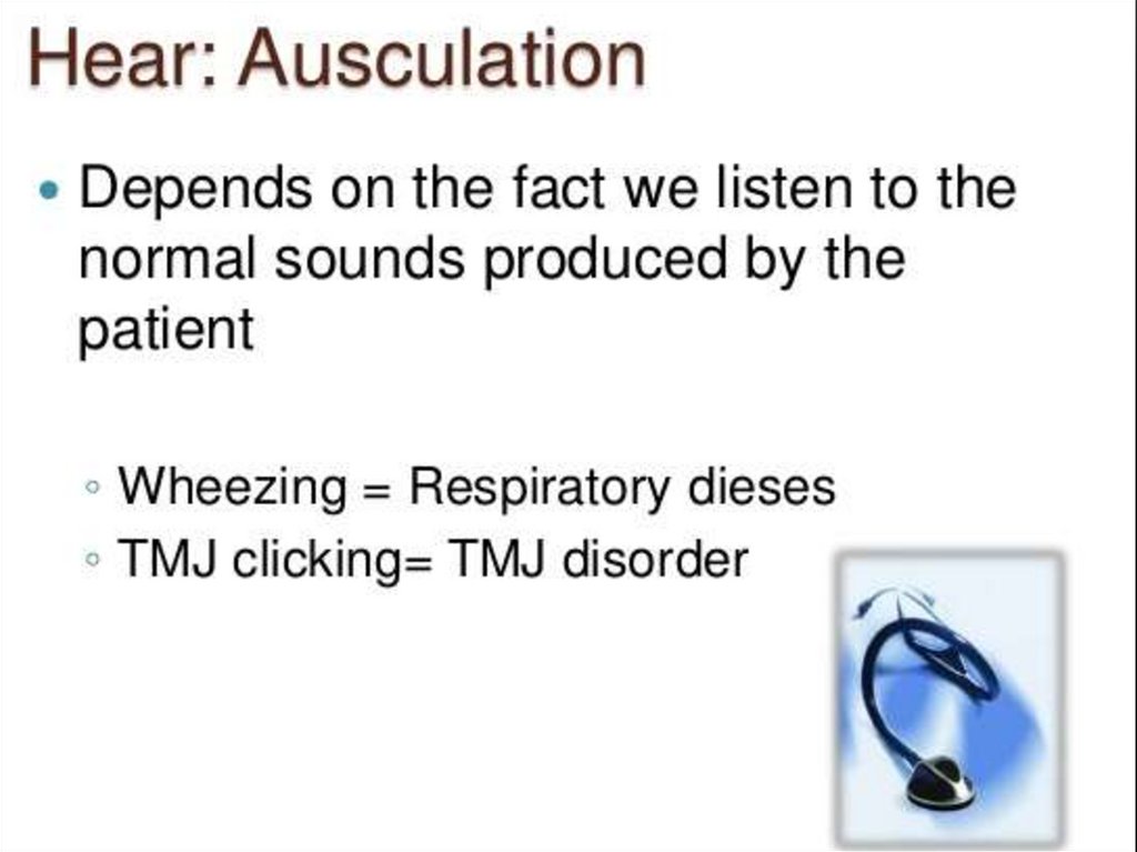
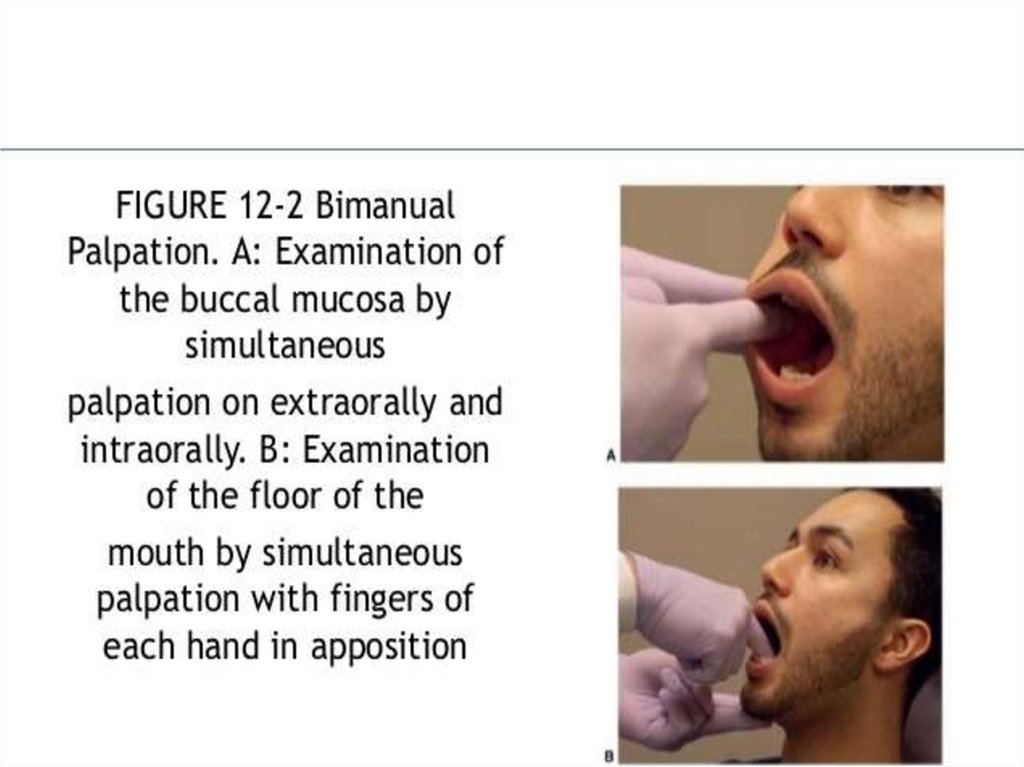
Inspection – palpation – percussion – probing auscultation
41.
42.
43.
44.
45.
46.
47.
48.
49.
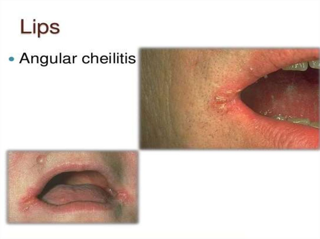
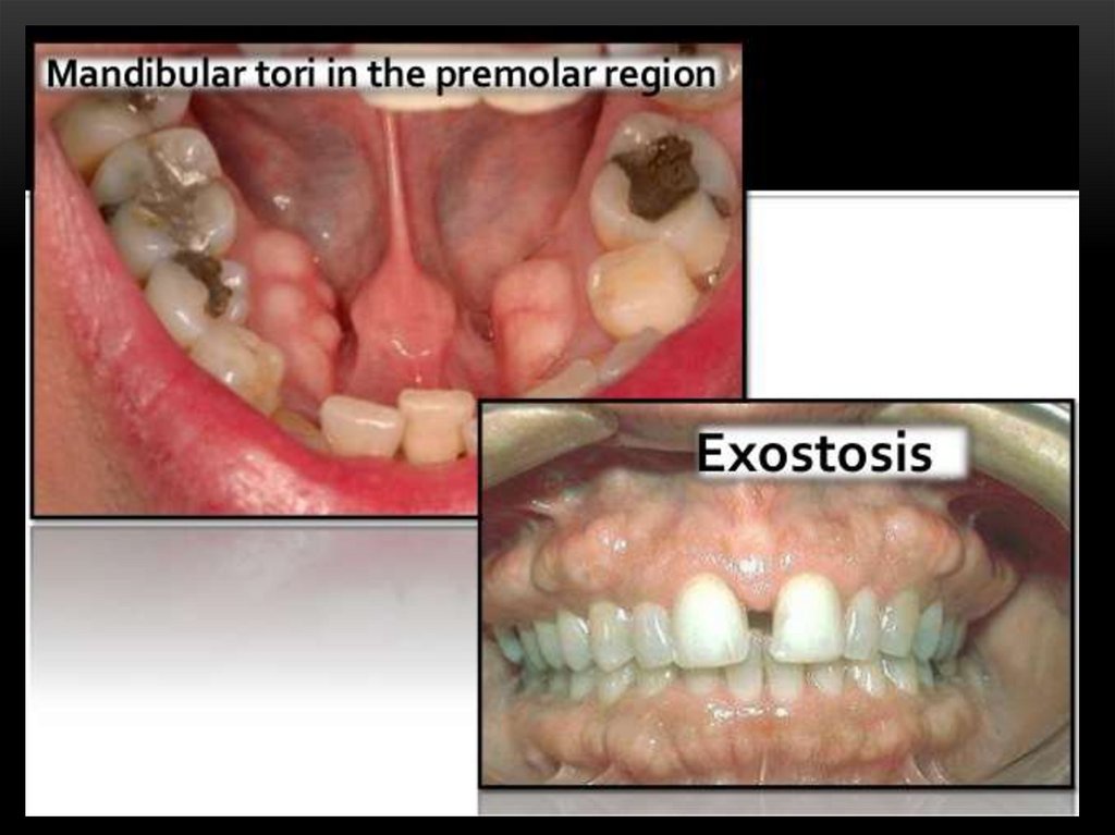
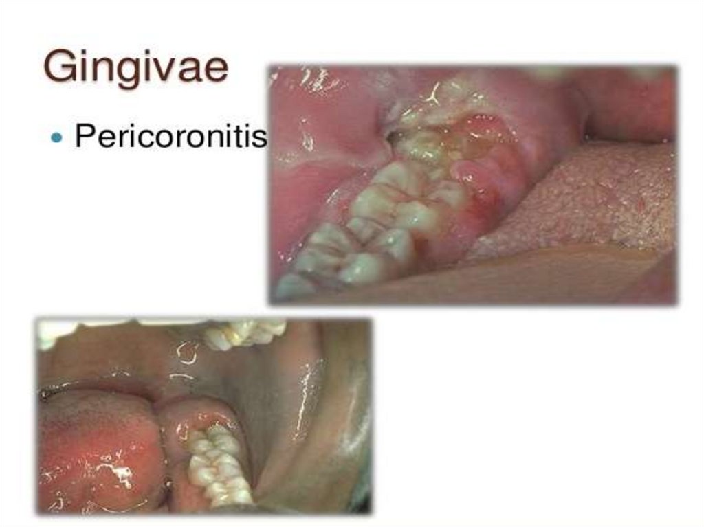
50. Angular cheilitis
ANGULAR CHEILITIS51.
52.
53.
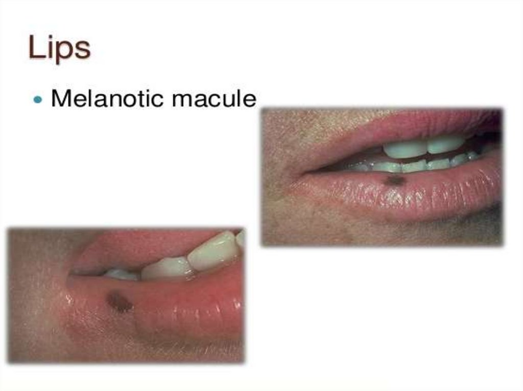
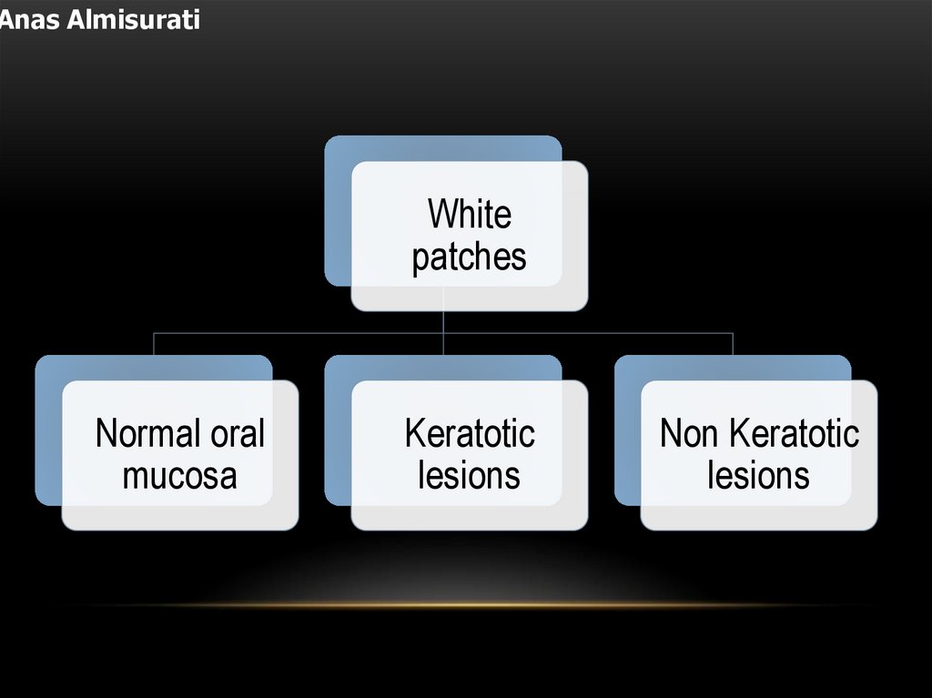
Anas AlmisuratiWhite
patches
Normal oral
mucosa
Keratotic
lesions
Non Keratotic
lesions
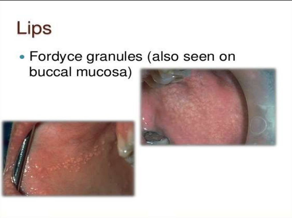
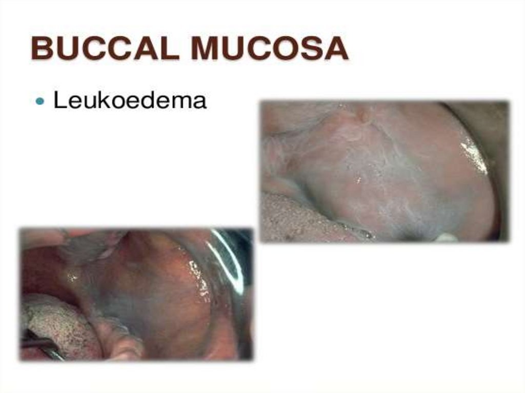
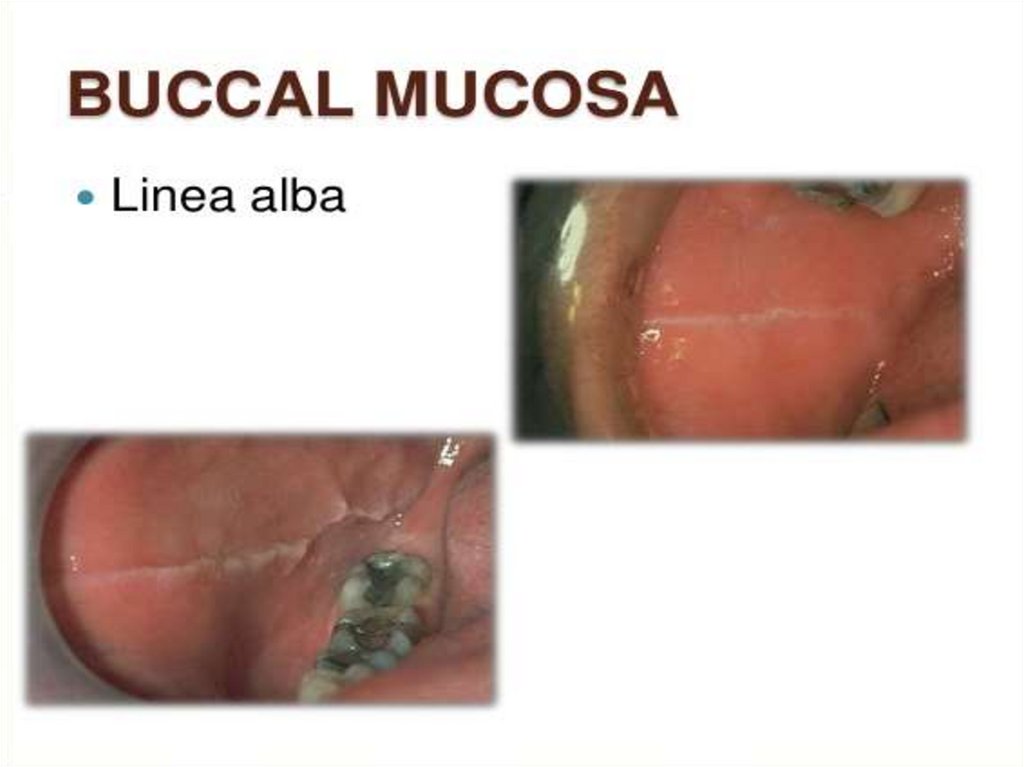
54. Normal oral mucosa
NORMAL ORAL MUCOSANormal oral mucosa with variation in structure and appearance :-
1- Fordyces granules
2- Linea alba
3- Leukodema
55.
56. linea alba
LINEA ALBA57.
58. Fordyce's granules
FORDYCE'S GRANULESDr.Anas Almisurati
59.
60. Keratotic lesion
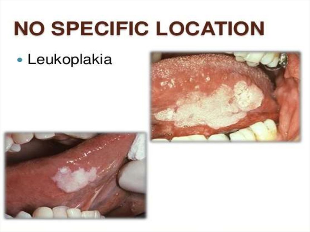
KERATOTIC LESIONKeratotic lesion (can’t rubbed off) :-
1- oral keratosis
2- leukoplakia
3- candidal leukoplakia
4- LP
5- DLE
6- White Spongy nevus
61. Oral keratosis (can’t Rubbed off )
ORAL KERATOSIS (CAN’T RUBBED OFF )reversible
Def. :IS a group of
the white keratotic
lesions which cannot
be rubbed off or
stripped off and have
definite
etiological
factors
irreversible
Frictional
keratosis
Smoker’s
keratosis
Traumatic
keratosis
Reverse
smoking
habit
Glass
blower’s
keratosis
Betel nut &
tobacco
chewer
Cigarette
smoking
(stomatitis
nicotina)
Oral sunff
keratosis
Pipe smoking
Syphilitic
keratosis
Actinic
keratosis
Dr.Anas Almisurati
62. Frictional keratotic
FRICTIONAL(reversible)KERATOTIC
Dr.Anas Almisurati
63. Smoker,s patches
SMOKER,S PATCHESWhite keratinized .a
patch on the
vermilion border of
the lips.
b. it may be flat, raised
or nodular.
c. lips and finger burns
may be associated.
(reversible)
64. Nicotinic Stomatitis
NICOTINIC STOMATITISEtiology the epithelial lining of the
ducts of the minor salivary glands often
shows squamous metaplasia
obstruction of the duct retention cyst
inflammation of the duct.
Site posterior part of the hard
palate.
Clinically the lesion appears as raised
yellowish white rings around the openings
of salivary gland ducts, which appear as
red dots (umbilicated appearance).
Dr.Anas Almisurati
65. Actinic keratosis
ACTINIC KERATOSIS(irreversible)
It is a premalignant
lesion due to
exposure to
ultraviolet rays.
Damaging effect due
to cumulative
exposure to UV rays
in white people
having little melanin.
66. Homogenous leukoplakia
HOMOGENOUS LEUKOPLAKIA• Flat
• Corrugated
• smooth & elevated
• wrinkled
67. Speckled Leukoplakia
SPECKLED LEUKOPLAKIAcorner of the mouth.
white patches (keratotic) on
erythematous base (atrophic
mucosa).
68.
69.
70. Duration 2 years maximum leaving some pigmentations on the skin.
DURATION2 YEARS MAXIMUM LEAVING SOME PIGMENTATIONS ON
THE SKIN.
Wickham's striae
Kobner phenomenon
71.
72.
73.
74.
75.
76.
77.
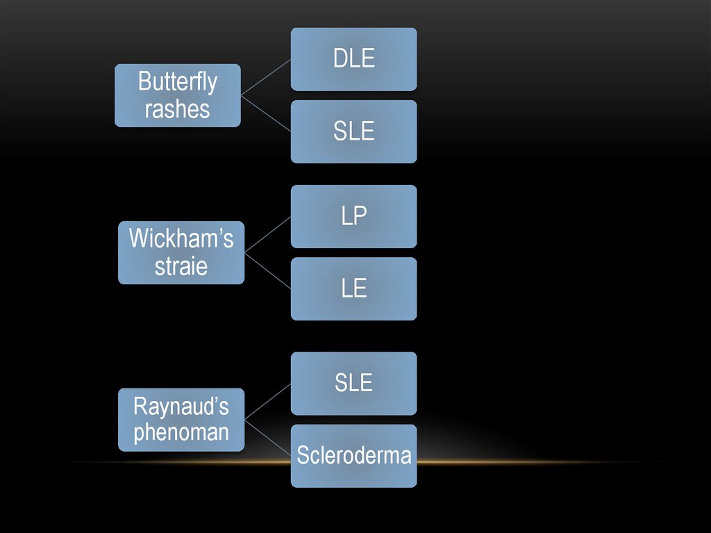
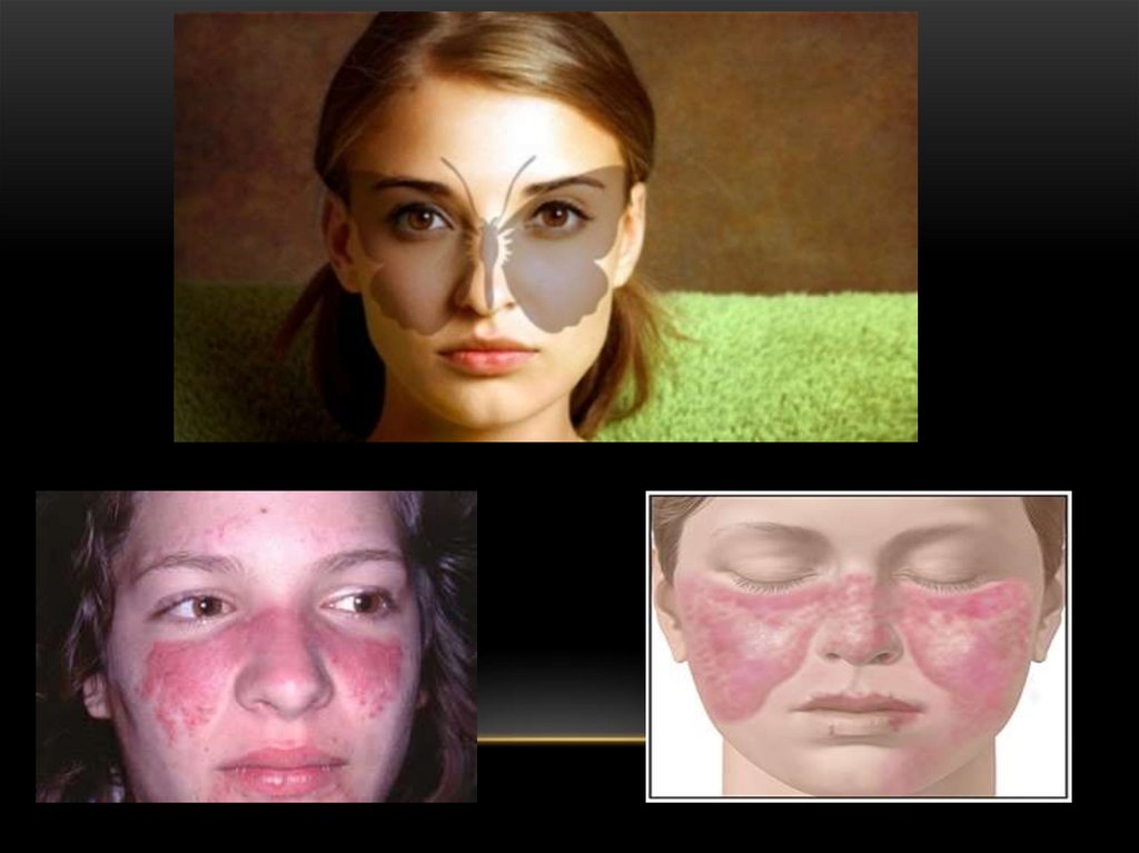
Butterflyrashes
Wickham’s
straie
Raynaud’s
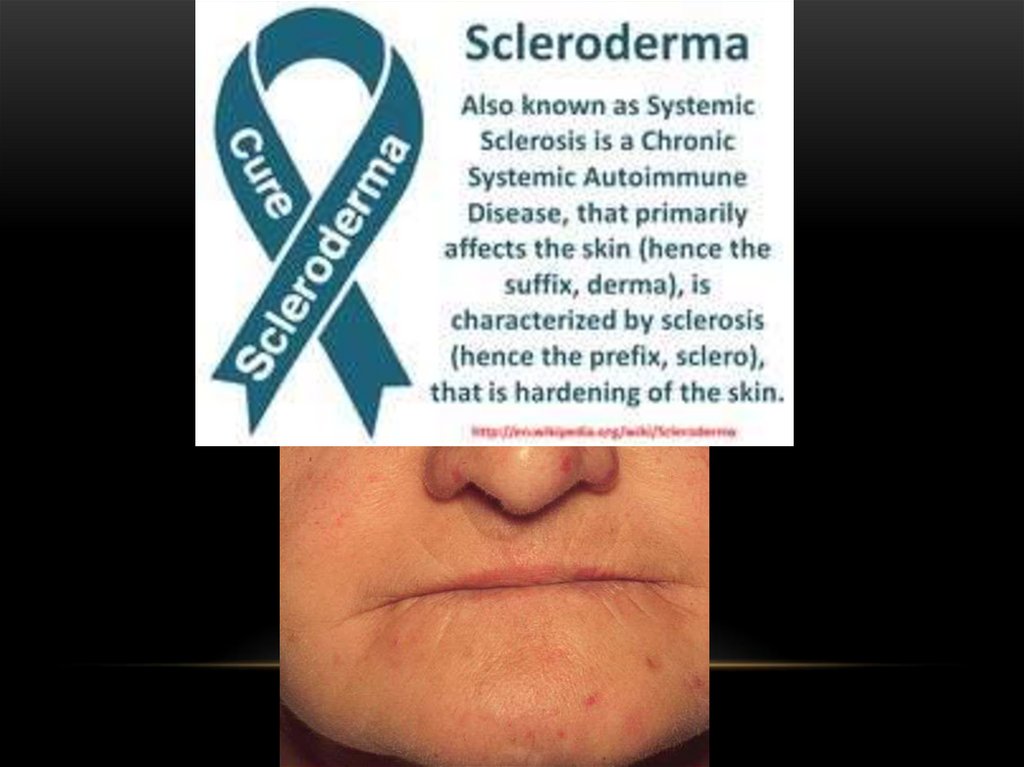
phenoman
DLE
SLE
LP
LE
SLE
Scleroderma
78.
79.
80.
81. Raynaud’s phenoman
RAYNAUD’S PHENOMAN• Is cyanosis and pain of finger and toes on exposure to cold
Common in
systemic LE
and
sclerodermas
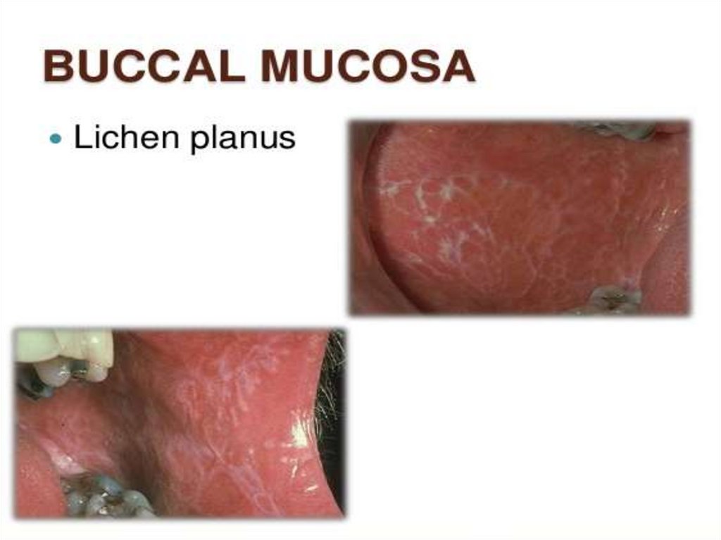
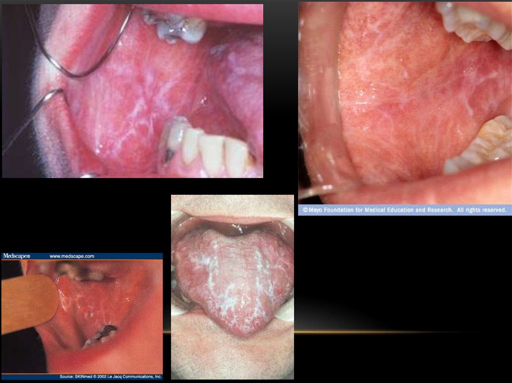
82. Stretching of white lesion may show:-
STRETCHING OF WHITE LESION MAY SHOW:The lesionbecome
accentuated
Wickham stria
The lesion
disappear
The lesion may
reveal pinpoint
elevation
LP
leukoedema
Hereditary
benign intraepithelial
dyskeratosis
HBID
83.
84.
85.
86.
87.
88.
89.
90.
91.
92.
93.
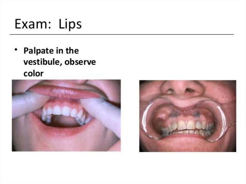
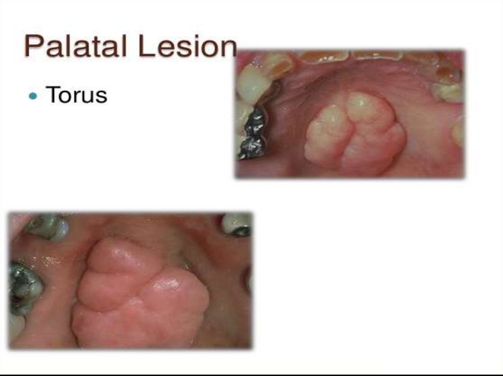
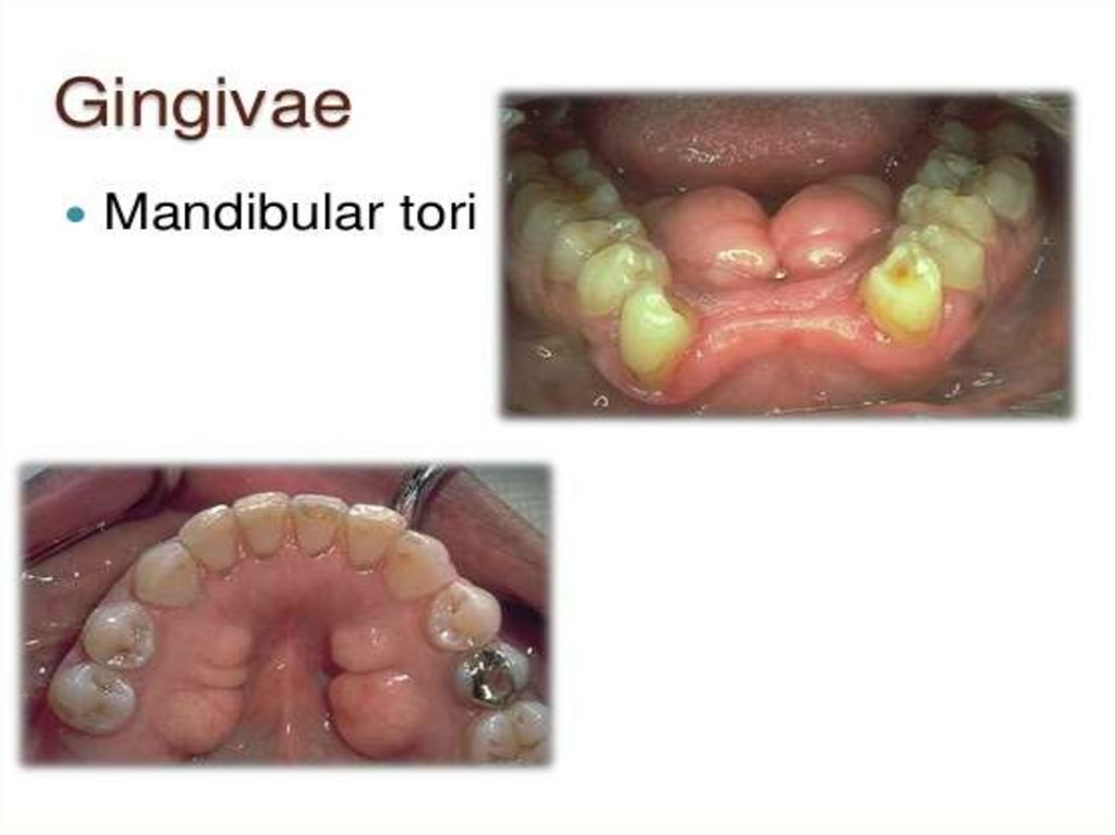
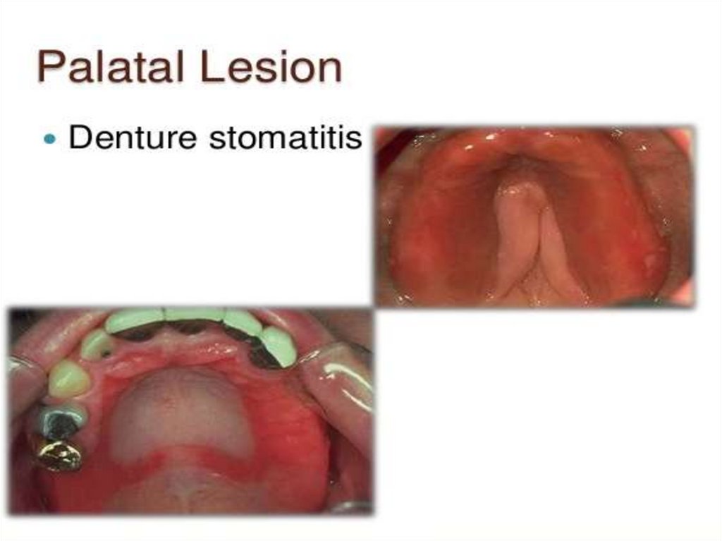
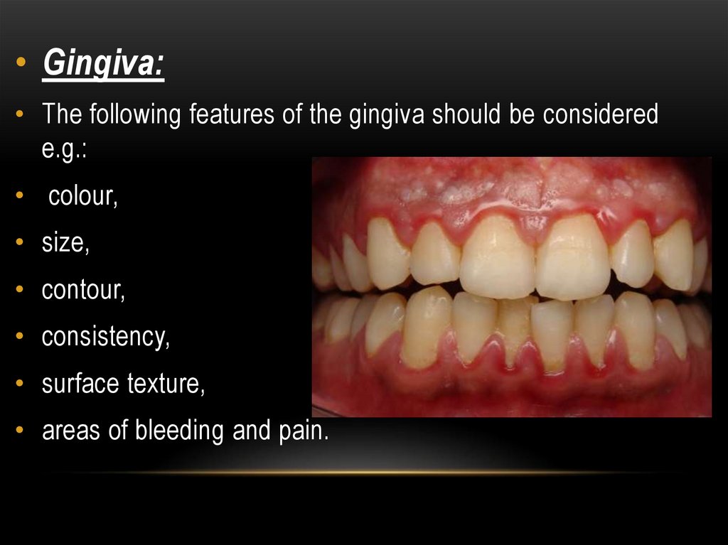
• Gingiva:• The following features of the gingiva should be considered
e.g.:
• colour,
• size,
• contour,
• consistency,
• surface texture,
• areas of bleeding and pain.
94. Marginal gingival inflammation
MARGINAL GINGIVAL INFLAMMATION95.
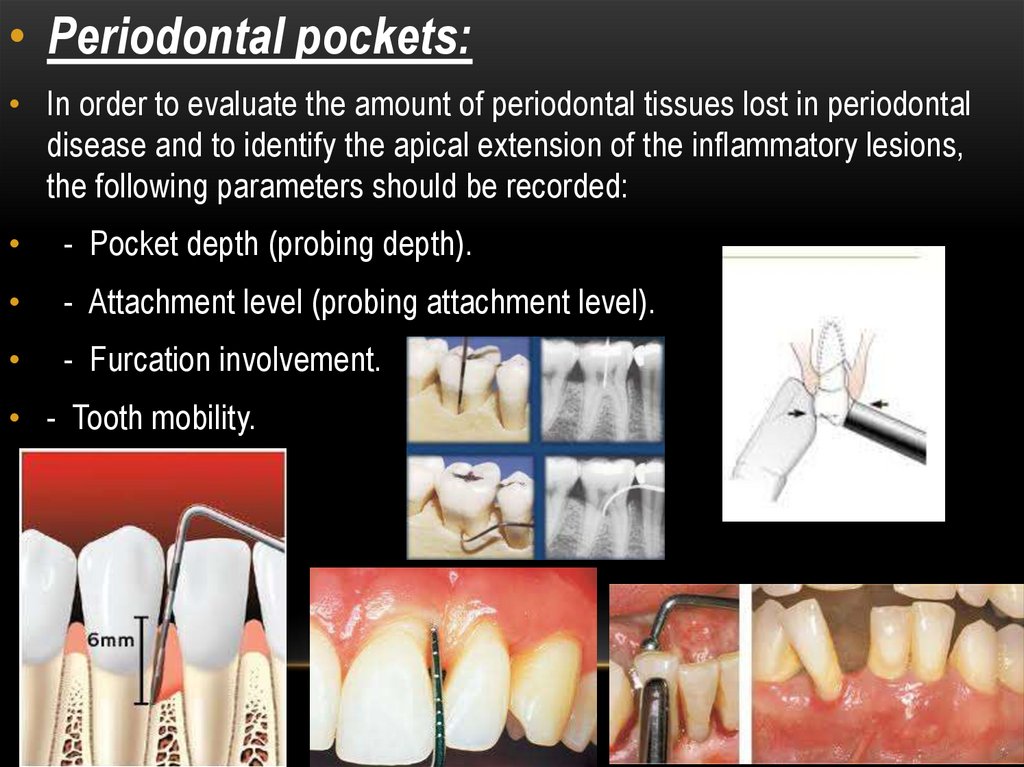
• Periodontal pockets:• In order to evaluate the amount of periodontal tissues lost in periodontal
disease and to identify the apical extension of the inflammatory lesions,
the following parameters should be recorded:
- Pocket depth (probing depth).
- Attachment level (probing attachment level).
- Furcation involvement.
• - Tooth mobility.
96.
97.
98.
99.
100.
101.
102.
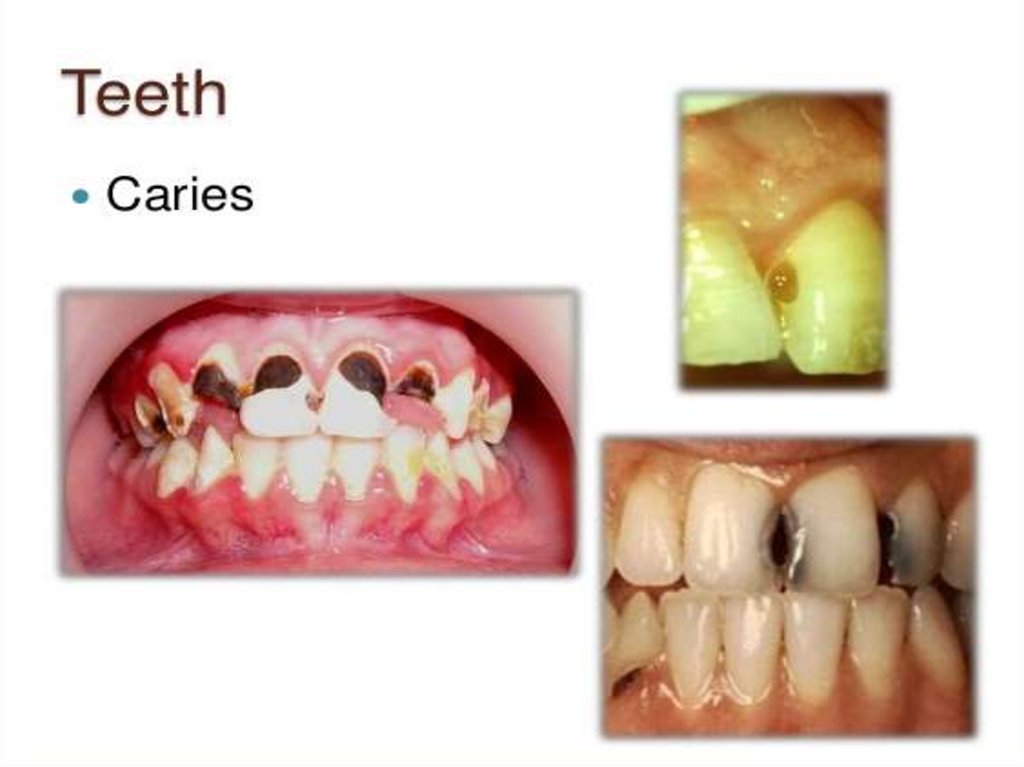
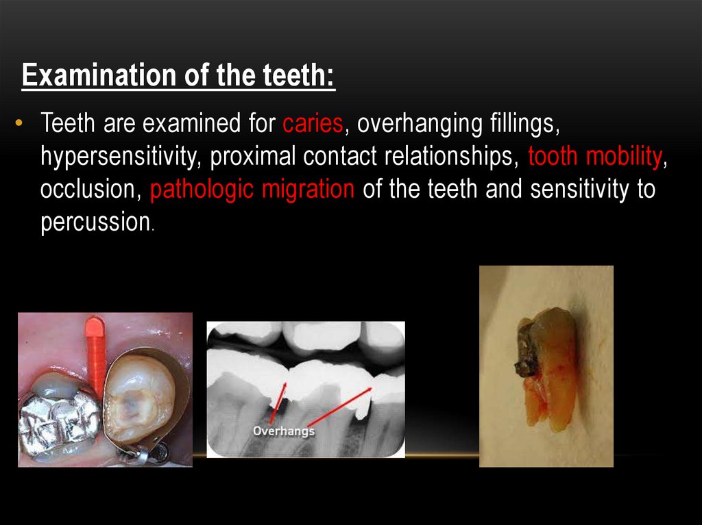
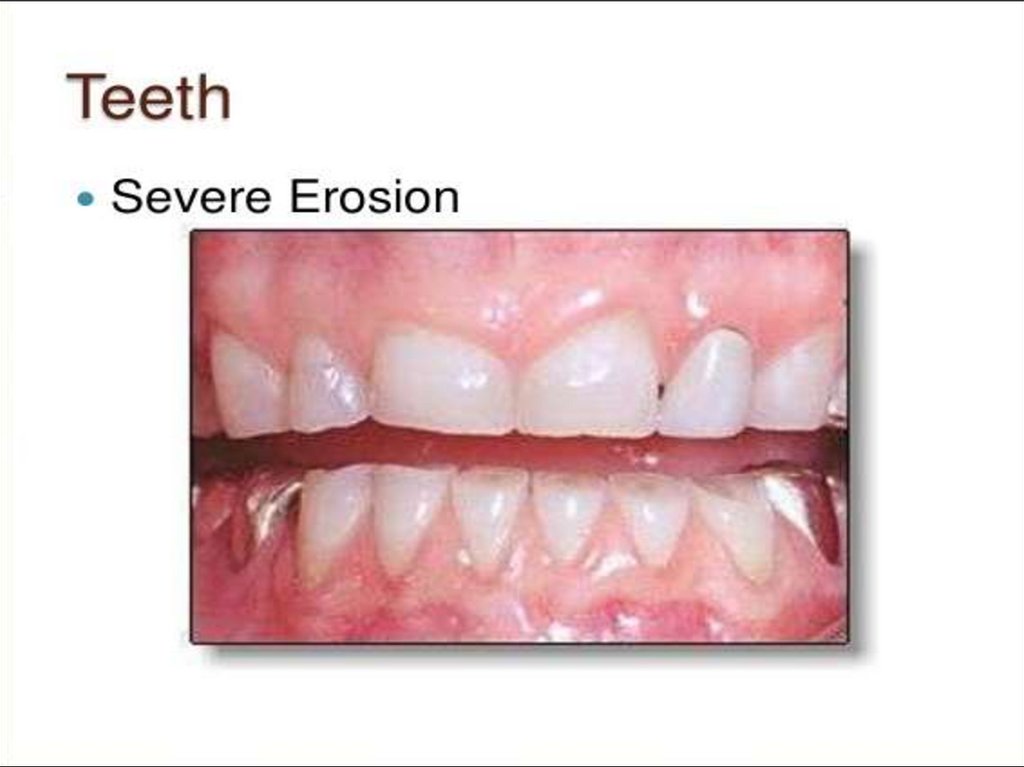
Examination of the teeth:• Teeth are examined for caries, overhanging fillings,
hypersensitivity, proximal contact relationships, tooth mobility,
occlusion, pathologic migration of the teeth and sensitivity to
percussion.
103.
104.
105.
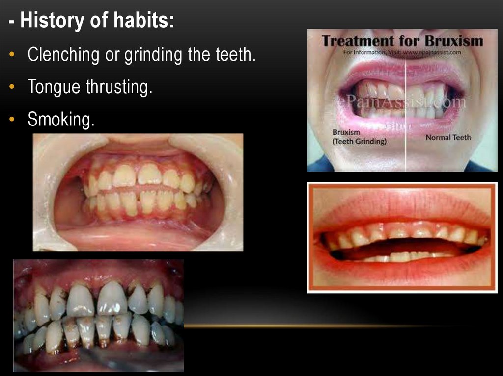
- History of habits:• Clenching or grinding the teeth.
• Tongue thrusting.
• Smoking.
106. EXTRA ORAL EXAMINATION
GENERAL APPRAISAL
SKIN
SKULL ( CRANIUM)
JAWS & TMJ
FACE
SALIVARY GLANDS
EYE
LYMPH NODES
NOSE
THYROID GLAND
HAIR
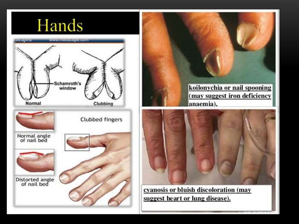
HANDS AND FINGERS
107. General appraisal
GENERAL APPRAISAL• Starts while patient entering the clinic.
• Performed without patient interruption.
Report, record, or observe the following:
108.
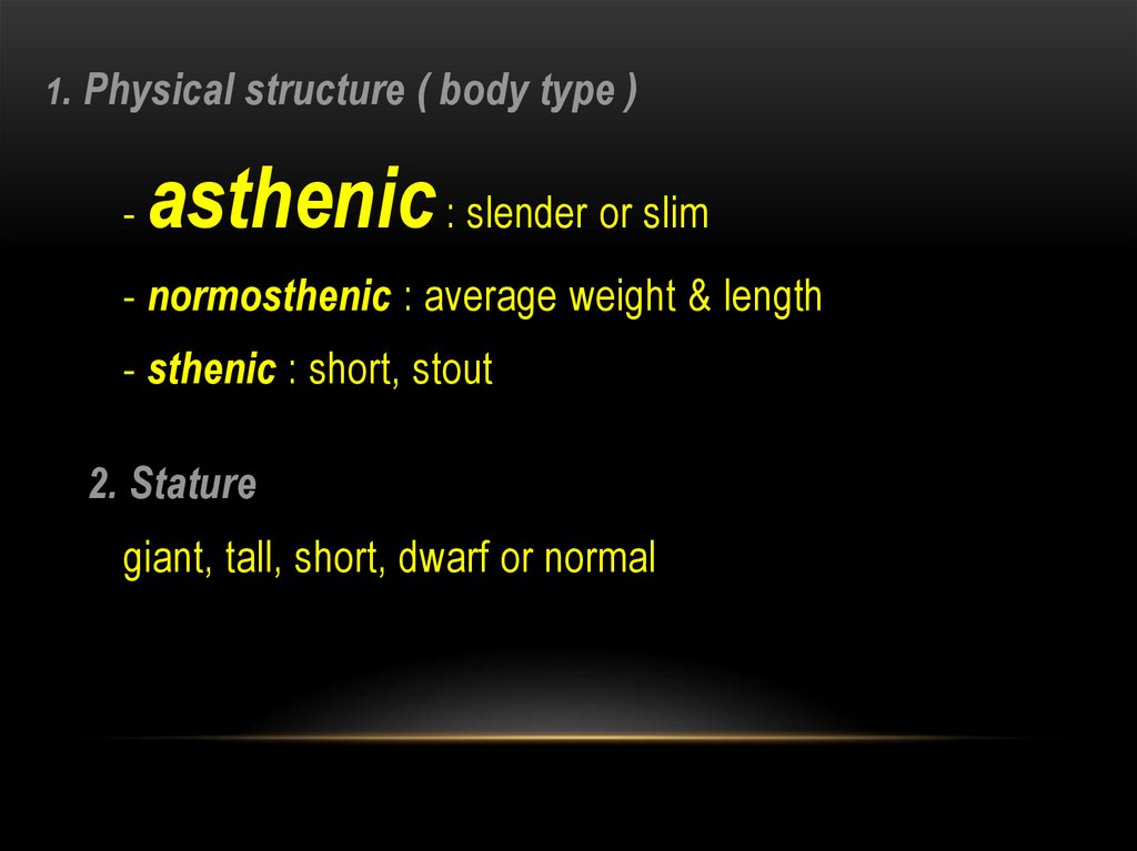
1. Physical structure ( body type )-
asthenic : slender or slim
- normosthenic : average weight & length
- sthenic : short, stout
2. Stature
giant, tall, short, dwarf or normal
109.
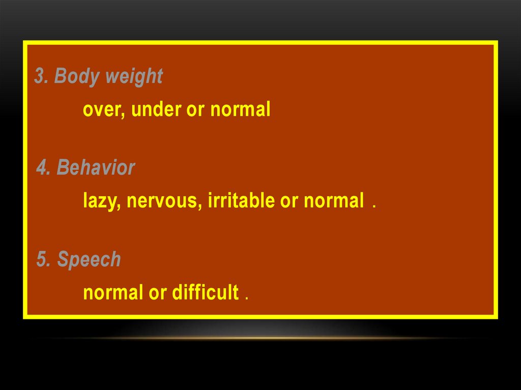
3. Body weightover, under or normal
4. Behavior
lazy, nervous, irritable or normal .
5. Speech
normal or difficult .
110.
111.
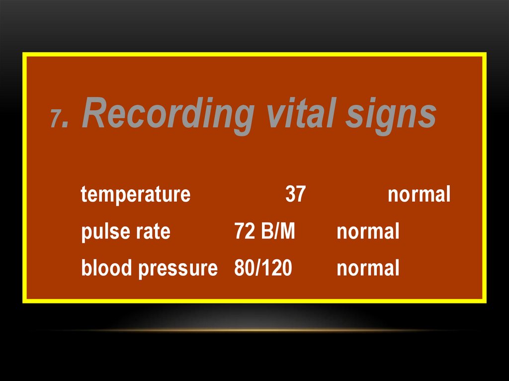
7. Recording vital signs
temperature
pulse rate
37
normal
72 B/M
normal
blood pressure 80/120
normal
112. Skull and Cranium
SKULL AND CRANIUM• Size : from supra orbital ridge to occipital protuberance.
- Small head (micro cephalus) brain under development
- Large head
paget
hydro cephalus
acromegalic
• Shape : prominent forehead
- rickets
- congenital syphilis
113. CONGENITAL SYPHILIS
114. PAGET,S disease
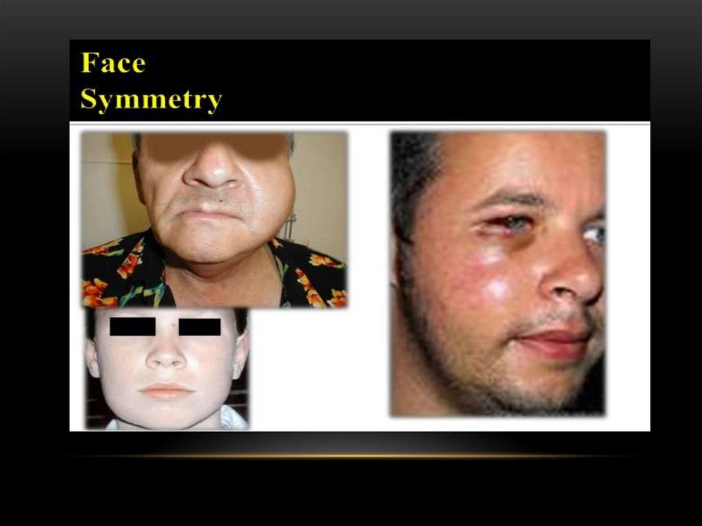
PAGET,S DISEASE115. The Face
THE FACECharacteristic face pattern
1. Acromegalic face: coarse features
prognathism prominent forehead.
2. Moon’s face: in Cushing disease the face
round, flushed & obese.
3. Hyper thyroid face: moist skin, protruded
ball and nervous muscle movement
4. Congenital syphilis face: saddle nose,
and interstitial keratitis.
eye
rhagades
116. ACROMEGALY
117. Acromegaly
ACROMEGALY118. Acromegaly
ACROMEGALY119. THyrotoxicosis
THYROTOXICOSIS120. Moon face
MOON FACE121. Cortisone therapy
CORTISONE THERAPY122. obesity
OBESITY123.
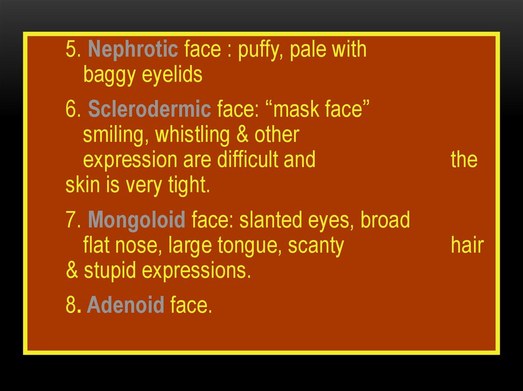
5. Nephrotic face : puffy, pale withbaggy eyelids
6. Sclerodermic face: “mask face”
smiling, whistling & other
expression are difficult and
skin is very tight.
7. Mongoloid face: slanted eyes, broad
flat nose, large tongue, scanty
& stupid expressions.
8. Adenoid face.
the
hair
124.
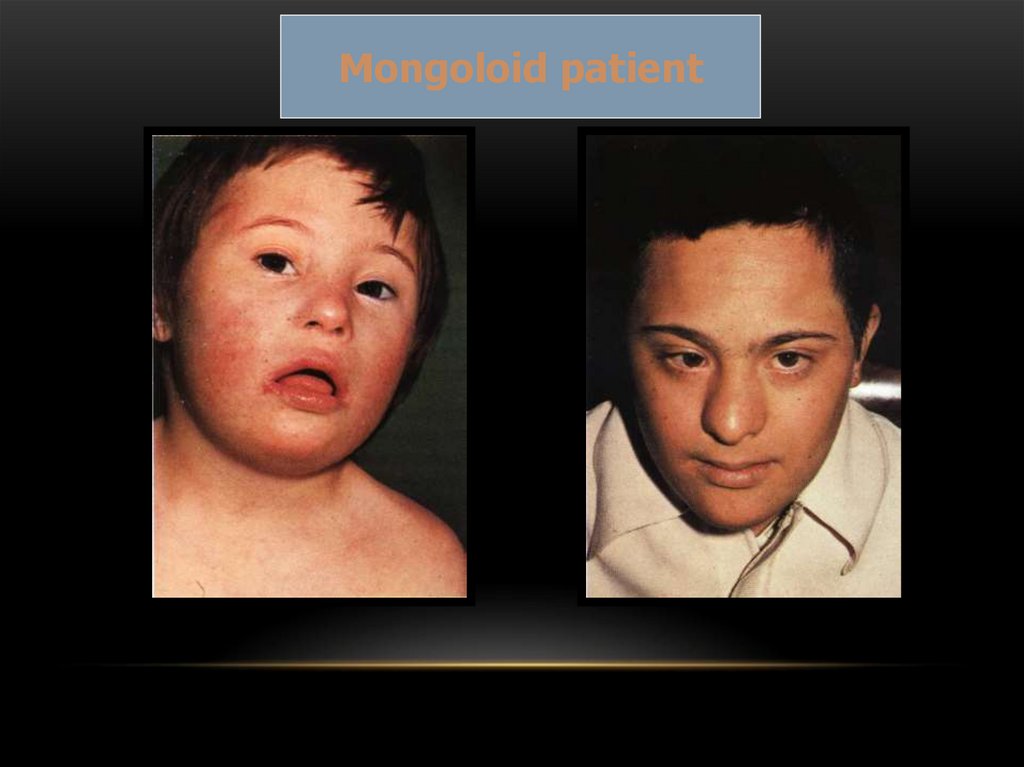
Mongoloid patient125. DOWN,S SYNDROME
126. Clinical findings of MONGOLS
CLINICAL FINDINGS OF MONGOLS• Mouth breather
• Cracked lips
• Macroglossia
• Fissured tongue
• Cleft lip or palate
• Poor oral hygiene
• Short roots lead to rapid
loss of teeth
• malocclusion
127.
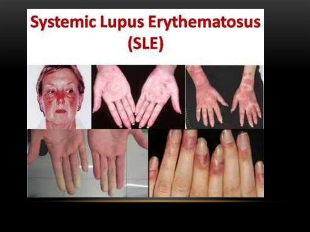
Clinical findings128. LUPUS ERYTHEMATOSIS
129. neurofibromatosis
NEUROFIBROMATOSIS• Mandibular canal
enlargement (lip
numbness).
• Macroglossia,fissuring
and precancerous
130. Angio edema
ANGIO EDEMAsever facial
swelling
131. Surgical trauma
SURGICAL TRAUMAThird molars
ext
Post operative
Two weeks later
132. Masseter hypertrophy
MASSETER HYPERTROPHY133. EMPHYSEMA
• AIR EMPHYSEMAis a compressible swelling
that produce crackling
sound upon palpation . It
is caused by air forced
under mucoperiosteal flap
from using high speed
hand piece during surgery.
134.
135. Ewing,s sarcoma
EWING,S SARCOMA• An aggressive and rapidly
growing malignant tumor
that has extended via
mandibular cortical plate .
136. Bell, s palsy
BELL, S PALSYLeft side paralysis
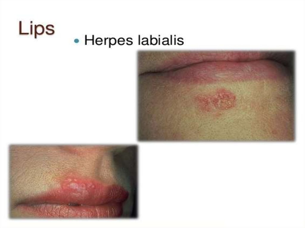
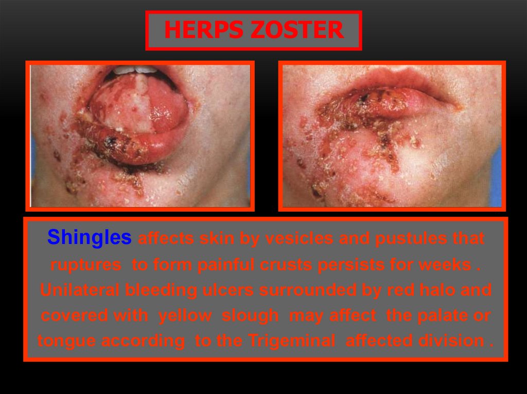
137. Herpes Zoster
• Chicken pox is the primaryinfection by Varicella – Zoster
herpetic virus. Papules, vesicles
and pustules as skin rash on the
trunk, neck, and face will be seen
for 7-10 days before spontaneous
resolving. Reactivation of
dormant varicella virus from
sensory ganglia and migration
along nerves will induce Herpes
Zoster ( Shingle).
HERPES
ZOSTER
Varivax is a life
time vaccine is
now available.
138.
HERPS ZOSTERShingles affects skin by vesicles and pustules that
ruptures to form painful crusts persists for weeks .
Unilateral bleeding ulcers surrounded by red halo and
covered with yellow slough may affect the palate or
tongue according to the Trigeminal affected division .
139. Infected cyst
INFECTED CYSTUPPER INCISORS
140. acute dento alveolar abscess
ACUTE DENTO ALVEOLAR ABSCESSLOWER INCISORS
141. ADAA
UPPER PRE MOLARSLOWER MOLAR
142. micrognathia
MICROGNATHIA143. Facial palsy
FACIAL PALSY144. PAROTID GLAND ENLARGEMENT
145. Salivary calculi
SALIVARY CALCULI146.
147. THE Nose
THE NOSENasal abnormalities may be interrelated to oral lesions.
The following might be affected:
- Shape: as saddle nose (depressed nasal bridge) in
congenital syphilis, myxodema, sickle cell anemia and
due to trauma.
148. Saddle nose
SADDLE NOSE149. Acromegaly
ACROMEGALYEnlarged nose
150. THE EYE
scleraIris
conjunctiva
pupil
151.
Eye lesions of dental relation1) Ptosis
- Dropping of upper eye lid
- Inability to open the eye completely
It is due to paralysis of levator muscle
supplied by third occulomotor N.
152. Congenital PTOSIS
CONGENITAL PTOSISbilateral
unilateral
153. Dentinogenesis imperfecta
DENTINOGENESIS IMPERFECTABlue sclera
Opalescent cracked
teeth
154. PEMPHIGUS VULGARIS
• Autoimmune vesiculobullous lesion affects skin and oral mucosa or othermucosal tissue.
• Clinically flaccid intraepithelial bullae easily rupture causing electrolytes
imbalance.
155.
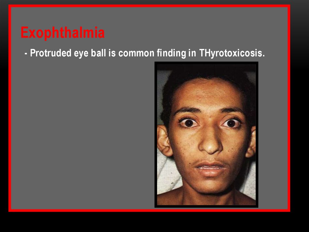
Exophthalmia- Protruded eye ball is common finding in THyrotoxicosis.
156. conjunctivitis
CONJUNCTIVITISREITER,S
Behcet,s
157.
SYNDROMES AND OTHERDISEASES
Muco Cutaneous Ocular Syndromes
1-
STEVEN JHONSON S
2- BEHCET S
3- RITTER S
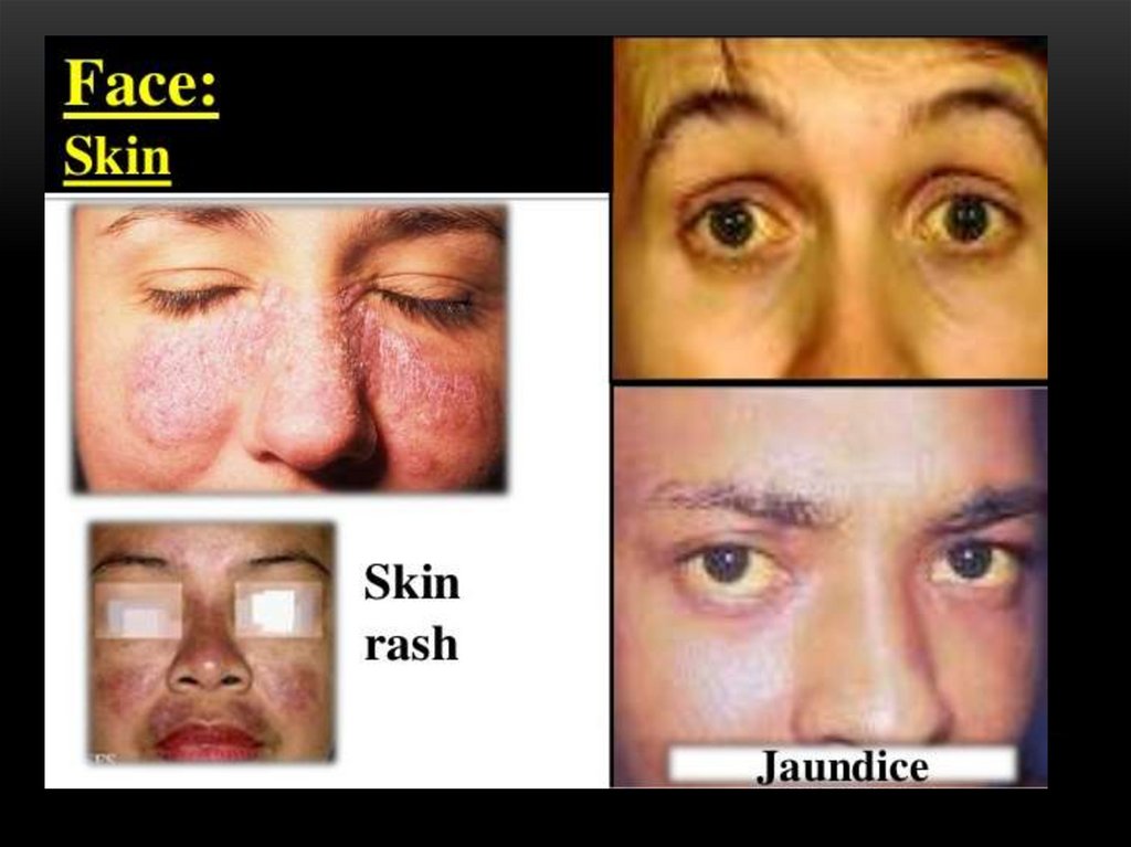
158. The Skin
THE SKINThe skin should be inspected for :
• color changes,
• pigmented lesions, and
• scars
159.
Palpation is used to examine surfacetexture changes and to check skin
temperature.
- Skin lesions in dermatologic diseases might
be used for differentiation between similar
oral lesions as erythema multiform, erosive
lichen planus and lupus erythematosis .
160.
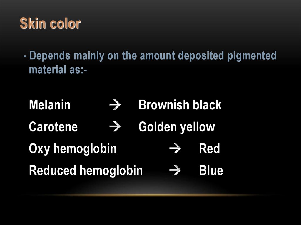
Skin color- Depends mainly on the amount deposited pigmented
material as:-
Melanin
Brownish black
Carotene
Golden yellow
Oxy hemoglobin
Red
Reduced hemoglobin
Blue
161.
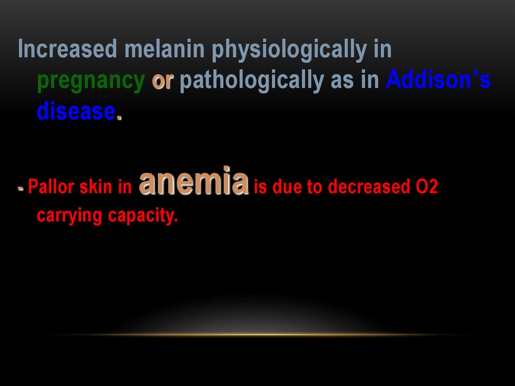
Increased melanin physiologically inpregnancy or pathologically as in Addison’s
disease.
anemia is due to decreased O2
- Pallor skin in
carrying capacity.
162.
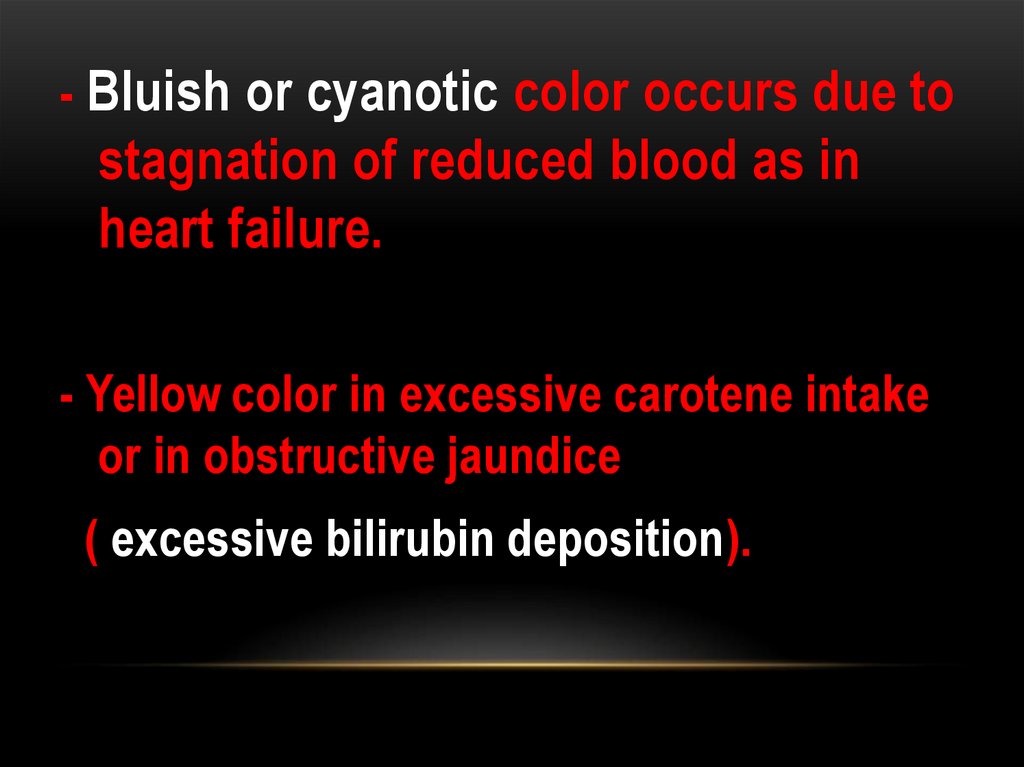
- Bluish or cyanotic color occurs due tostagnation of reduced blood as in
heart failure.
- Yellow color in excessive carotene intake
or in obstructive jaundice
( excessive bilirubin deposition).
163.
164.
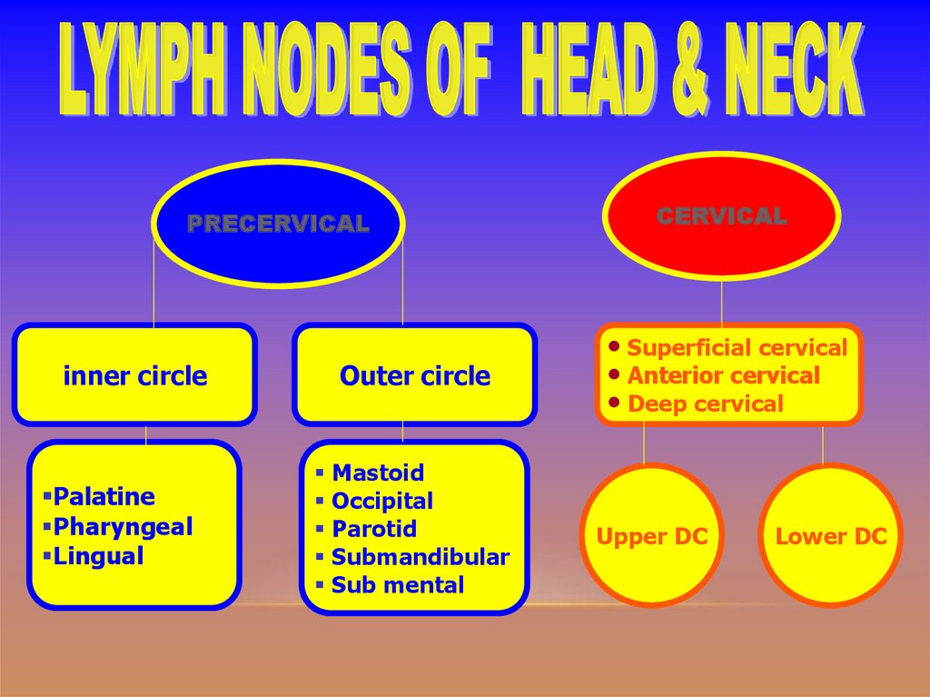
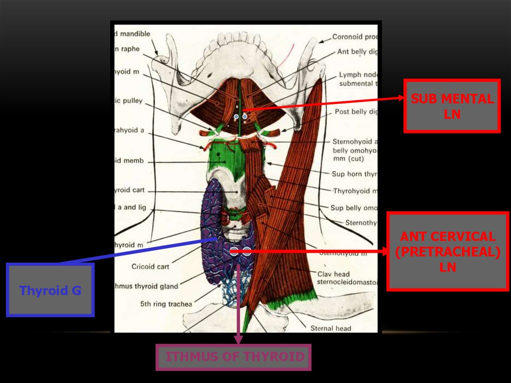
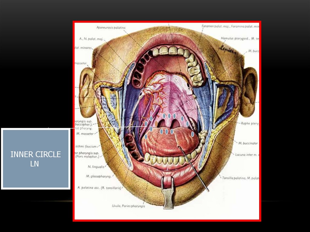
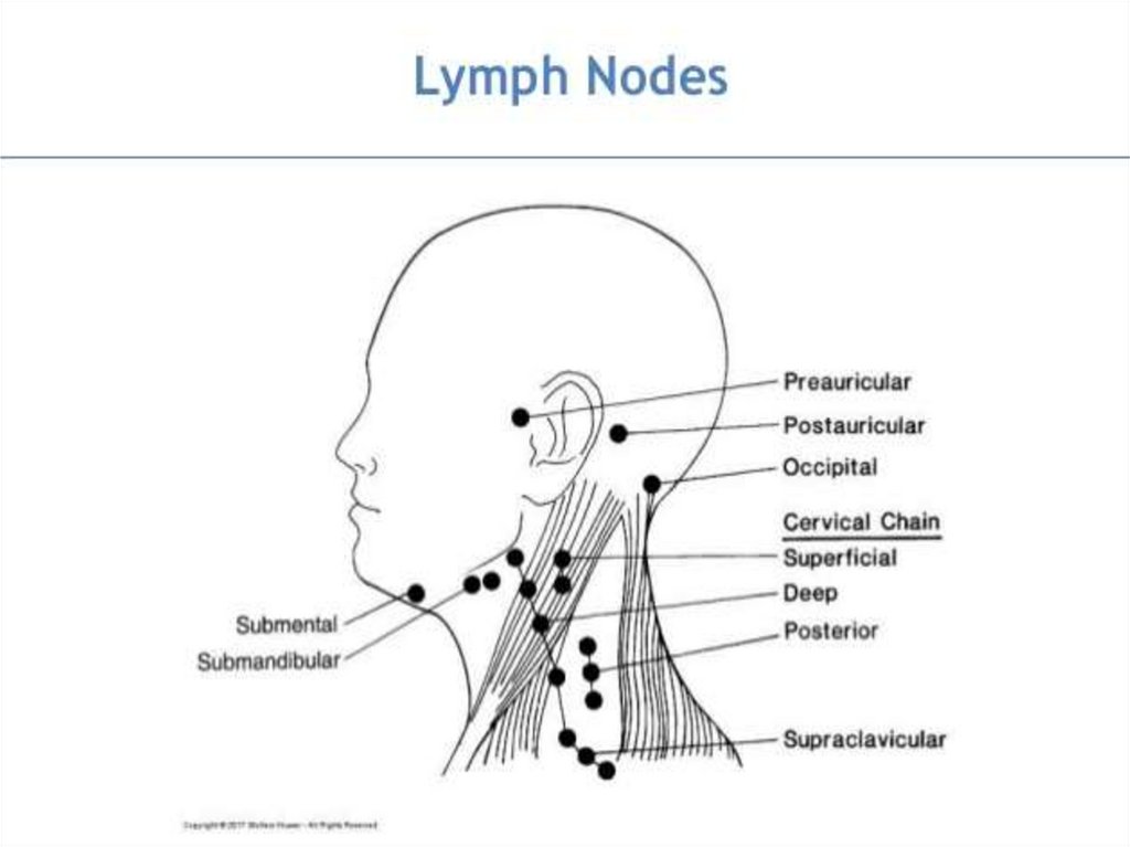
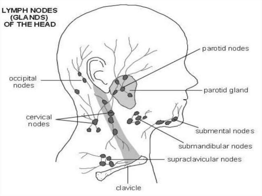
PRECERVICALinner circle
Palatine
Pharyngeal
Lingual
Outer circle
Mastoid
Occipital
Parotid
Submandibular
Sub mental
CERVICAL
• Superficial cervical
• Anterior cervical
• Deep cervical
Upper DC
Lower DC
165.
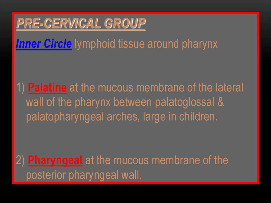
PRE-CERVICAL GROUPInner Circle lymphoid tissue around pharynx
1) Palatine at the mucous membrane of the lateral
wall of the pharynx between palatoglossal &
palatopharyngeal arches, large in children.
2) Pharyngeal at the mucous membrane of the
posterior pharyngeal wall.
166.
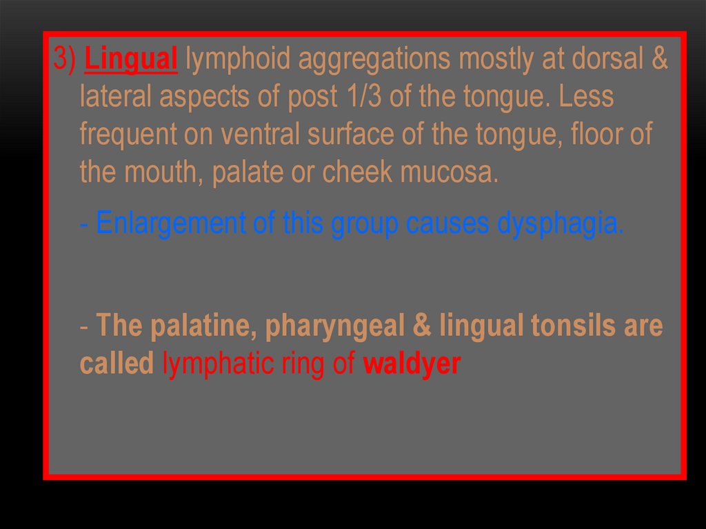
3) Lingual lymphoid aggregations mostly at dorsal &lateral aspects of post 1/3 of the tongue. Less
frequent on ventral surface of the tongue, floor of
the mouth, palate or cheek mucosa.
- Enlargement of this group causes dysphagia.
- The palatine, pharyngeal & lingual tonsils are
called lymphatic ring of waldyer
167.
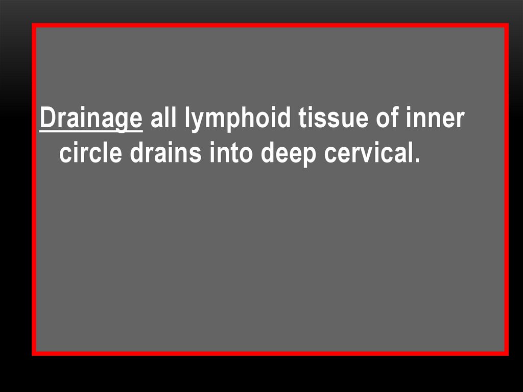
Drainage all lymphoid tissue of innercircle drains into deep cervical.
168.
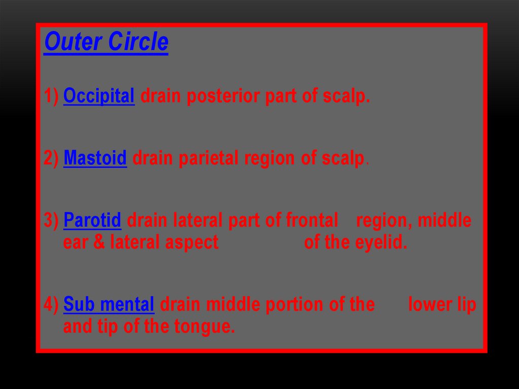
Outer Circle1) Occipital drain posterior part of scalp.
2) Mastoid drain parietal region of scalp.
3) Parotid drain lateral part of frontal region, middle
ear & lateral aspect
of the eyelid.
4) Sub mental drain middle portion of the
and tip of the tongue.
lower lip
169.
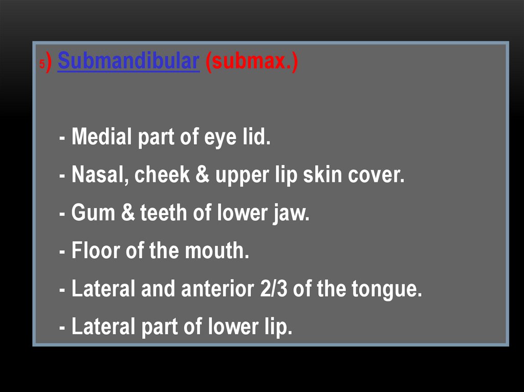
5) Submandibular (submax.)
- Medial part of eye lid.
- Nasal, cheek & upper lip skin cover.
- Gum & teeth of lower jaw.
- Floor of the mouth.
- Lateral and anterior 2/3 of the tongue.
- Lateral part of lower lip.
170.
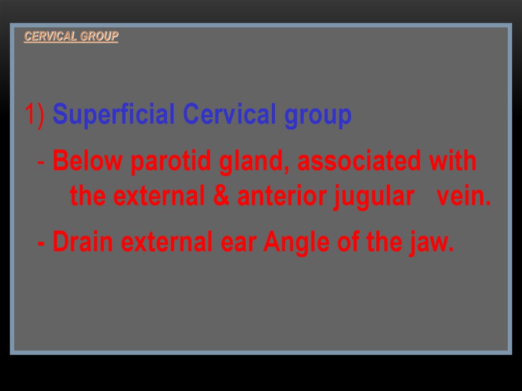
CERVICAL GROUP1) Superficial Cervical group
- Below parotid gland, associated with
the external & anterior jugular vein.
- Drain external ear Angle of the jaw.
171.
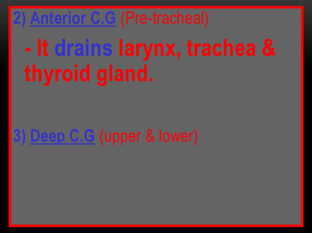
2) Anterior C.G (Pre-tracheal)- It drains larynx, trachea &
thyroid gland.
3) Deep C.G (upper & lower)
172.
N.B.Deep cervical drains
- Maxillary teeth, gum, hard palate and post 1/3 of
tongue.
- all pre cervical & superficial cervical
L.N.
173.
SUBMANDIBULAR
Upper deep
Cervical
LN
LN
LOWER deep
Cervical
LN
174.
SUB MENTALLN
ANT CERVICAL
(PRETRACHEAL)
LN
Thyroid G
ITHMUS OF THYROID
175.
176.
INNER CIRCLELN
177.
178.
179.
180.
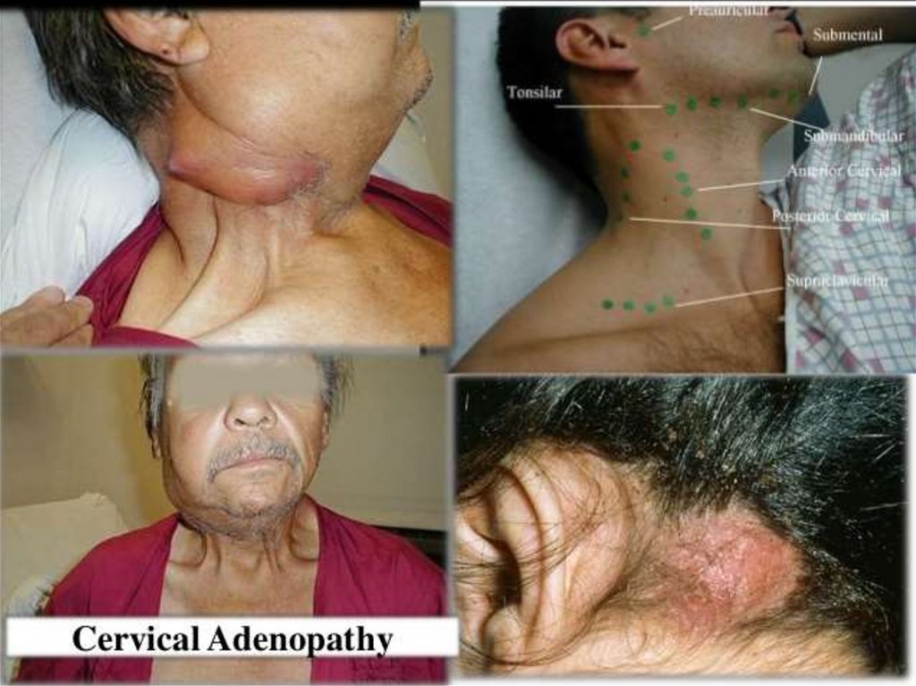
Lymph node enlargementLocalized factors
1. Infection
a) Acute: NUG, ADAA, AHGS, Chancre
b) Chronic: Scrofula (T.B. Lymph Nodes)
2. Neoplastic metastasis
181.
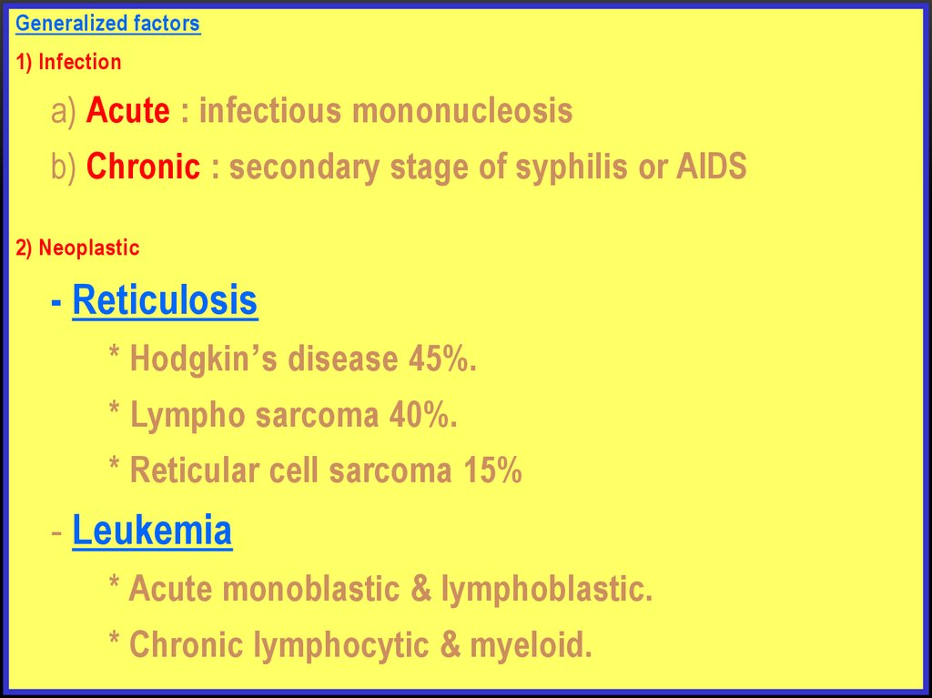
Generalized factors1) Infection
a) Acute : infectious mononucleosis
b) Chronic : secondary stage of syphilis or AIDS
2) Neoplastic
- Reticulosis
* Hodgkin’s disease 45%.
* Lympho sarcoma 40%.
* Reticular cell sarcoma 15%
- Leukemia
* Acute monoblastic & lymphoblastic.
* Chronic lymphocytic & myeloid.
182.
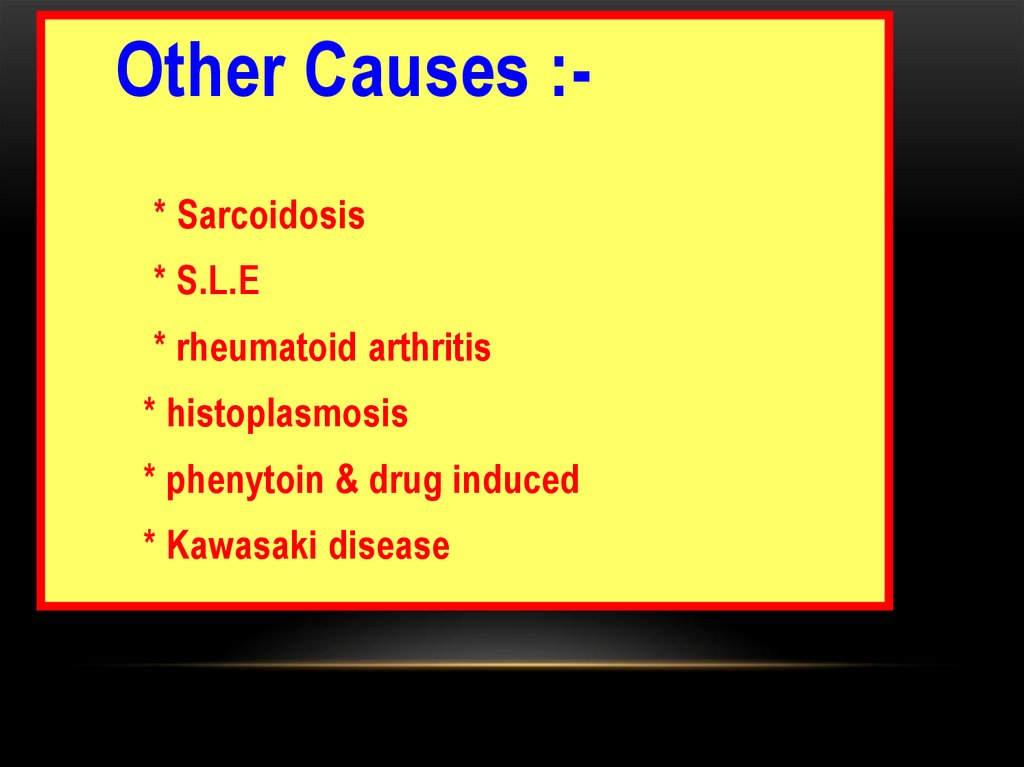
Other Causes :* Sarcoidosis* S.L.E
* rheumatoid arthritis
* histoplasmosis
* phenytoin & drug induced
* Kawasaki disease
183.
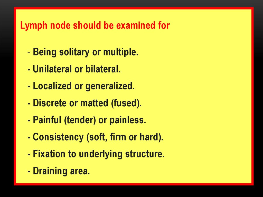
Lymph node should be examined for- Being solitary or multiple.
- Unilateral or bilateral.
- Localized or generalized.
- Discrete or matted (fused).
- Painful (tender) or painless.
- Consistency (soft, firm or hard).
- Fixation to underlying structure.
- Draining area.
184.
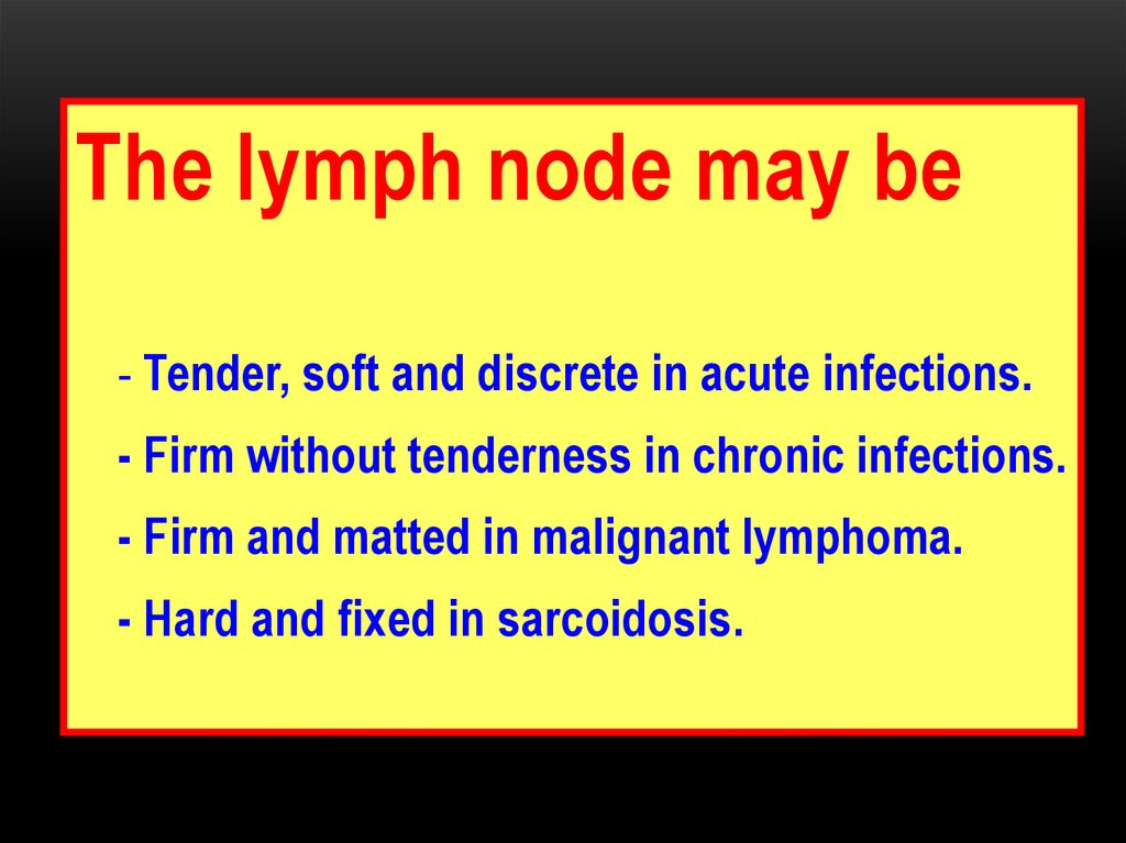
The lymph node may be- Tender, soft and discrete in acute infections.
- Firm without tenderness in chronic infections.
- Firm and matted in malignant lymphoma.
- Hard and fixed in sarcoidosis.
185.
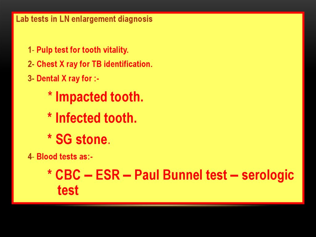
Lab tests in LN enlargement diagnosis1- Pulp test for tooth vitality.
2- Chest X ray for TB identification.
3- Dental X ray for :-
* Impacted tooth.
* Infected tooth.
* SG stone.
4- Blood tests as:-
* CBC – ESR – Paul Bunnel test – serologic
test
186.
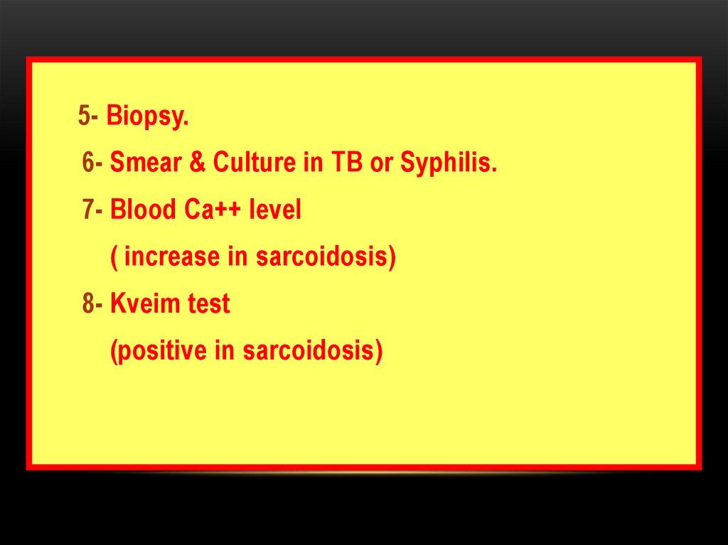
5- Biopsy.6- Smear & Culture in TB or Syphilis.
7- Blood Ca++ level
( increase in sarcoidosis)
8- Kveim test
(positive in sarcoidosis)
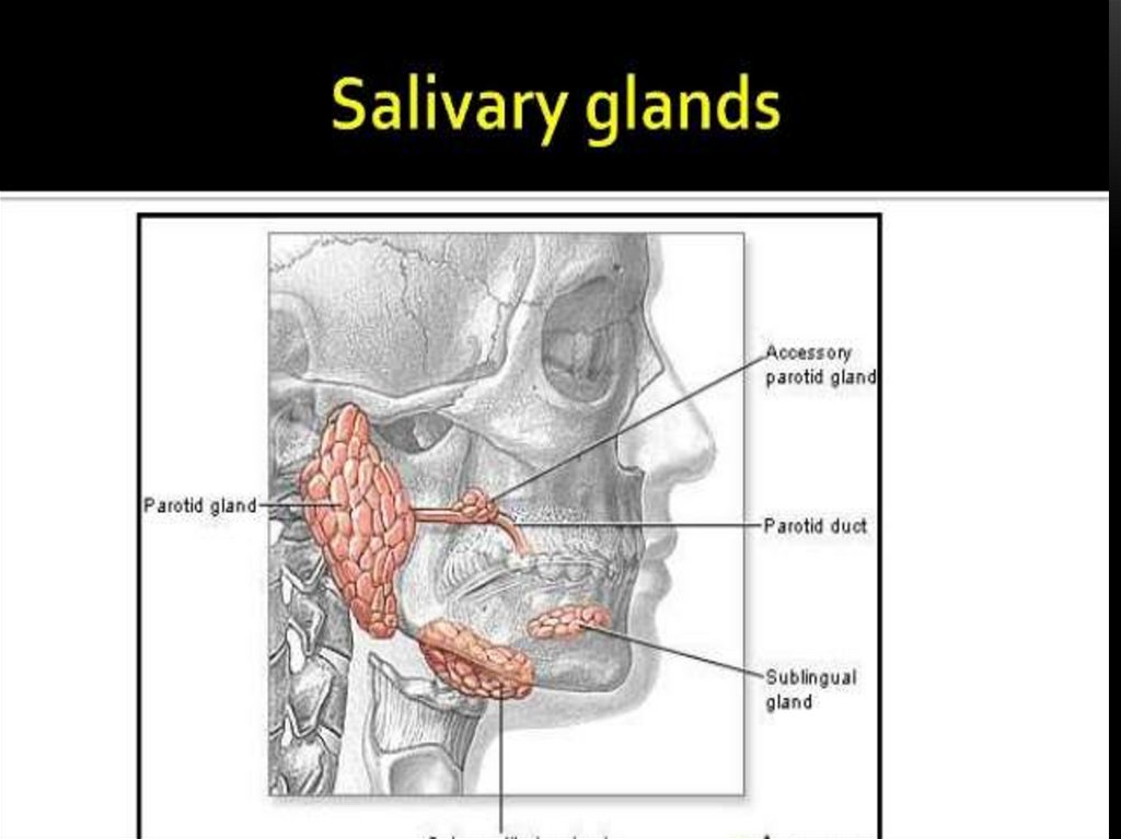
187. Salivary Glands
SALIVARY GLANDSEnlargement of major salivary glands may
be due to :
1) Infection (viral or bacterial)
2) Mechanical (Stone in main duct)
3) Systemic disease as diabetes, malnutrition,
liver cirrhosis, sarcoidosis, Sjogren
disease.
4) Neoplasm (benign or malignant).
5) drugs as antihypertensive (diuretics)
188.
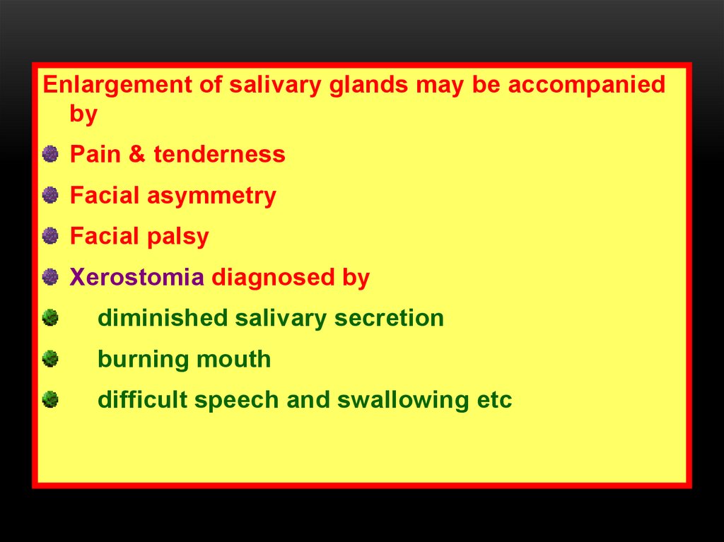
Enlargement of salivary glands may be accompaniedby
Pain & tenderness
Facial asymmetry
Facial palsy
Xerostomia diagnosed by
diminished salivary secretion
burning mouth
difficult speech and swallowing etc
189.
190.
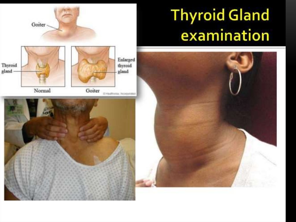
191. Thyroid Glands
THYROID GLANDSNormally the gland is usually palpable as two lobes
connected by isthmus at the level of 2,3 & 4 tracheal
rings.
Examination could be done by:
Inspection
The head is extended and the patient is observed during
swallowing. Any mobile swelling related to the gland
should be reported.
192.
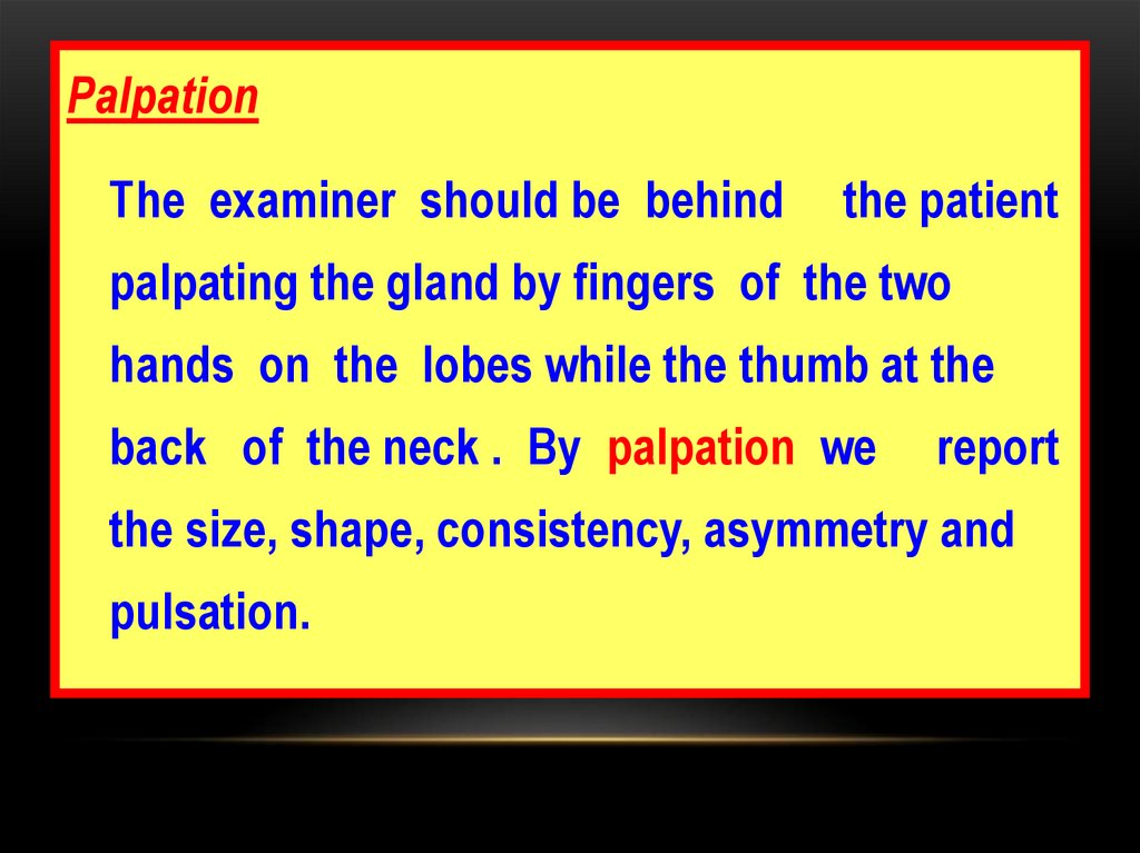
PalpationThe examiner should be behind
the patient
palpating the gland by fingers of the two
hands on the lobes while the thumb at the
back of the neck . By palpation we
report
the size, shape, consistency, asymmetry and
pulsation.
193.
194.
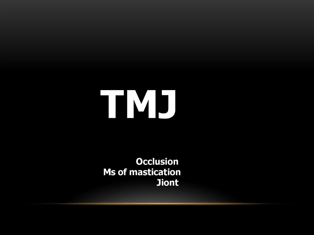
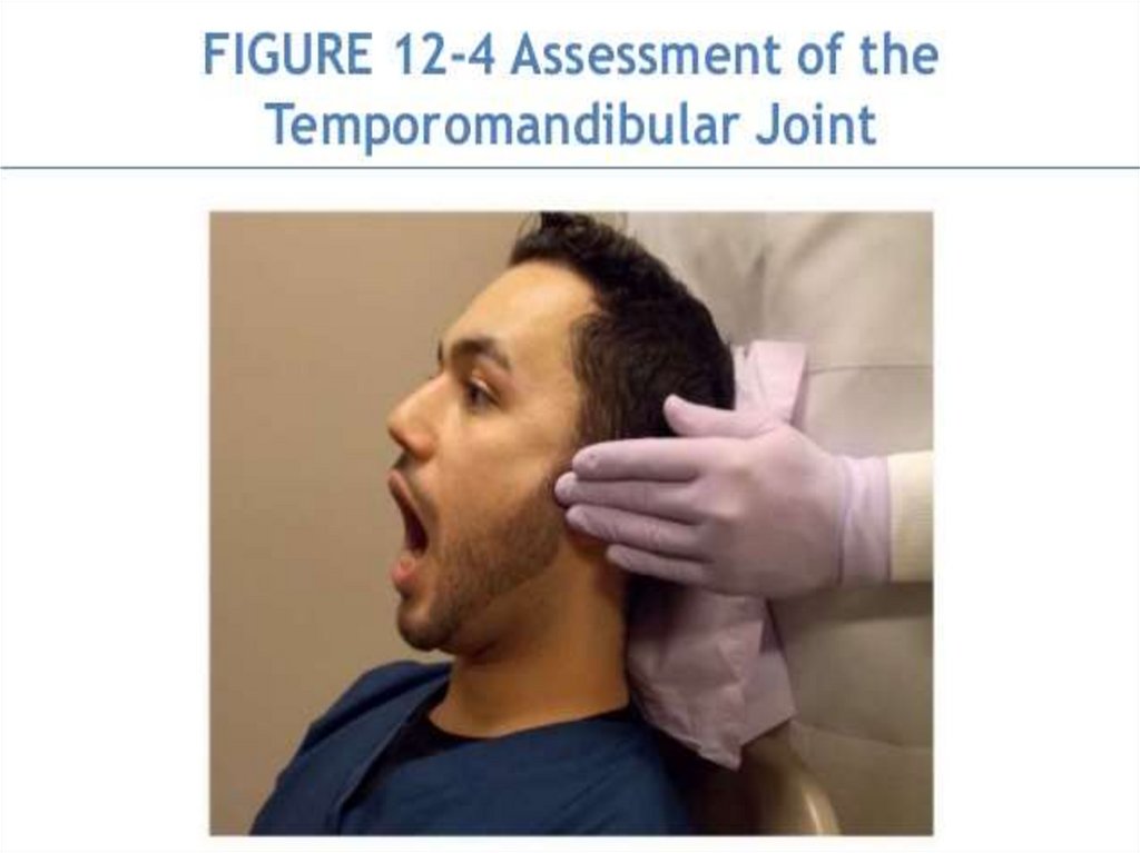
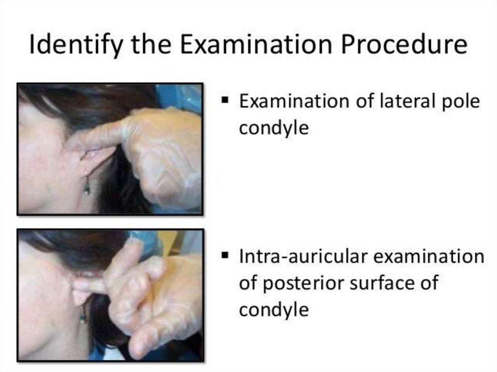
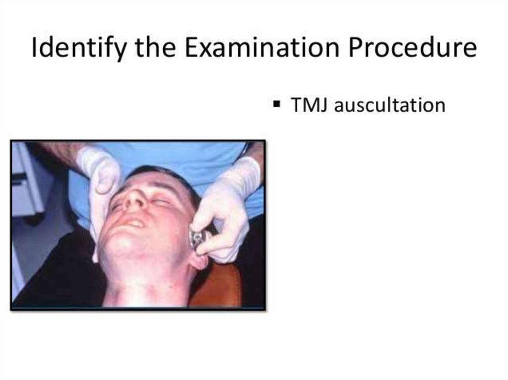
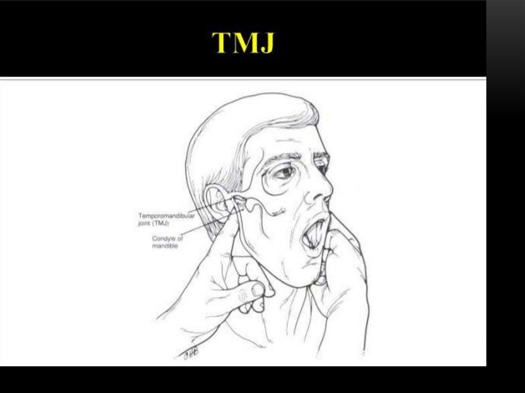
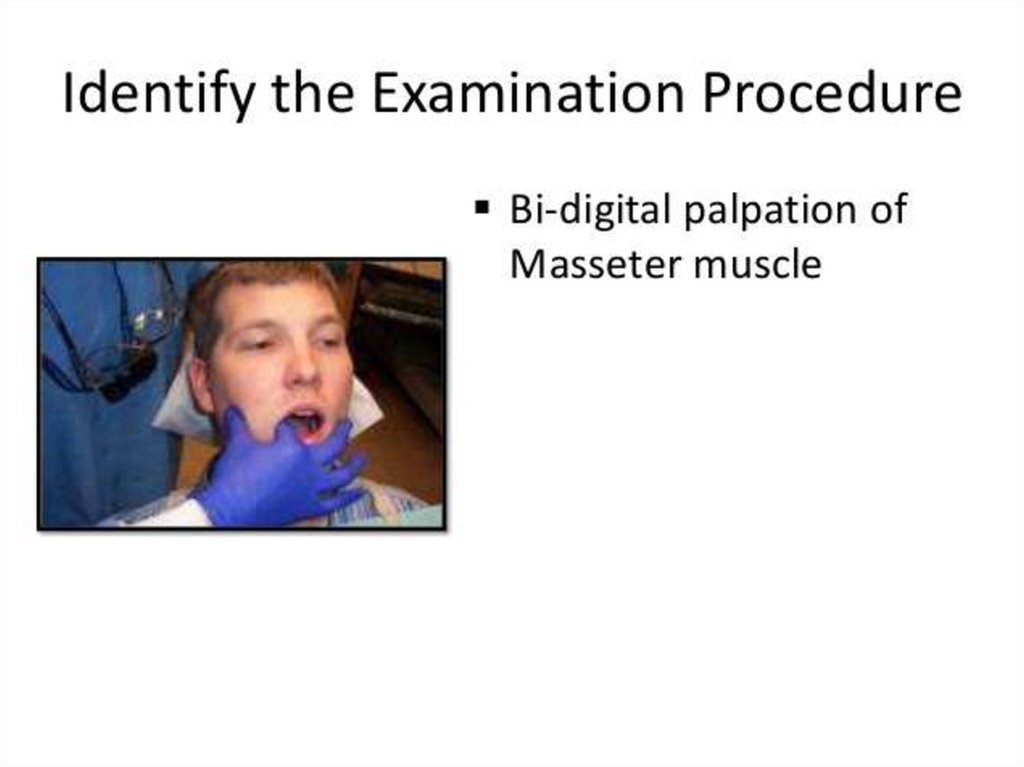
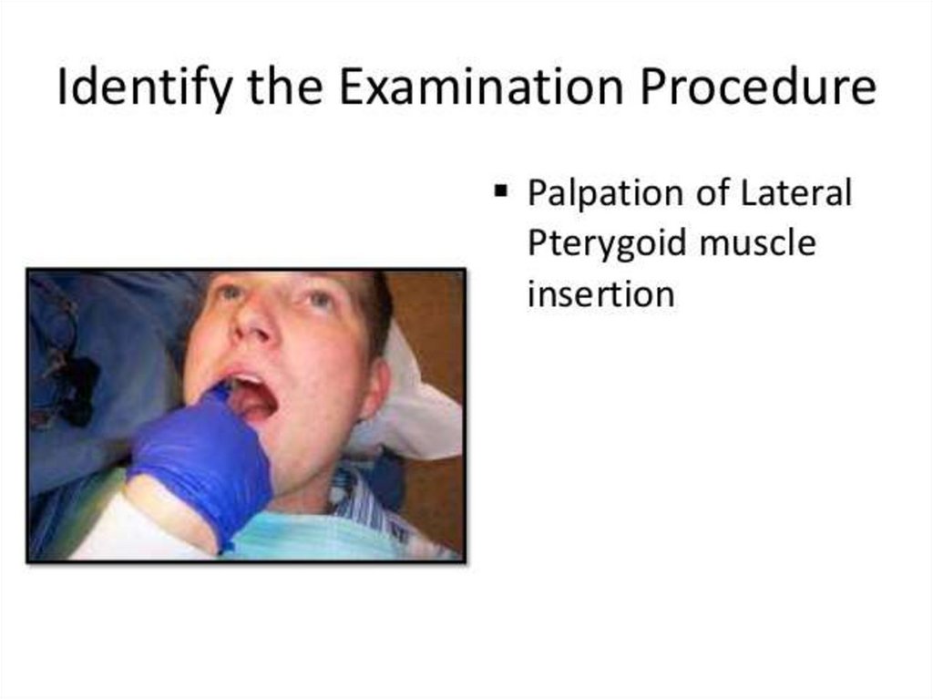
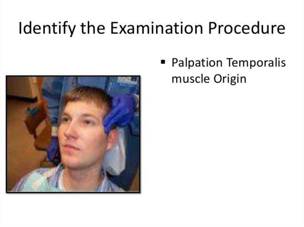
TMJOcclusion
Ms of mastication
Jiont