
")


")

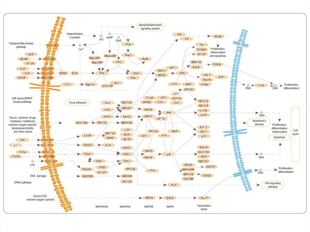
 kinases")
")

 Медицина
Похожие презентации:


 identification")
Antigen recognition and activation of T-cells
1. Antigen recognition and activation of T-cells
2. T-cells can be distinguished from other lymphocyte types, such as B cells and NK cells by the presence of T cell receptors (TCR)
• The TCR is a molecule found on the surface ofT cells that is, in general, responsible for
recognizing antigens bound to major
histocompatibility complex (MHC) molecules
3. Subsets of T cells:
• T helper cell (TH cells) assist other white blood cells in immunologicprocesses, including maturation of B cells into plasma cells and memory B
cells, and activation of cytotoxic T cells and macrophages, among other
functions (CD4+)
• Cytotoxic T cells (TC cells, or CTLs) destroy virally infected cells and tumor
cells, and are also implicated in transplant rejection (CD8+)
• Regulatory T cells (Treg cells), formerly known as suppressor T cells, are
crucial for the maintenance of immunological tolerance
(CD4+CD25+FoxP3+)
• Natural killer T cells (NKT cells) are a special kind of lymphocyte that that
recognize glycolipid antigen presented by a molecule called CD1d
• γδ T cells (gamma delta T cells) represent a small subset of T cells (2% of
total T cells) that possess a distinct T cell receptor (TCR) on their surface
and rapidly respond to a set of non-peptidic phosphorylated isoprenoid
precursors, collectively named phosphoantigens (isopentenyl
pyrophosphate (IPP) and its isomer dimethylallyl pyrophosphate (DMAPP))
• Memory T cells are a subset of antigen-specific T cells that comprise two
subtypes: central memory T cells (TCM cells) and effector memory T cells
(TEM cells).
4. TCR and CD3 complex
ITAM - immunoreceptor tyrosine-based activation motifs5. TCR co-receptors:
• CD4 – for Th(that is specific for class II MHC).
• CD8 – for CTL
(that is specific for class I MHC).
6. CD4
CD4 uses its D1 domain to interact with theβ2-domain of MHC class II molecules.
7. CD8
The extracellular domain of CD8-α interacts withto the α3 portion of the Class I MHC molecule
8. T-cell activation
• The mechanism by which a T-cell elicits thisresponse upon contact with its unique antigen
is termed T-cell activation
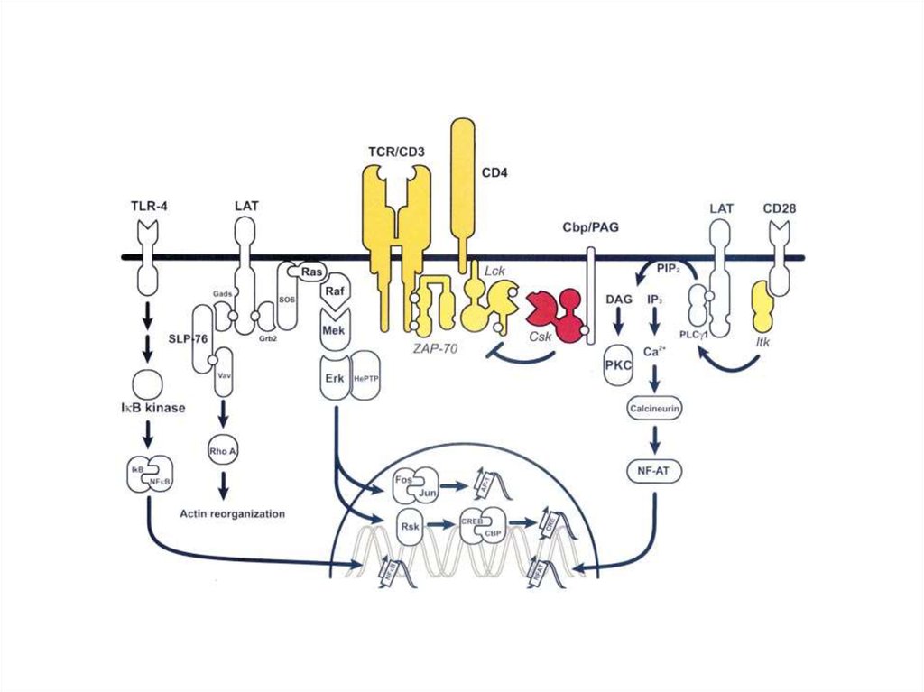
9. Early signaling steps implicate the following molecules:
Lck - Associated with the transmembrane tail
of CD4
• Fyn - Associated with ITAMs of the TCR
complex
• CD45 - The transmembrane tail of which
functions as a Tyrosine phosphatase
• Zap70 - Binds to ITAM sequences upon
phosphorylation by Lck and Fyn
10.
• Lck (lymphocyte-specific protein tyrosinekinase) and Fyn are members of the Src family
of tyrosine kinases.
11.
• Src (pronounced "sarc" as it is short forsarcoma) is a proto-oncogene encoding a
tyrosine kinase originally discovered by
J.Michael Bishop and Harold E. Varmus, for
which they won the 1989 Nobel Prize in
Physiology or Medicine
• This gene is similar to the v-src gene of Rous
sarcoma virus
• v-src lacks the C-terminal inhibitory
phosphorylation site (tyrosine-527), and is
therefore constitutively active
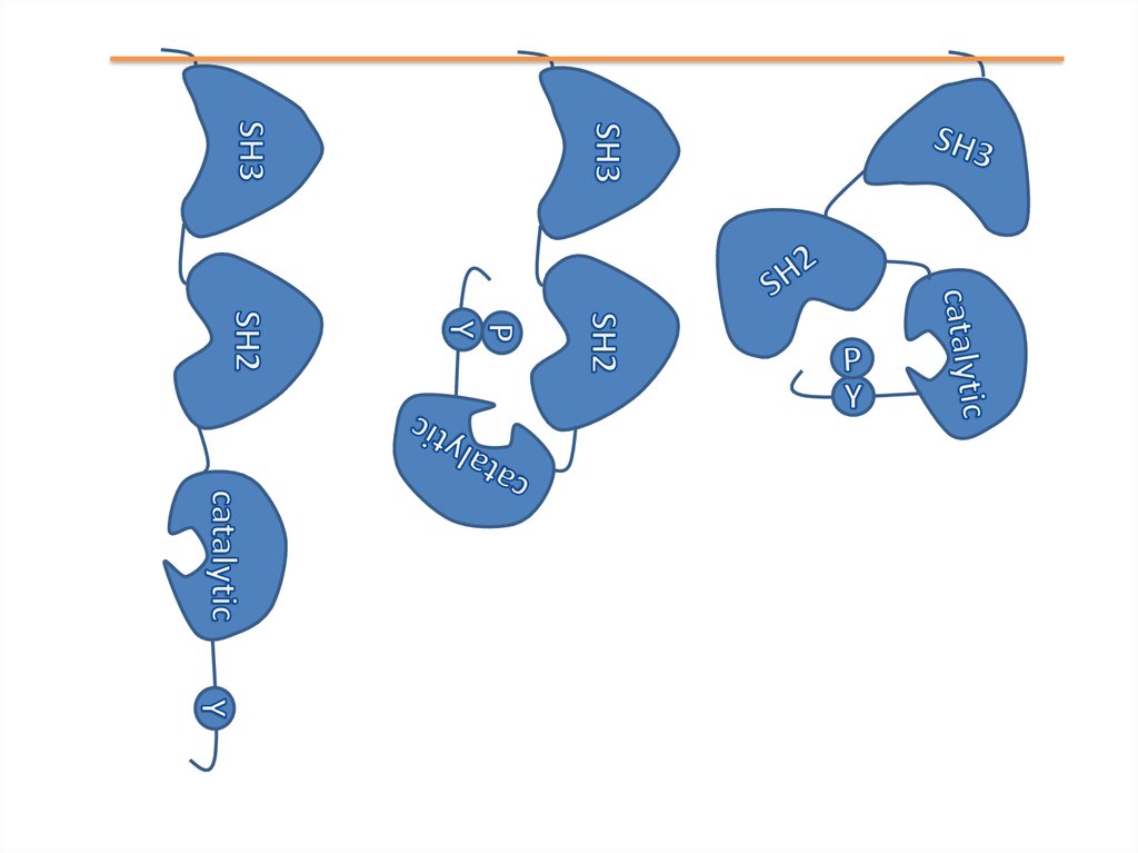
12. Structure of Src kinase
13.
14.
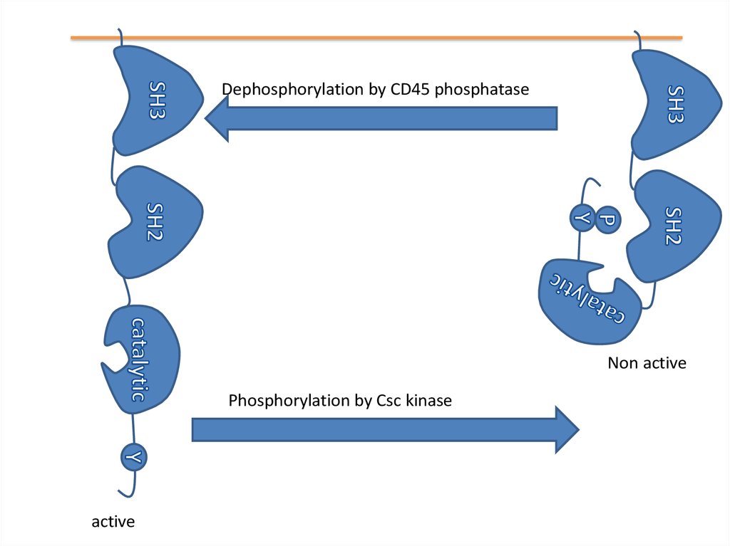
Dephosphorylation by CD45 phosphataseNon active
Phosphorylation by Csc kinase
active
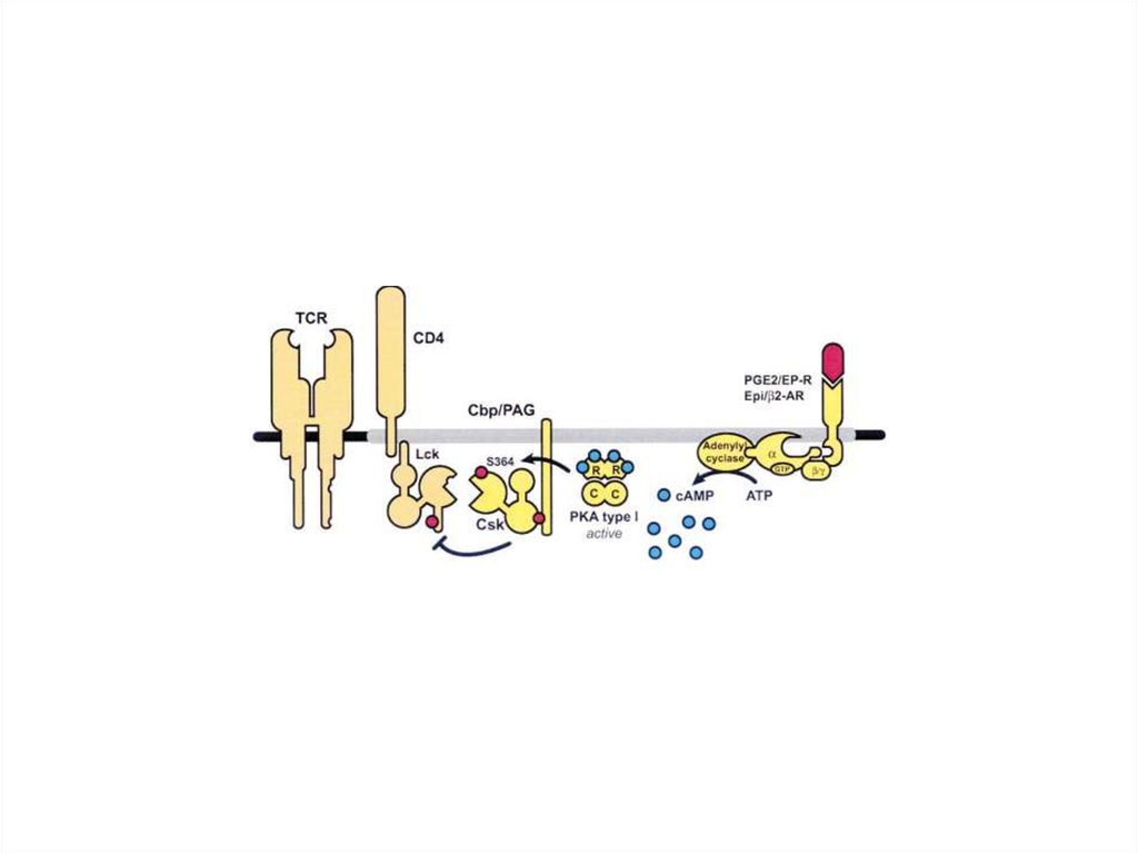
15.
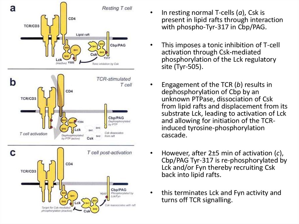
In resting normal T-cells (a), Csk is
present in lipid rafts through interaction
with phospho-Tyr-317 in Cbp/PAG.
This imposes a tonic inhibition of T-cell
activation through Csk-mediated
phosphorylation of the Lck regulatory
site (Tyr-505).
Engagement of the TCR (b) results in
dephosphorylation of Cbp by an
unknown PTPase, dissociation of Csk
from lipid rafts and displacement from its
substrate Lck, leading to activation of Lck
and allowing for initiation of the TCRinduced tyrosine-phosphorylation
cascade.
However, after 2±5 min of activation (c),
Cbp/PAG Tyr-317 is re-phosphorylated by
Lck and/or Fyn thereby recruiting Csk
back into lipid rafts.
this terminates Lck and Fyn activity and
turns off TCR signalling.
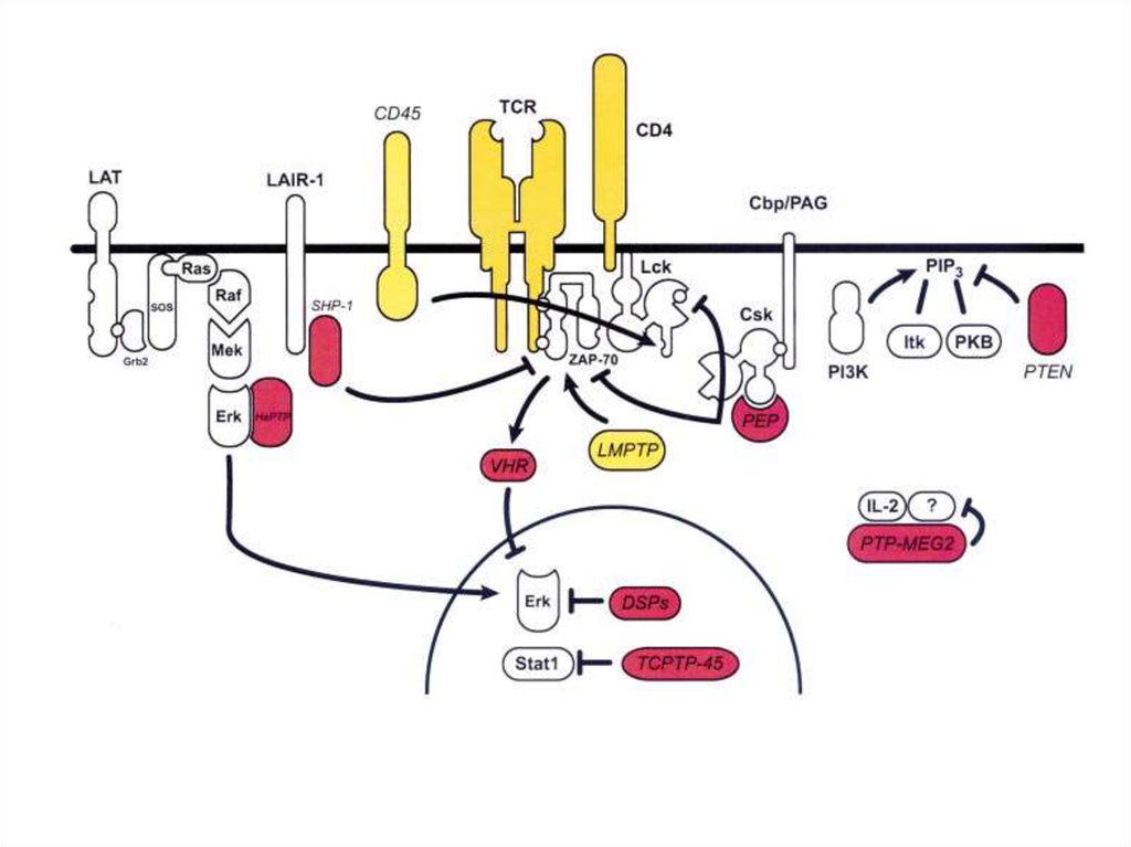
16. SH2 and SH3 domains were found in several other protein families
Kinases
Phosphatases
Phospholipases
Adaptor proteins
Cytoskeleton proteins
Transcription factors etc.
17. ZAP-70 kinase
• ZAP-70 is an abbreviation for Zeta-chainassociated protein kinase 70 (70 is themolecular weight in kDa).
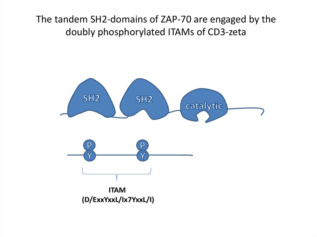
The tandem SH2-domains of ZAP-70 are engaged by the
doubly phosphorylated ITAMs of CD3-zeta
18. 3D view of Zap-70
19.
The tandem SH2-domains of ZAP-70 are engaged by thedoubly phosphorylated ITAMs of CD3-zeta
ITAM
(D/ExxYxxL/Ix7YxxL/I)
20.
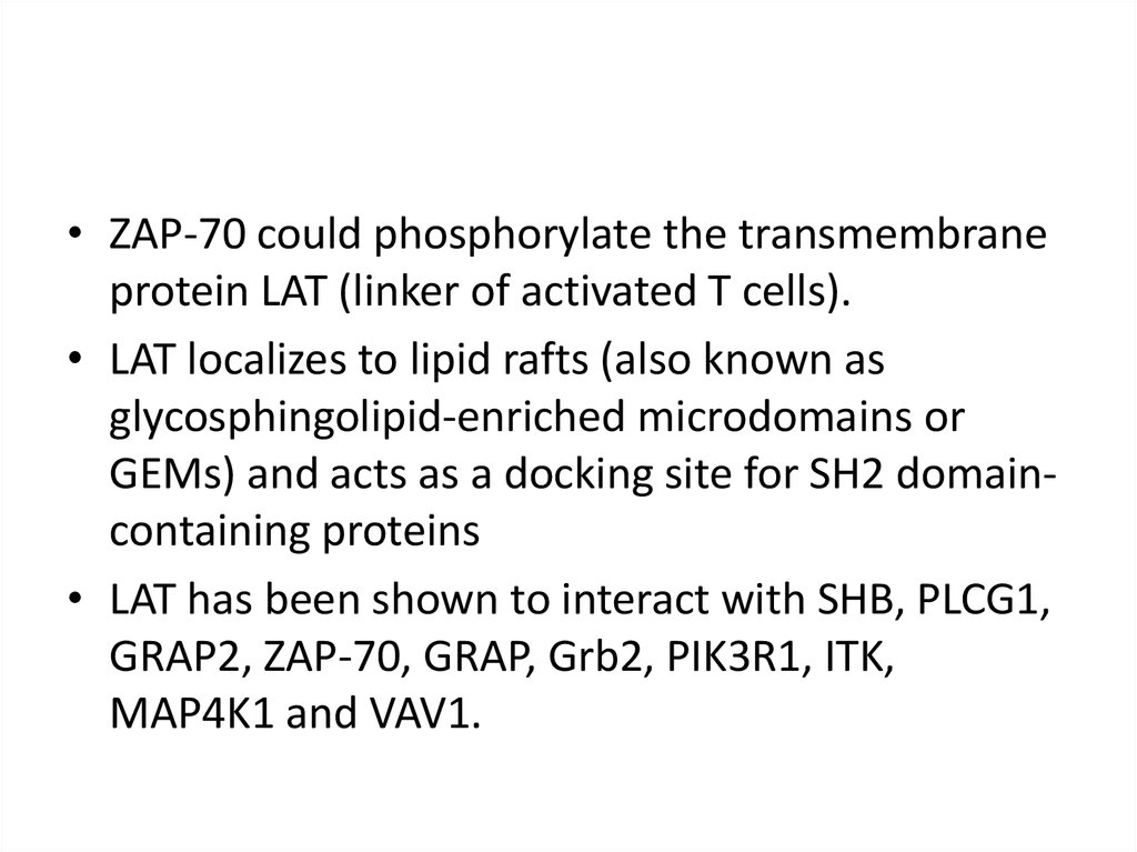
• ZAP-70 could phosphorylate the transmembraneprotein LAT (linker of activated T cells).
• LAT localizes to lipid rafts (also known as
glycosphingolipid-enriched microdomains or
GEMs) and acts as a docking site for SH2 domaincontaining proteins
• LAT has been shown to interact with SHB, PLCG1,
GRAP2, ZAP-70, GRAP, Grb2, PIK3R1, ITK,
MAP4K1 and VAV1.
21. Lipid raft
• The plasma membrane of cells is made of acombination of glycosphingolipids and protein
receptors organized in glycolipoprotein
microdomains termed lipid rafts.
22. Sphingomyelin
23. T-cell signaling in lipid rafts
24. Human LAT is a 233 amino-acid type III transmembrane protein
25. A model of the signaling complexes assembled through LAT in T cells
26. “LAT signalosome”
27.
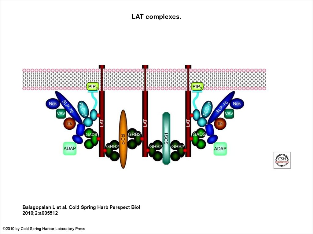
LAT complexes.Balagopalan L et al. Cold Spring Harb Perspect Biol
2010;2:a005512
©2010 by Cold Spring Harbor Laboratory Press
28.
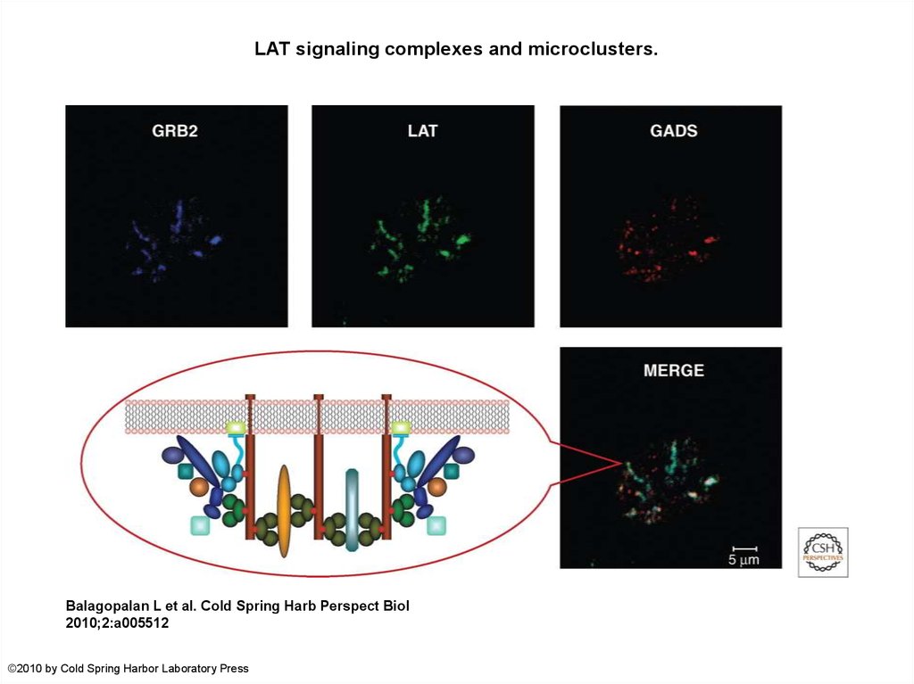
LAT signaling complexes and microclusters.Balagopalan L et al. Cold Spring Harb Perspect Biol
2010;2:a005512
©2010 by Cold Spring Harbor Laboratory Press
29.
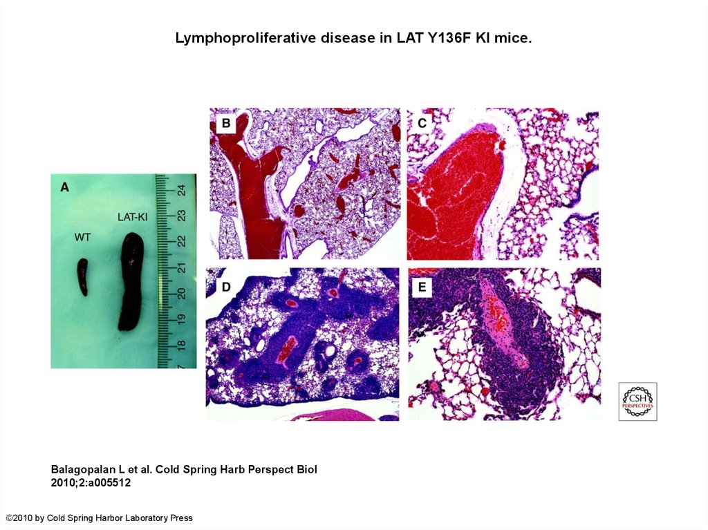
Lymphoproliferative disease in LAT Y136F KI mice.Balagopalan L et al. Cold Spring Harb Perspect Biol
2010;2:a005512
©2010 by Cold Spring Harbor Laboratory Press
30.
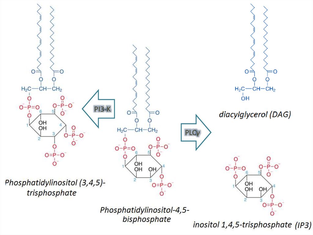
PI3-KPLCγ
31.
PIP2 cleavage to IP3 and DAG initiates intracellular calcium release and PKC activation.32.
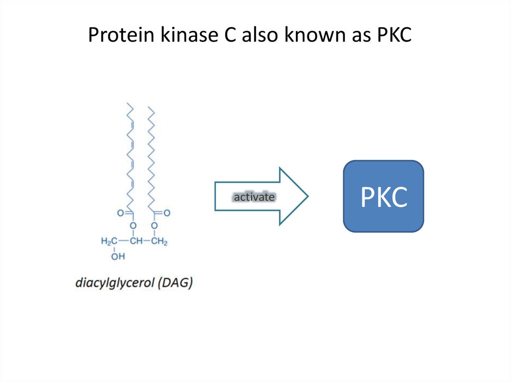
Protein kinase C also known as PKCactivate
PKC
33. Pleckstrin homology domain (PH domain)
• is a protein domain of approximately 120amino acids
• can bind Phosphatidylinositol (3,4,5)trisphosphate within biological membranes