


 by fibrous attachments. The gingival fibers (H)")

 Медицина
Похожие презентации:


Dental anatomy
1. Dental anatomy
DENTALANATOMY
2.
For the general description and information on human teeth, see Teeth (human). Forother uses, see Tooth (disambiguation).
Adult and "Baby" teeth diagram. Note that this diagram uses non-standard dental
notation.
Dental anatomy is a field of anatomy dedicated to the study of human tooth
structures. The development, appearance, and classification of teeth fall within its
purview. (The function of teeth as they contact one another falls elsewhere, under
dental occlusion.) Tooth formation begins before birth, and teeth's eventual
morphology is dictated during this time. Dental anatomy is also a taxonomical
science: it is concerned with the naming of teeth and the structures of which they
are made, this information serving a practical purpose in dental treatment.
Usually, there are 20 primary ("baby") teeth and 28 to 32 permanent teeth, the last
four being third molars or "wisdom teeth", each of which may or may not grow in.
Among primary teeth, 10 usually are found in the maxilla (upper jaw) and the other
10 in the mandible (lower jaw). Among permanent teeth, 16 are found in the maxilla
and the other 16 in the mandible. Most of the teeth have distinguishing features.
3.
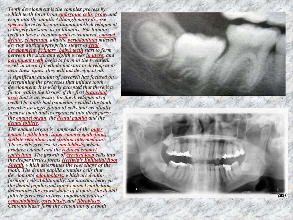
Tooth development is the complex process bywhich teeth form from embryonic cells, grow, and
erupt into the mouth. Although many diverse
species have teeth, non-human tooth development
is largely the same as in humans. For human
teeth to have a healthy oral environment, enamel,
dentin, cementum, and the periodontium must all
develop during appropriate stages of fetal
development. Primary (baby) teeth start to form
between the sixth and eighth weeks in utero, and
permanent teeth begin to form in the twentieth
week in utero.If teeth do not start to develop at or
near these times, they will not develop at all.
A significant amount of research has focused on
determining the processes that initiate tooth
development. It is widely accepted that there is a
factor within the tissues of the first branchial
arch that is necessary for the development of
teeth.The tooth bud (sometimes called the tooth
germ) is an aggregation of cells that eventually
forms a tooth and is organized into three parts:
the enamel organ, the dental papilla and the
dental follicle.
The enamel organ is composed of the outer
enamel epithelium, inner enamel epithelium,
stellate reticulum and stratum intermedium.
These cells give rise to ameloblasts, which
produce enamel and the reduced enamel
epithelium. The growth of cervical loop cells into
the deeper tissues forms Hertwig's Epithelial Root
Sheath, which determines the root shape of the
tooth. The dental papilla contains cells that
develop into odontoblasts, which are dentinforming cells.Additionally, the junction between
the dental papilla and inner enamel epithelium
determines the crown shape of a tooth. The dental
follicle gives rise to three important entities:
cementoblasts, osteoblasts, and fibroblasts.
Cementoblasts form the cementum of a tooth
4. . Nomenclature Teeth are named by their sets and also arch, class, type, and side. Teeth can belong to one of two sets of
.NOMENCLATURE
TEETH ARE NAMED BY THEIR SETS AND ALSO ARCH, CLASS, TYPE, AND SIDE. TEETH CAN BELONG TO ONE OF TWO SETS OF TEETH:
PRIMARY ("BABY") TEETH OR PERMANENT TEETH. OFTEN, "DECIDUOUS" MAY BE USED IN PLACE OF "PRIMARY", AND "ADULT" MAY
BE USED FOR "PERMANENT". "SUCCEDANEOUS" REFERS TO THOSE TEETH OF THE PERMANENT DENTITION THAT REPLACE PRIMARY
TEETH (INCISORS, CANINES, AND PREMOLARS OF THE PERMANENT DENTITION). SUCCEDANEOUS WOULD REFER TO THESE TEETH AS
A GROUP. FURTHER, THE NAME DEPENDS UPON WHICH ARCH THE TOOTH IS FOUND IN. THE TERM, "MAXILLARY", IS GIVEN TO TEETH
IN THE UPPER JAW AND "MANDIBULAR" TO THOSE IN THE LOWER JAW. THERE ARE FOUR CLASSES OF TEETH: INCISORS, CANINES,
PREMOLARS, AND MOLARS. PREMOLARS ARE FOUND ONLY IN PERMANENT TEETH; THERE ARE NO PREMOLARS IN DECIDUOUS TEETH.
WITHIN EACH CLASS, TEETH MAY BE CLASSIFIED INTO DIFFERENT TRAITS. INCISORS ARE DIVIDED FURTHER INTO CENTRAL AND
LATERAL INCISORS. AMONG PREMOLARS AND MOLARS, THERE ARE 1ST AND 2ND PREMOLARS, AND 1ST, 2ND, AND 3RD MOLARS. THE
SIDE OF THE MOUTH IN WHICH A TOOTH IS FOUND MAY ALSO BE INCLUDED IN THE NAME. FOR EXAMPLE, A SPECIFIC NAME FOR A
TOOTH MAY BE "PERMANENT MAXILLARY LEFT LATERAL INCISOR."
5. The tooth is attached to the surrounding gingival tissue and alveolar bone (C) by fibrous attachments. The gingival fibers (H)
The term "crown" of a tooth can be used in two ways. The term"anatomic crown" of a tooth refers to the area above the
cementoenamel junction (CEJ) or "neck" of the tooth. It is
completely covered in enamel. The term "clinical crown" often
is convenient in referring to any part of the tooth visible in the
mouth, but as a rule the unqualified term "crown" refers to the
anatomic crown. The bulk of the crown is composed of dentin,
with the pulp chamber within. The crown is enclosed within
bone before the tooth erupts, but after eruption the crown is
almost always visible in an anatomically normal and clinically
healthy mouth.
The anatomic root is found below the cementoenamel
junction and is covered with cementum, whereas the clinical
root is any part of a tooth not visible in the mouth. Similarly,
the anatomic root is assumed in most circumstances. Dentin
composes most of the root, which normally has pulp canals.
The roots of teeth may be single in number (single-rooted
teeth) or multiple. Canines and most premolars, except for
maxillary first premolars, usually have one root. Maxillary first
premolars and mandibular molars usually have two roots.
Maxillary molars usually have three roots. The tooth is
supported in bone by an attachment apparatus, known as the
periodontium, which interacts with the root.
THE TOOTH IS ATTACHED TO THE SURROUNDING GINGIVAL TISSUE AND ALVEOLAR BONE (C) BY FIBROUS
ATTACHMENTS. THE GINGIVAL FIBERS (H) RUN FROM THE CEMENTUM (B) INTO THE GINGIVA IMMEDIATELY
APICAL TO THE JUNCTIONAL EPITHELIAL ATTACHMENT AND THE PERIODONTAL LIGAMENT FIBERS (I), (J) AND (K)
RUN FROM THE CEMENTUM INTO THE ADJACENT CORTEX OF THE ALVEOLAR BONE.
6.
SurfacesSurfaces that are nearest the cheeks or lips are referred to as facial, and those nearest the tongue
are known as lingual. Facial surfaces can be subdivided into buccal (when found on posterior
teeth nearest the cheeks) and labial (when found on anterior teeth nearest the lips). Lingual
surfaces can also be described as palatal when found on maxillary teeth beside the hard palate.
Surfaces that aid in chewing are known as occlusal on posterior teeth and incisal on anterior
teeth. Surfaces nearest the junction of the crown and root are referred to as cervical, and those
closest to the apex of the root are referred to as apical. The tissue surrounding apex is called
periapical. The words mesial and distal are also used as descriptions. "Mesial" signifies a
surface closer to the median line of the face, which is located on a vertical axis between the
eyes, down the nose, and between the contact of the central incisors. Surfaces further away
from the median line are described as distal.
Cusp
A cusp is an elevation on an occlusal surface of posterior teeth and canines. It contributes to a
significant portion of the tooth's surface. Canines have one cusp. Maxillary premolars and the
mandibular first premolars usually have two cusps. Mandibular second premolars frequently
have three cusps--- one buccal and two lingual. Maxillary molars have two buccal cusps and
two lingual cusps. A fifth cusp that may form on the maxillary first molar is known as the cusp
of Carabelli. Mandibular molars may have five or four cusps
7.
Distinguishing characteristics of teethIncisor
8 incisors are anterior teeth, 4 in the upper arch and
4 in the lower. Their function is for shearing or
cutting food during chewing. There are no cusps on
the teeth. Instead, the surface area of the tooth used
in eating is called the incisal ridge or incisal edge.
Though similar, there are some minor differences
between the primary and permanent incisors.
Maxillary central incisor
The maxillary central incisors are usually the most
visible teeth, since they are the top center two teeth
in the front of a mouth, and they are located mesial
to the maxillary lateral incisor.The overall length of
the deciduous maxillary central incisor is 16 mm on
average, with the crown being 6 mm and the root
being 10 mm.In comparison to the permanent
maxillary central incisor, the ratio of the root length
to the crown length is greater in the deciduous
tooth. The diameter of the crown mesiodistally is
greater than the length cervicoincisally, which
makes the tooth appear wider rather than taller from
a labial viewpoint.
The permanent maxillary central incisor is the
widest tooth mesiodistally in comparison to any
other anterior tooth. It is larger than the neighboring
lateral incisor and is usually not as convex on its
labial surface. As a result, the central incisor
appears to be more rectangular or square in shape.
The mesial incisal angle is sharper than the distal
incisal angle. When this tooth is newly erupted into
the mouth, the incisal edges have three rounded
features called mammelons.Mammelons disappear
with time as the enamel wears away by friction.