


 Медицина
Похожие презентации:


The Immune-Brain Connection
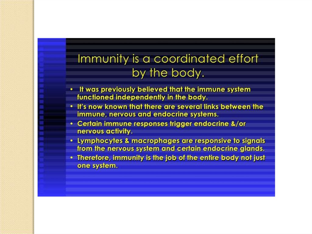
1. The Immune-Brain Connection
2.
3.
Early Concepts, Pre 1950.Fifty years ago, biomedical scientists were certain that there was an impervious barrier
between the immune system and the brain. The barrier, called the blood-brain barrier,
was thought to protect the brain from any effects of the immune system. It was not
thought possible for immune cells to migrate from the blood, through the blood-brain
barrier and into the brain. According to the prevailing view of the time, immune cells
did not reside in the brain either. The concept of the immune system releasing chemical
messengers which traveled through the blood and into the brain had absolutely no
scientific support. The notion of immune cells secreting chemical messengers of any
sort was immunological heresy. As a result, a model of the immune system
communicating with the brain was never proposed or discussed because it was
considered biologically impossible.
Communicating in the other direction, that is, from the brain to the immune system, was
considered impossible also. For one thing, neuroanatomists could not find nerves
extending from the brain to cells or structures of the immune system. For another, there
were no reports of the brain secreting chemical messengers which could regulate the
immune system. Without chemical messengers or nerve connections, the brain could not
send vital information to the immune system.
Consequently, before 1950, there were no biological concepts of a functional connection
between the immune system and the brain. The biological dogma of the time was: 1).
The immune system cannot communicate with the brain or control any brain function;
2). The brain cannot communicate with the immune system or control any immune
system functions. In short, there was no hypothesis of an immune-brain connection
before 1950.
4.
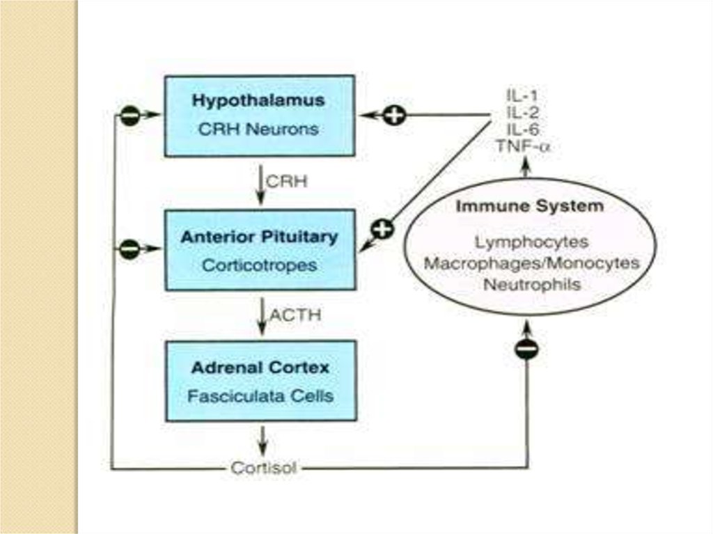
In the 1950's, biologists discovered that hormones help regulate the immunesystem. Cortisol (also called hydrocortisone) is the best known example of
hormonal control of the immune system. It is an anti-inflammatory and
immunosuppressive hormone made by the adrenal cortex. The production of
cortisol is governed by the pituitary and the pituitary is controlled by the
hypothalamus. Therefore another revolutionary conclusion: the brain, via its
control over cortisol and other hormone secretions, helps regulate the immune
system.
Many hormones influence the immune system. In fact, most of them do. The
hypothalamus, for example, secretes many potent hormones and a number of
them help control immune cells. The pituitary secretes many different
hormones and these hormones help regulate immune cells. In like manner,
sex hormones secreted by the ovaries and testes influence immune cells. So
do thyroid hormones.
The brain and peripheral nerves release numerous neurotransmitters and
other chemicals called neuropeptides. Neurotransmitters and neuropeptides
are not usually called hormones, but they do have hormone like properties,
that is, they are chemical messengers. Most neurotransmitters and
neuropeptides influence immune cell activities. As you can see, there are
many hormones, neurotransmitters and neuropeptides released by the brain
or by structures controlled by the brain which regulate the immune system.
5.
Neurotransmitters and hormones are chemicals secreted inside our brain and are largely responsible for our behaviorand attitude. There are many similarities in the two compounds that make people think they are one and the same whereas
in reality there are great differences between a neurotransmitter and a hormone that need to be appreciated.
The most notable difference between a neurotransmitter and a hormone pertains to the point of its release inside the
body. A hormone is a compound produced by endocrine gland and is released directly into the bloodstream where it easily
finds its target cells at a small distance from the point of release. On the other hand, a neurotransmitter is a compound
released by a nerve terminal when the nerve is triggered by an electrical impulse. As this electrical impulse reaches the end
of the nerve, it secretes a chemical compound at a special place in between the nerve cells called synapse. In comparison to
hormones that take time to have their effect; these nerve cells are in direct apposition with the target cells which ensures
quick delivery of the signal.
There are receptors for both hormones as well as neurotransmitters in the target cells and these receptors induce
biochemical responses from the individual depending upon the type of hormone or neurotransmitter. Thus the difference
between a neurotransmitter and a hormone boils down to the release mechanism and this mechanism alone decides
whether the released molecule is a hormone or a neurotransmitter. Thus adrenaline is a hormone secreted by adrenaline
gland directly into the bloodstream which goes to heart and the lungs. On the other hand serotonin is a neurotransmitter as
it is released by a stimulated presynaptic nerve cell and acts on its neighboring postsynaptic cell.
Both hormones and neurotransmitters are vital for human beings as they play an important role in many bodily processes
such as digestion, metabolism, reproduction etc. They are also important in mood control. Some people are more
aggressive than others and it is a result of secretion of higher amounts of some hormones and neurotransmitters inside the
body.
Difference between Neurotransmitters and Hormones
• Both neurotransmitters and hormones are chemicals secreted inside our bodies.
• While hormones are produced by endocrine gland, neurotransmitters are produced at
nerve terminals when triggered by an electrical impulse.
• Hormones are secreted directly into the bloodstream whereas neurotransmitters are
secreted at nerve synapses.
• Hormones can be synthesized whereas it is impossible to make neurotransmitters.
They are made inside the body only.
6.
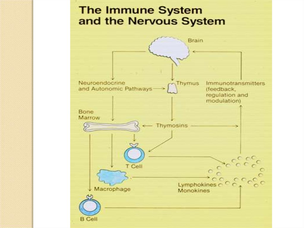
In addition to the extensive ability to chemically (i.e. via hormones, neurotransmittersand neuropeptides) regulate the immune system, the brain can also directly control
important parts of the immune system through its network of nerves. Starting in the
1960's, neuroanatomical investigations began finding direct nervous links between the
brain and the immune system. There are nerves going directly from the brain to
important immune organs like the thymus, bone marrow, spleen, lymph nodes
and gut associated lymphoid tissue. By having nerves connected to these important
immune organs, the brain is able to directly regulate immune system activities.
Extensive animal studies have shown that the brain, via its nerve connections, does
exert significant control over these immune organs.
Thus, from 1950 to 1978, a radically changed view of the immune-brain connection
developed. Massive hormonal and neuroanatomical evidence made it clear that there
was a connection between the brain and the immune system. The brain, through its
direct nerve connections to the immune system and its control over the extensive
hormone network, helped govern the immune system. A new biomedical discipline,
called psychoneuroimmunology, grew up around these discoveries.
In 1978, the paradigm for the immune-brain connection was: 1). The brain, in a very
complex way, regulated the immune system. The direction of the control was from the
brain to the immune system, that is, Brain→Immune System. 2). There was no
evidence the immune system could control the brain, therefore the immune-brain
connection was a one-way street, Brain→Immune System, and not a two-way street,
Brain↔Immune System.
7.
Before the remarkable discoveries on cytokines, it was assumedto be impossible for the immune system to communicate with the
brain.
Like
no
other
previous
discovery,
cytokines
have
revolutionized our understanding of the communications link
between the immune system and the brain. The one direction
pathway model is now untenable. It is simply wrong. Instead, we
now know the communications pathway is bi-directional, that is, it
is a two-way street: Immune system↔Brain. There is a continuous
information loop going from the immune system to the brain and
from the brain back to the immune system. Thus, the immune
system can control the brain and the brain can control the
immune system.
8.
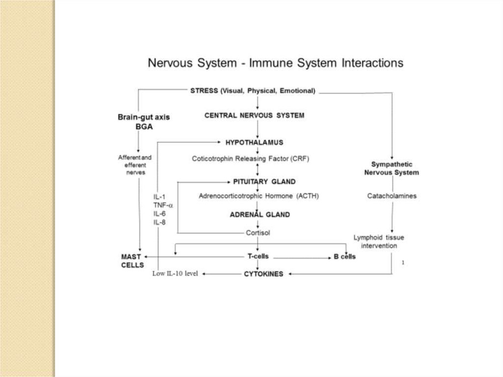
Human body maintains its homeostasis under different stress conditions with the helpof central nervous system (through neurotransmitter), endocrine system (through
hormones) and immune system (through antibodies and specialised cells). All the
three major systems work in synchrony to regulate the body function smoothly under all
diverse situations of fight or flight. Hormones and neurotransmitters are two separate
chemical messengers with some similarities as some molecules can act as both
hormones and neurotransmitters as well. One example of this overlap
is norepinephrine which can be released into the bloodstream by the adrenal
glands as a hormone or can be released by sympathetic nerve endings as a
neurotransmitter.
9.
10.
vagus nerve –блуждающий нерв
Schematic
illustration
of
connections between the
nervous and immune
systems. Signalling between
the immune system and the
central nervous system (CNS)
through systemic routes, the
vagus nerve, the hypothalamic–
pituitary–adrenal (HPA) axis,
the sympathetic nervous system
(SNS) and the peripheral
nervous system (PNS) are
shown.
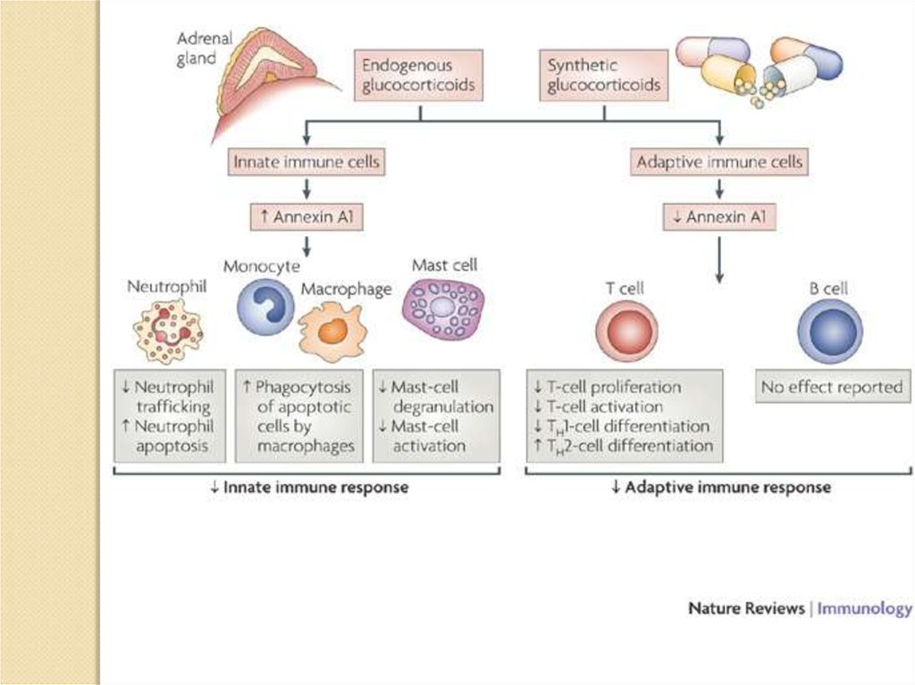
Glucocorticoids can cause a
shift in adaptive immune
responses from a T helper 1
(TH1) type to a TH2 type,
largely through inhibiting the
production of the TH1-cellinducing cytokine interleukin-12
(IL-12)
by
DCs
and
macrophages.
11.
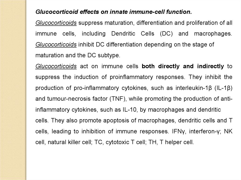
Glucocorticoid effects on innate immune-cell function.Glucocorticoids suppress maturation, differentiation and proliferation of all
immune cells, including Dendritic Cells (DC) and macrophages.
Glucocorticoids inhibit DC differentiation depending on the stage of
maturation and the DC subtype.
Glucocorticoids act on immune cells both directly and indirectly to
suppress the induction of proinflammatory responses. They inhibit the
production of pro-inflammatory cytokines, such as interleukin-1β (IL-1β)
and tumour-necrosis factor (TNF), while promoting the production of antiinflammatory cytokines, such as IL-10, by macrophages and dendritic
cells. They also promote apoptosis of macrophages, dendritic cells and T
cells, leading to inhibition of immune responses. IFNγ, interferon-γ; NK
cell, natural killer cell; TC, cytotoxic T cell; TH, T helper cell.
12.
13.
14.
15.
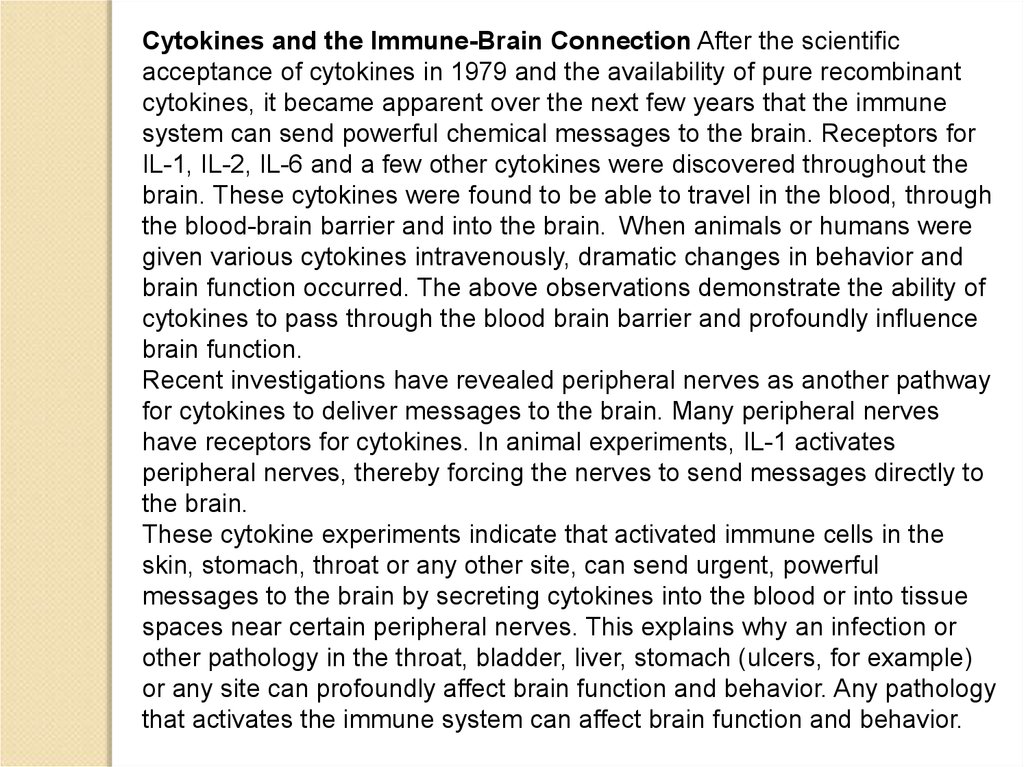
Cytokines and the Immune-Brain Connection After the scientificacceptance of cytokines in 1979 and the availability of pure recombinant
cytokines, it became apparent over the next few years that the immune
system can send powerful chemical messages to the brain. Receptors for
IL-1, IL-2, IL-6 and a few other cytokines were discovered throughout the
brain. These cytokines were found to be able to travel in the blood, through
the blood-brain barrier and into the brain. When animals or humans were
given various cytokines intravenously, dramatic changes in behavior and
brain function occurred. The above observations demonstrate the ability of
cytokines to pass through the blood brain barrier and profoundly influence
brain function.
Recent investigations have revealed peripheral nerves as another pathway
for cytokines to deliver messages to the brain. Many peripheral nerves
have receptors for cytokines. In animal experiments, IL-1 activates
peripheral nerves, thereby forcing the nerves to send messages directly to
the brain.
These cytokine experiments indicate that activated immune cells in the
skin, stomach, throat or any other site, can send urgent, powerful
messages to the brain by secreting cytokines into the blood or into tissue
spaces near certain peripheral nerves. This explains why an infection or
other pathology in the throat, bladder, liver, stomach (ulcers, for example)
or any site can profoundly affect brain function and behavior. Any pathology
that activates the immune system can affect brain function and behavior.
16.
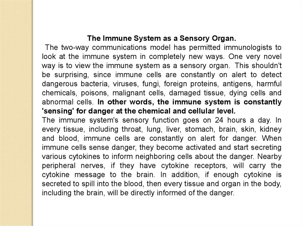
The Immune System as a Sensory Organ.The two-way communications model has permitted immunologists to
look at the immune system in completely new ways. One very novel
way is to view the immune system as a sensory organ. This shouldn't
be surprising, since immune cells are constantly on alert to detect
dangerous bacteria, viruses, fungi, foreign proteins, antigens, harmful
chemicals, poisons, malignant cells, damaged tissue, dying cells and
abnormal cells. In other words, the immune system is constantly
'sensing' for danger at the chemical and cellular level.
The immune system's sensory function goes on 24 hours a day. In
every tissue, including throat, lung, liver, stomach, brain, skin, kidney
and blood, immune cells are constantly on alert for danger. When
immune cells sense danger, they become activated and start secreting
various cytokines to inform neighboring cells about the danger. Nearby
peripheral nerves, if they have cytokine receptors, will carry the
cytokine message to the brain. In addition, if enough cytokine is
secreted to spill into the blood, then every tissue and organ in the body,
including the brain, will be directly informed of the danger.
17.
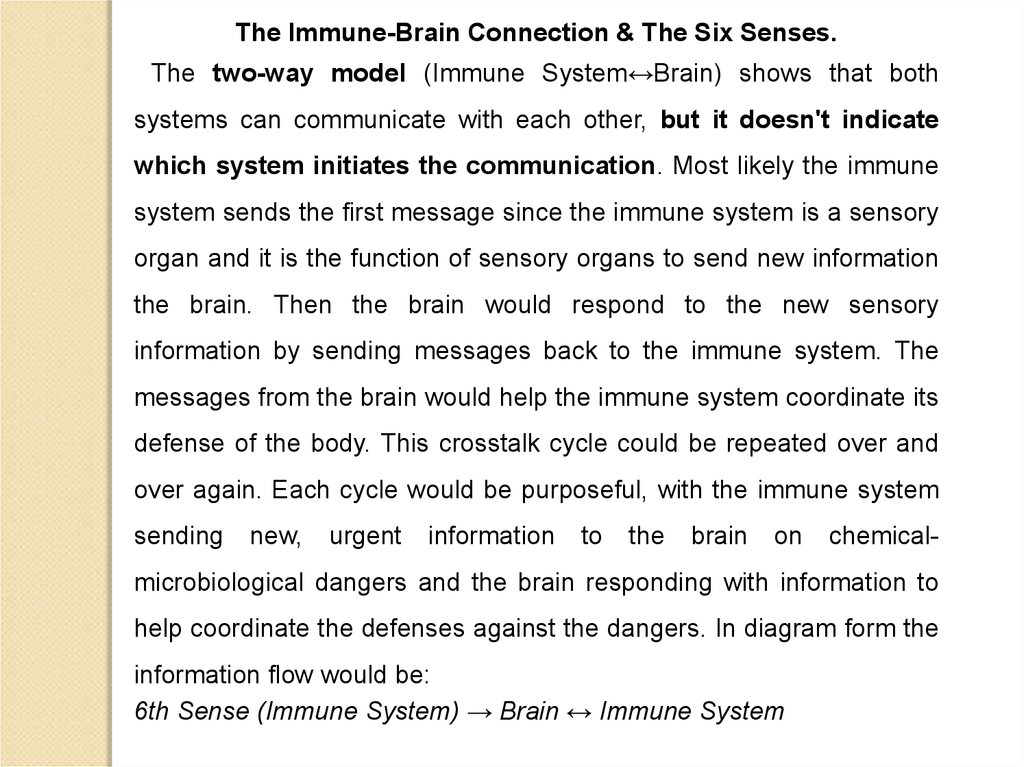
The Immune-Brain Connection & The Six Senses.The two-way model (Immune System↔Brain) shows that both
systems can communicate with each other, but it doesn't indicate
which system initiates the communication. Most likely the immune
system sends the first message since the immune system is a sensory
organ and it is the function of sensory organs to send new information
the brain. Then the brain would respond to the new sensory
information by sending messages back to the immune system. The
messages from the brain would help the immune system coordinate its
defense of the body. This crosstalk cycle could be repeated over and
over again. Each cycle would be purposeful, with the immune system
sending
new,
urgent
information
to
the
brain
on
chemical-
microbiological dangers and the brain responding with information to
help coordinate the defenses against the dangers. In diagram form the
information flow would be:
6th Sense (Immune System) → Brain ↔ Immune System
18.
19.
20.
21.
Fig. 1. Activated T-cells may release opioid peptides such as methionineenkephalin that modulate T-cells via autocrine and paracrine interactions
with opioid receptors (e.g., opioid receptor or DOR).
22.
Activation-dependent expression and intracellular signalling by DORs on T-cells. Tcell receptor (TCR) activation and cell–cell interactionsboth stimulate the expression of DORs, resulting in a greater percentage of mature Tcells that express DOR and in higher levels per cell within a
subpopulation of T-cells. Enhanced DOR expression occurs in both naпve and
memory T-cells.