


 Медицина
Похожие презентации:


Radiation diagnosis of acute and chronic heart failure
1. Radiation diagnosis of acute and chronic heart failure
2.
• Heart failure• • Acute heart failure
• - develops very quickly (from several minutes
to several hours). It appears in the form of
pulmonary edema, cardiac asthma and
cardiogenic shock. The main causes of acute
heart failure are myocardial infarction, rupture
of the walls of the left ventricle, acute failure
of the aortic and mitral valves.
3.
• • Chronic heart failure• - the formation of pathology is gradual and
develops throughout the weeks, months or
even years). Causes of chronic heart failure
include diseases such as heart disease,
hypertension, chronic respiratory failure,
prolonged anemia.
4.
There are three types of lesion localization:
1. Left ventricular heart failure
2. Right ventricular heart failure
3. Mixed heart failure
5. X-ray examination.
In acute or uncompensated chronic heart failure,
chest radiographic examination of the chest may have
purple alveolar pulmonary edema, interstitial lung
edema, basal pleural effusion, or venous congestion in
the lungs. In some patients, especially the elderly, it is
possible to identify the expansion of the heart. The
presence of cardiomegaly indicates a serious heart
disease, but the determination of the size of the heart
by chest radiograph is not entirely informative, since
sometimes they can be normal even in patients with
proven heart failure.
6.
• Chest X-ray can help in diagnosingaugmentation of the left atrium with mitral
valve defects, calcification of valve flaps or
pericarditis, left ventricular aneurysm or
pericardial effusion, which looks like a general
increase in the heart.
7.
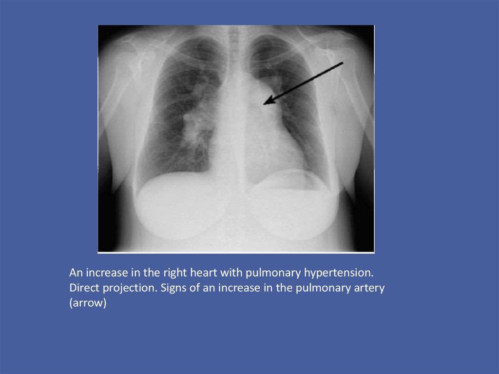
An increase in the right heart with pulmonary hypertension.Direct projection. Signs of an increase in the pulmonary artery
(arrow)
8.
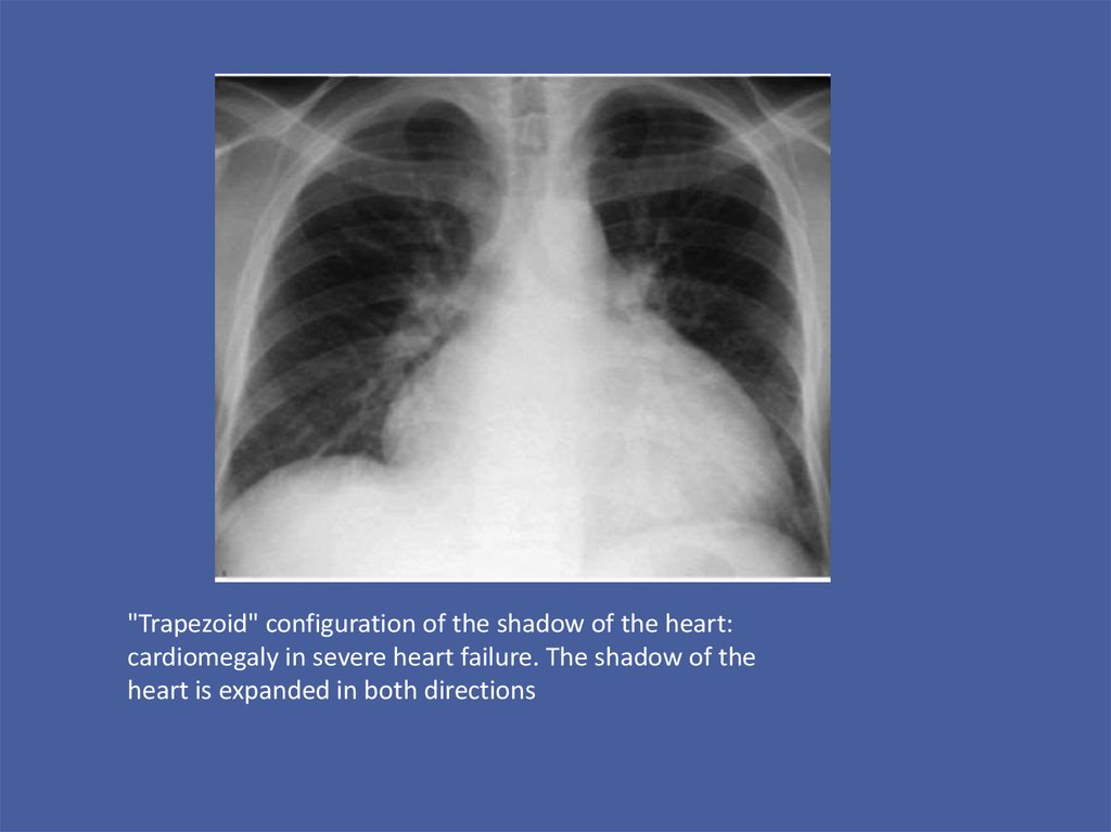
"Trapezoid" configuration of the shadow of the heart:cardiomegaly in severe heart failure. The shadow of the
heart is expanded in both directions
9.
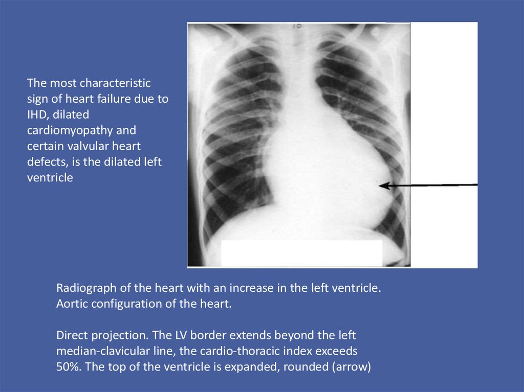
The most characteristicsign of heart failure due to
IHD, dilated
cardiomyopathy and
certain valvular heart
defects, is the dilated left
ventricle
Radiograph of the heart with an increase in the left ventricle.
Aortic configuration of the heart.
Direct projection. The LV border extends beyond the left
median-clavicular line, the cardio-thoracic index exceeds
50%. The top of the ventricle is expanded, rounded (arrow)
10.
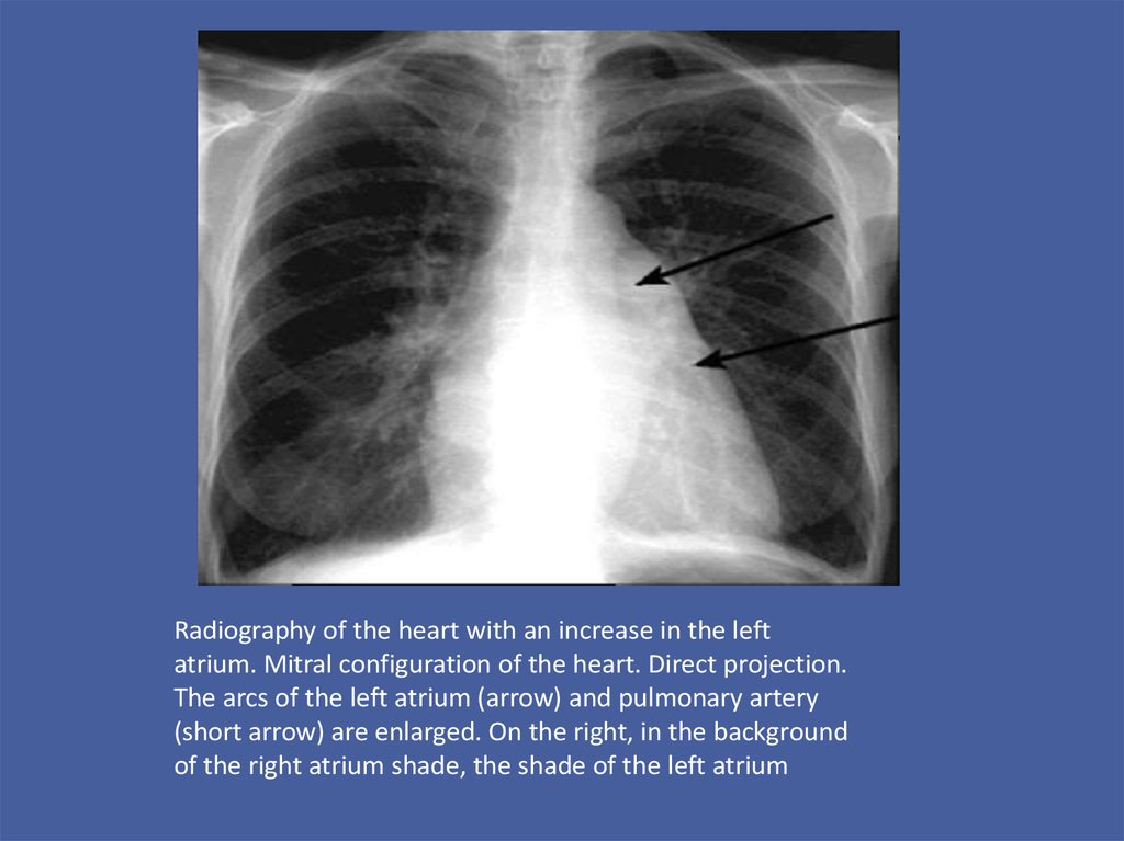
Radiography of the heart with an increase in the leftatrium. Mitral configuration of the heart. Direct projection.
The arcs of the left atrium (arrow) and pulmonary artery
(short arrow) are enlarged. On the right, in the background
of the right atrium shade, the shade of the left atrium
11. Echocardiography.
ECHO-KG refers to
the main methods of
diagnosing heart failure
and monitoring its
treatment. The method
makes it possible to
directly diagnose the
dysfunction of the heart
muscle and to identify
its cause.
12.
• Doppler echocardiography allows to identifyand assess valve stenosis and regurgitation,
congenital heart defects, valvular vegetations,
intracardial tumors and intracavitary thrombi.
13.
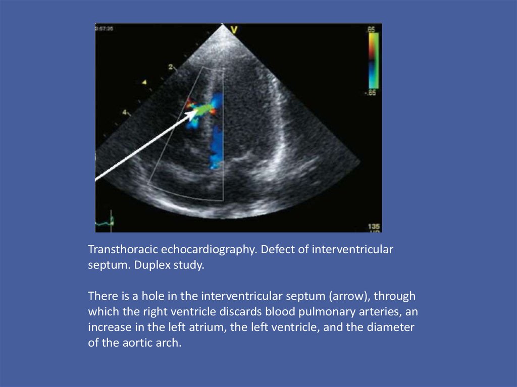
Transthoracic echocardiography. Defect of interventricularseptum. Duplex study.
There is a hole in the interventricular septum (arrow), through
which the right ventricle discards blood pulmonary arteries, an
increase in the left atrium, the left ventricle, and the diameter
of the aortic arch.
14.
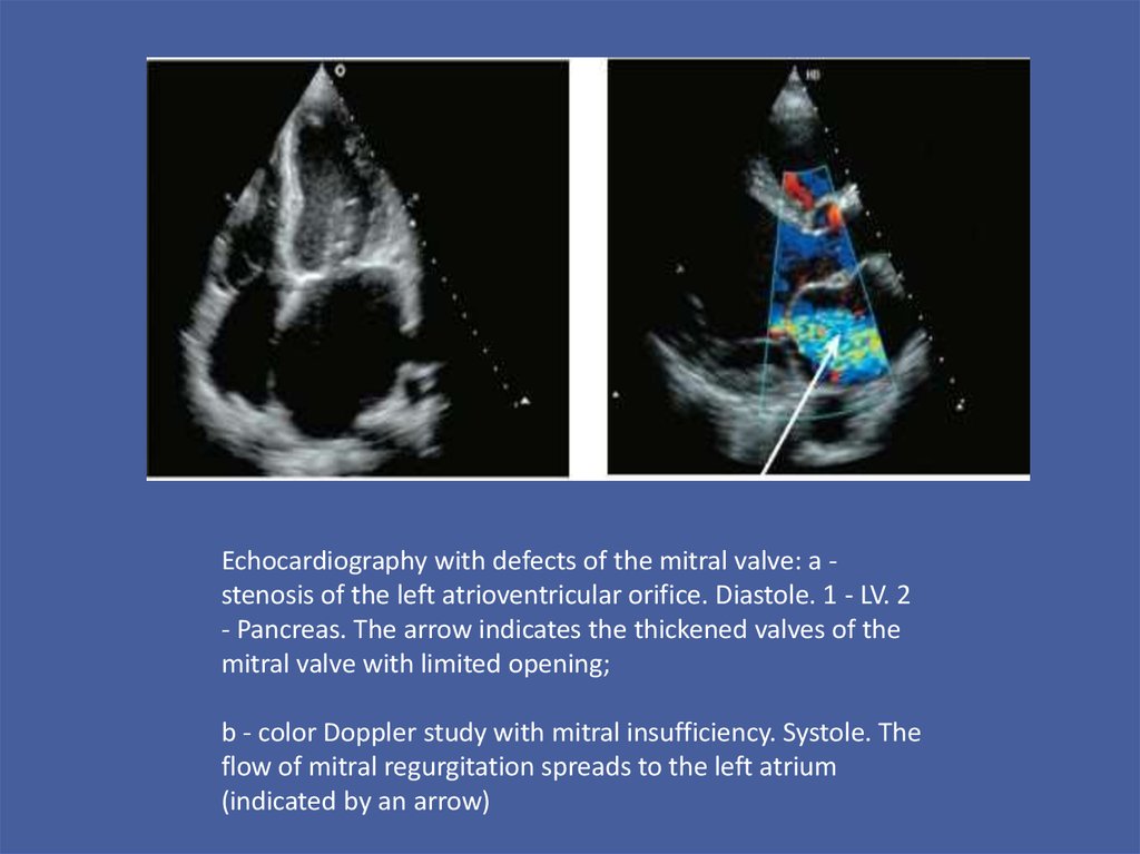
Echocardiography with defects of the mitral valve: a stenosis of the left atrioventricular orifice. Diastole. 1 - LV. 2- Pancreas. The arrow indicates the thickened valves of the
mitral valve with limited opening;
b - color Doppler study with mitral insufficiency. Systole. The
flow of mitral regurgitation spreads to the left atrium
(indicated by an arrow)