


 - hemangioma, arterial blood vessels from growing type.")


 Медицина
Похожие презентации:


")


Hemangioma
1. HEMANGIOMA
KAZAKH NATIONAL MEDICAL UNIVERSITYNAMED AFTER S.D.ASFENDIYAROV
HEMANGIOMA
MADE BY KALAMBEKOV MEREY
CHECKED SAPARGALIEVA A.D.
ALMATY 2015
2. PLAN
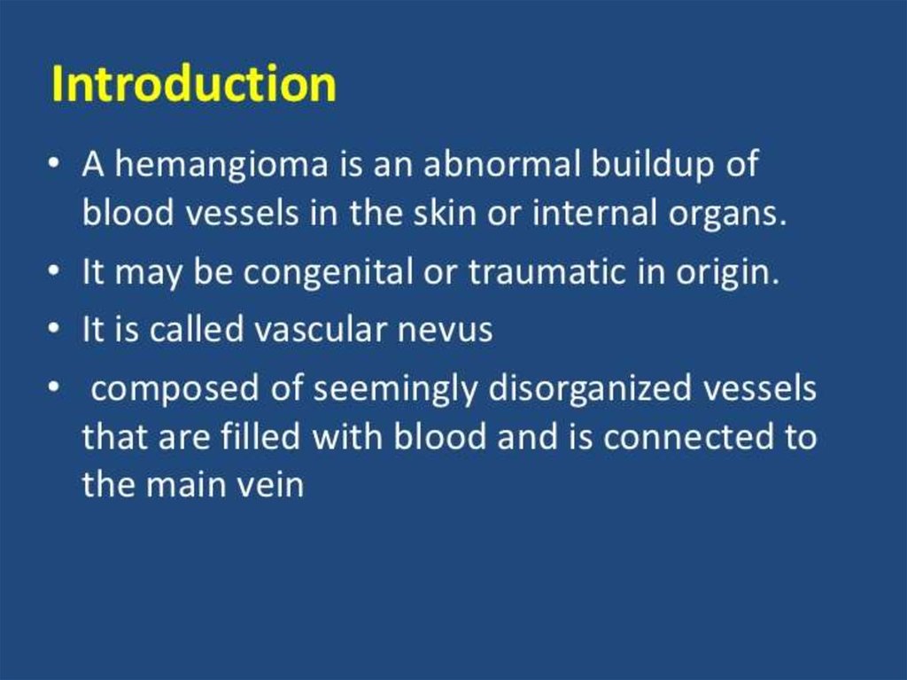
• Introduction• The main part
• Reference
3.
4.
5. Simple hemangioma
• is usually red or blue-purple color, is locatedon the surface, clearly delineated boundaries,
affects the skin and a few millimeters of
subcutaneous fat layer, usually grows in hand.
Rare hemangioma uneven, slightly protruding
above the skin (usually smooth). One
symptom of hemangiomas is that if you push
on it, it is for a short period of time fades,
then again takes its color.
6. Cavernous hemangioma
– is usually located under the skin, is a limitednodular formation, soft-elastic consistency.
Consists of various cavities - caverns filled with
blood. Look cavernous hemangioma as tumor
formation, on top of the usual skin color,
sometimes bluish. With the growth of the tumor
skin becomes blue-purple color. When pressed on
the hemangioma she falls and thus a bit pale (due
to the outflow of blood). When you cry and cough
hemangioma increases.
7. Combined hemangioma
– usually a combination of surface andsubcutaneous hemangiomas (simple and
cavernous). Detected by the prevalence of one or
the other of the tumor vasculature. Appearance
and consistency, again, depends on its constituent
tissues.
8. Extensive capillary-cavernous hemangioma of the left half of the head with a pronounced exophytic growth
Cavernous hemangioma9. Capillary hemangioma of the child
Hemangioma of the humerus10. Hemangioma arterial (h. Arteriale) - hemangioma, arterial blood vessels from growing type.
Hemangioma arterial (h. Arteriale) hemangioma, arterial blood vesselsfrom growing type.
11. HEMANGIOMA CAN BE
LIVER
KIDNEY
VERTEBRAE
LIPS
12.
13.
• Microscopic structure of capillary hemangioma. The wall ofthe capillaries represented two-three-layer endothelium
(tissue atypia), the cavity often filled with blood.
14.
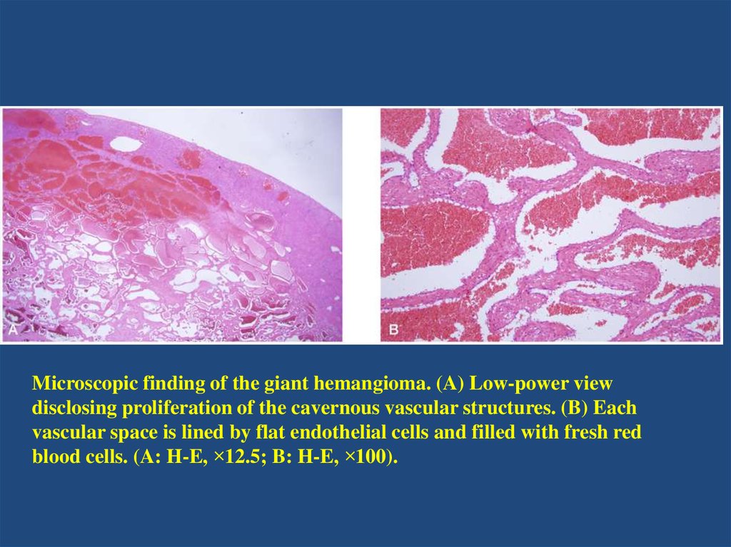
Microscopic finding of the giant hemangioma. (A) Low-power viewdisclosing proliferation of the cavernous vascular structures. (B) Each
vascular space is lined by flat endothelial cells and filled with fresh red
blood cells. (A: H-E, ×12.5; B: H-E, ×100).
15. Cavernous Hemangioma of the Maxillary andEthmoid Sinus
16.
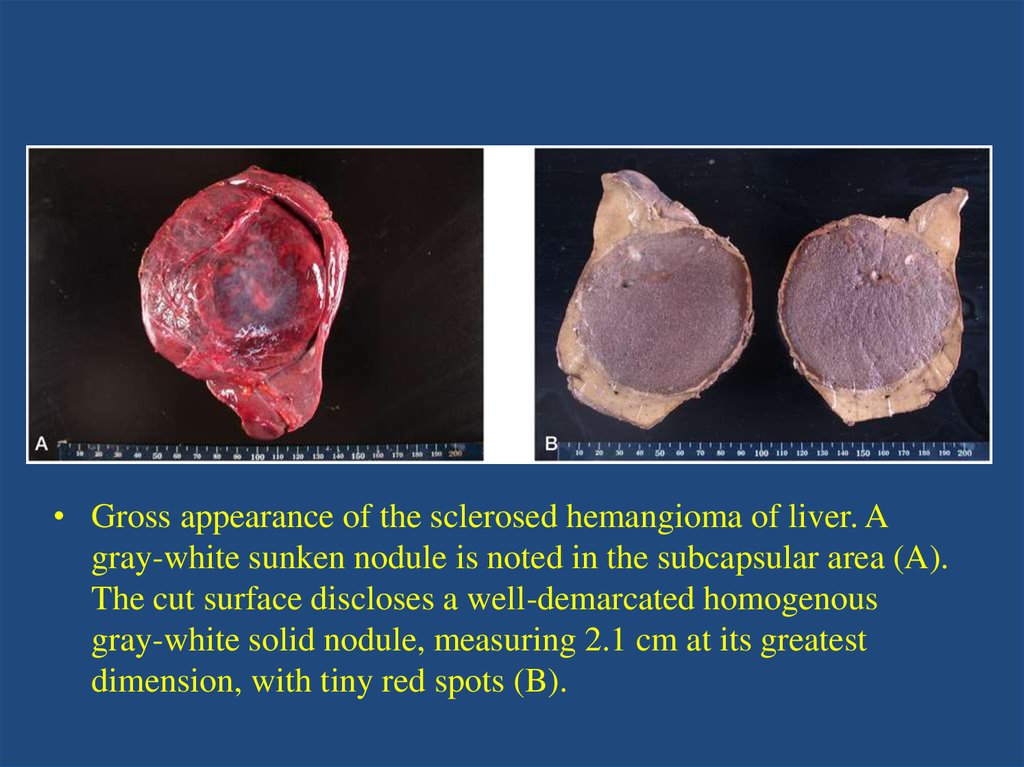
• Gross appearance of the sclerosed hemangioma of liver. Agray-white sunken nodule is noted in the subcapsular area (A).
The cut surface discloses a well-demarcated homogenous
gray-white solid nodule, measuring 2.1 cm at its greatest
dimension, with tiny red spots (B).
17.
18.
19. REFERENCE
1.2.
3.
4.
5.
http://www.ayzdorov.ru/lechenie_gemangioma_chto.php
http://razvitie-krohi.ru/zdorove-rebenka/vse-o-gemangiomah-unovorozhdennyih-detey.html
https://ru.wikipedia.org/wiki/%D0%93%D0%B5%D0%BC%D0%B0
%D0%BD%D0%B3%D0%B8%D0%BE%D0%BC%D0%B0
https://www.google.kz/search?q=Microscopic+structure+of+capill
ary+hemangioma&es_sm=93&source=lnms&tbm=isch&sa=X&ei=
xrA2VaOcF-ehyAPooLIBA&ved=0CAcQ_AUoAQ&biw=1366&bih=643#tbm=isch&q=Mi
croscopic+structure+hemangioma
http://synapse.koreamed.org/DOIx.php?id=10.3350/kjhep.2010.1
6.4.410&vmode=PUBREADER#!po=91.6667